University Protein 4TVO Assignment: Structure, Function, and Analysis

Verified

Added on  2022/08/26

|5
|757
|26
Homework Assignment
AI Summary
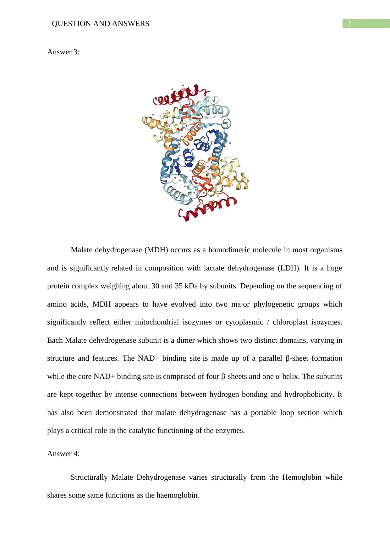
This assignment delves into the analysis of protein 4TVO, specifically Malate Dehydrogenase (MDH) from Mycobacterium tuberculosis. The solution begins by identifying the protein and its function, which involves inter-conversion between malate and oxaloacetate, playing a crucial role in oxidative mechanisms. The structural features of MDH, including its homodimeric nature, the presence of NAD+ binding sites, and the role of hydrogen bonds in subunit interactions, are thoroughly described. The assignment further compares and contrasts the structure and function of MDH with hemoglobin, highlighting both similarities and differences in their structural components and roles as transport proteins. Finally, the solution addresses the protein's thermal stability, correlating it with the hydrogen bonding within the protein's structure and the influence of Michaelis constants on temperature alterations.
Document Page
Running Head: QUESTION AND ANSWERS
Protein Structure Determination and Answering
Name of the Student:
Name of the University:
Author Note:
tabler-icon-diamond-filled.svg

Paraphrase This Document

Need a fresh take? Get an instant paraphrase of this document with our AI Paraphraser
Document Page
1QUESTION AND ANSWERS
Answer 1:
The protein ID: 4TVO describes Malate Dehydrogenase (MDH) in the organism
Mycobacterium Tuberculosis (Rcsb.org, 2020).
Answer 2:
MDH is a fundamental protein for the oxidative mechanisms and an enzyme that is
widely spread throughout living organisms. Using NAD+ or NADP+ as electron acceptor, it
induces the inter-conversion between malate and oxaloacetate (Takahashi-Íñiguez et al.
2016). Amazingly there are few studies in prokaryotes about this enzyme was noted this
enzyme has been studied extensively in eukaryotes only. In addition, the sequencing and
activity of this enzyme are intensively controlled because of the significance of its
mechanism. Through information regarding its activity, cofactor attachment power, substrate
sensitivity, and thermostability, this structural knowledge can help to make direct alterations
to enhance the enzyme reaction kinetics. This reaction plays a significant role within the
mitochondrial matrix in the malate / aspartate transfer throughout the mitochondrial
membrane, and even in the tricarboxylic acid cycle.
Document Page
2QUESTION AND ANSWERS
Answer 3:
Malate dehydrogenase (MDH) occurs as a homodimeric molecule in most organisms
and is significantly related in composition with lactate dehydrogenase (LDH). It is a huge
protein complex weighing about 30 and 35 kDa by subunits. Depending on the sequencing of
amino acids, MDH appears to have evolved into two major phylogenetic groups which
significantly reflect either mitochondrial isozymes or cytoplasmic / chloroplast isozymes.
Each Malate dehydrogenase subunit is a dimer which shows two distinct domains, varying in
structure and features. The NAD+ binding site is made up of a parallel β-sheet formation
while the core NAD+ binding site is comprised of four β-sheets and one α-helix. The subunits
are kept together by intense connections between hydrogen bonding and hydrophobicity. It
has also been demonstrated that malate dehydrogenase has a portable loop section which
plays a critical role in the catalytic functioning of the enzymes.
Answer 4:
Structurally Malate Dehydrogenase varies structurally from the Hemoglobin while
shares some same functions as the haemoglobin.
Document Page
3QUESTION AND ANSWERS
The structure of Malate Dehydrogenase holds four units of β-sheets and one unit of α-helix,
whereas the structure of haemoglobin have two units of α-helix and two units of β-sheets.
However, the proteins are structurally different from each other but functions as a transport
protein. The haemoglobin functions in transfer of oxygen for respiration whereas Malate
Dehydrogenase acts in electron acceptor molecule for the inter-conversion of malate and
oxaloacetate. Thus, Malate Dehydrogenase shows similarity as well as differences with the
haemoglobin protein.
Answer 5:
Vmax, activation energy, and values of thermal stabilization of the total enzyme or of
isozymes alone were not properly correlated with alterations in temperature during the life
cycle of MDH. In comparison, the Michaelis constants (Kma) values was directly correlated
(Minarik et al. 2002).
The sheets are tightly held together by the hydrogen bonds. Thus, enhanced bonding
with hydrogen can bring up the most specific reason for thermal stability in the protein. In
two-thirds of the families tested, the amount of ion sets was also discovered to rise with
thermal stability; however, their level of addition was only about one-sixth that of the protein
atoms for internal hydrogen bonds (Shevkunov., 2015).
tabler-icon-diamond-filled.svg

Paraphrase This Document

Need a fresh take? Get an instant paraphrase of this document with our AI Paraphraser
Document Page
4QUESTION AND ANSWERS
References
Minarik, P., Tomaskova, N., Kollarova, M. and Antalik, M., 2002. Malate dehydrogenases-
structure and function. General physiology and biophysics, 21(3), pp.257-266.
Shevkunov, S.V., 2015. Structure and stability of hydrogen bonds under conditions of heating
in nanopores High Temperature, 53(2), pp.259-271.
Takahashi-Íñiguez, T., Aburto-Rodríguez, N., Vilchis-González, A.L. and Flores, M.E.,
2016. Function, kinetic properties, crystallization, and regulation of microbial malate
dehydrogenase. Journal of Zhejiang University-SCIENCE B, 17(4), pp.247-261.
www.rcsb.org (2020). RCSB PDB - 4TVO: Structure of Malate Dehydrogenase from
Mycobacterium tuberculosis. [online] Rcsb.org. Available at:
https://www.rcsb.org/3d-view/4TVO/1 [Accessed 14 Jan. 2020].
chevron_up_icon
1 out of 5
circle_padding
hide_on_mobile
zoom_out_icon
[object Object]