Protein Technology Report: GLUT1 Protein and Proteomic Methods
VerifiedAdded on 2022/10/10
|21
|3678
|499
Report
AI Summary
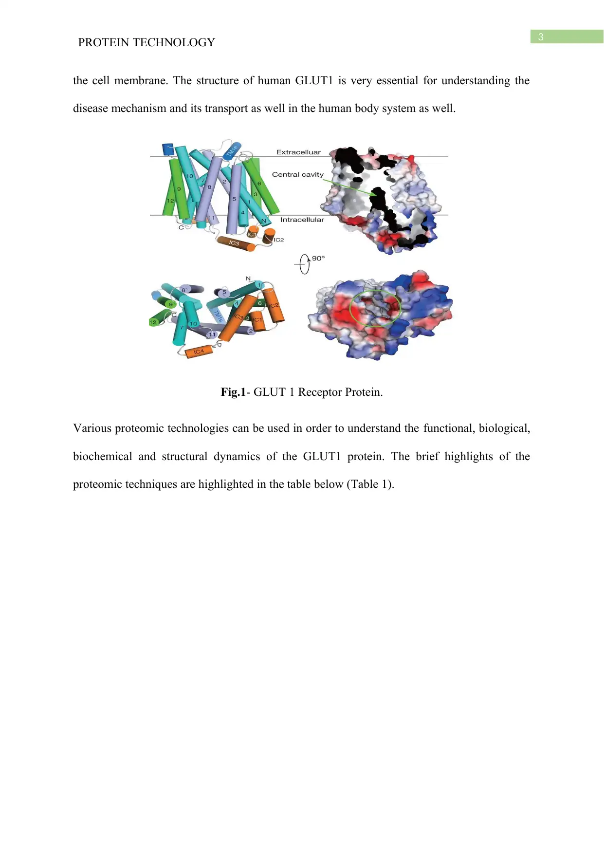
This report delves into the analysis of the Glucose Transporter 1 (GLUT1) protein, a crucial transmembrane protein responsible for glucose transport. The report begins with an overview of GLUT1's structure, function, and its significance in maintaining blood glucose levels and its role in various diseases, including cancer. The core of the report focuses on five key proteomic techniques employed to study GLUT1: X-ray crystallography, Bioluminescence Resonance Energy Transfer (BRET), Enzyme-Linked Immuno sorbent Assay (ELISA), High Performance Liquid Chromatography-Mass Spectroscopy (HPLC-MS), and Stable isotope labelling by amino acids in cell culture (SILAC). Each technique is described in detail, including its principles, applications, and advantages in revealing the molecular and chemical structure of the protein. The report also includes relevant figures and tables, along with peer-reviewed literature references, to support the discussion and provide a comprehensive understanding of the proteomic techniques used to analyze the GLUT1 protein.

Running head: PROTEIN TECHNOLOGY
Protein Technology
Name of the Student
Name of the University
Author Note
Protein Technology
Name of the Student
Name of the University
Author Note
Paraphrase This Document
Need a fresh take? Get an instant paraphrase of this document with our AI Paraphraser

1
PROTEIN TECHNOLOGY
Abstract
The report discusses about the protein identification techniques named as proteomic
techniques. The report addresses the structure of GLUT 1 protein that is one of the crucial
protein for transporting the glucose molecule inside the body. It is a transmembrane protein
present in almost each and every cell membrane. The report discusses about different
proteomic techniques in detecting the molecular and chemical structure of the protein. In this
report, five different techniques of protein detection is discussed. They are mainly the X-ray
crystallography, Bioluminescence Resonance Energy Transfer (BRET), Enzyme-Linked
Immuno sorbent Assay (ELISA), High Performance Liquid Chromatography- Mass
Spectroscopy, Stable isotope labelling by amino acids in cell culture (SILAC). However,
while discussing the principles of these techniques, it is observed that, a few principles are
correlated with each other.

2
PROTEIN TECHNOLOGY
Introduction
The protein molecule that is discussed in this report is the Glucose Transporter 1 or
GLUT 1 which is one of the most common transporter protein present involved in the
transportation of the glucose in a unidirectional manner. The protein is coded by the gene
named SLC2A1 and it is involved in the uptake of glucose molecule in order to maintain the
blood glucose level at the level of almost 5mMol concentration. In case of the endothelial
cells of the body, it is reported that the GLUT1 has a crucial role in maintain the proper
supply of glucose molecule across the cell membrane to the cells of brain and other organs as
well. The presence of GLUT 1 in the cell membrane of the different portion of the body.
According to the study of Deng et al. (2014), it is reported that the nonsense mutation of the
GLUT1 gene can cause alteration of the glucose transportation across the cell membrane.
Due to the absence of GLUT1 transporter in the cell membrane various disease such as
GLUT1 deficiency syndrome or De Vivo syndrome may take place. Apart from that, it is also
reported that, due to the malformation or absence of the GLUT 1 in the cell membrane,
disease such as early onset of seizures, lack of microcephaly development may happen. In
another study it is reported that, in case of cancer cells, in the body, the more amount of
glucose is required as the cells are less efficient in producing energy by the anaerobic
glycolysis that is referred to as Warburg effect4. The over expression of GLUT 1 is reported
in case of cancer patients and thus it is reported that GLUT1 expression is one of the
important factors in tumorigenesis. GLUT1 is the member of sugar porter subfamily of the
major facilitator superfamily (MFS) that is one of the largest and most ubiquitous secondary
transporter super families. According to the study of Yan (2016), the understanding of the
molecular structure of the GLUT1 protein is very crucial as this uniporter protein channel is
associated with the translocation of the glucose along with its concentration gradient across
⊘ This is a preview!⊘
Do you want full access?
Subscribe today to unlock all pages.

Trusted by 1+ million students worldwide

3
PROTEIN TECHNOLOGY
the cell membrane. The structure of human GLUT1 is very essential for understanding the
disease mechanism and its transport as well in the human body system as well.
Fig.1- GLUT 1 Receptor Protein.
Various proteomic technologies can be used in order to understand the functional, biological,
biochemical and structural dynamics of the GLUT1 protein. The brief highlights of the
proteomic techniques are highlighted in the table below (Table 1).
Paraphrase This Document
Need a fresh take? Get an instant paraphrase of this document with our AI Paraphraser

4
PROTEIN TECHNOLOGY
Proteomic technique Brief
description
Citation
1 X-ray crystallography (X-ray diffraction) It is the most
broadly used
proteomic
method to
inspect the
molecular and
atomic structure
of protein and
other types of
biological
constituents
such as the
GLUT1 protein
that is described
above. The
process includes
crystallization
of the protein
sample in high
concentration
Crystal structure of a
glucose/H+ symporter
and its mechanism of
action, Proceeding of
the National
Academy of Sciences
of the United States
by Iancu et al. (2013).
In meso in situ serial
X-ray crystallography
of soluble and
membrane proteins.
Acta
Crystallographica
Section D: Biological
Crystallography by
Huang et al. (2015).

5
PROTEIN TECHNOLOGY
and exposure to
high energy x-
ray beam is also
required along
with this
technique. The
electronic
waves of the x-
rays are
disseminated by
the electrons of
the sample
constituent and
the diversion
array of x-ray is
recognized.
2 Bioluminescence Resonance Energy
Transfer ( BRET)
Bioluminescene
Resonance
Energy Transfer
(BRET) is
another
essential
progressive cell
based assay
Bioluminescence
resonance energy
transfer to detect
protein-protein
interactions in live
cells. In Protein-
⊘ This is a preview!⊘
Do you want full access?
Subscribe today to unlock all pages.

Trusted by 1+ million students worldwide

6
PROTEIN TECHNOLOGY
mechanism that
is used in the
research
procedure of the
receptor
proteins and
also this
technique is
used for the
plotting of the
signal
transduction
paths in the
molecular level.
This BRET
method is
completely
relied on the
Resonance
Energy Transfer
(RET) between
a fluorescent
acceptor moiety
and
bioluminescent
Protein Interactions
By Brown, Blumer, &
Hepler (2015).
Humana Press, New
York, NY.
Fundamentals of
protein interaction
network mapping by
Snider et al. (2015).
Molecular systems
biology
Paraphrase This Document
Need a fresh take? Get an instant paraphrase of this document with our AI Paraphraser

7
PROTEIN TECHNOLOGY
donor moiety.
3 Enzyme-Linked Immuno sorbent Assay
(ELISA)
Enzyme-Linked
Immuno sorbent
Assay (ELISA)
denotes to a
plate based
assay system
that is usually
used for the
revealing and
quantification
of the
constituents
such as
proteins,
antibodies
peptides, and
hormones and
being a
transmembrane
protein, the
GLUT1 can
certainly be
identified by
A short history,
principles, and types
of ELISA, and our
laboratory experience
with peptide/protein
analyses using
ELISA. Peptides by
Aydin (2015).
Peptides
Plasmonic-ELISA:
expanding horizons
by Satija et al. (2016).
Rsc Advances.
ELISA in the
multiplex era:
potentials and pitfalls
by Tighe et al.
(2015).
PROTEOMICS–
Clinical

8
PROTEIN TECHNOLOGY
using this
procedure. For
identifying the
GLUT1 protein,
usually
sandwich
ELISA method
is performed by
using the
ELISA kit. The
method is
generally
executed by
using a 96-well
or 384 well
ELISA kit that
can attach with
antibodies and
the proteins in a
passive method.
Applications
4 High Performance Liquid Chromatography-
Mass Spectroscopy
High
Performance
Liquid
Chromatograph
Separation
techniques:
chromatography by
⊘ This is a preview!⊘
Do you want full access?
Subscribe today to unlock all pages.

Trusted by 1+ million students worldwide

9
PROTEIN TECHNOLOGY
y- Mass
Spectroscopy is
also enormously
delicate
proteomic
technique which
is usually used
on protein
samples. This
method
comprises of a
collective
application of a
high liquid
chromatography
and mass
spectrometry
techniques,
improving the
efficiency and
the sensitivity
of the method.
High
Performance
Liquid
Coskun (2016).
Northern clinics of
Istanbul
High-performance
liquid
chromatography of
peptides and proteins:
separation, analysis,
and conformation by
Mant & Hodges
(2017). CRC press
Quantitation of the
immunodominant 33-
mer peptide from α-
gliadin in wheat
flours by liquid
chromatography
tandem mass
spectrometry by
Schalk et al. (2017).
Paraphrase This Document
Need a fresh take? Get an instant paraphrase of this document with our AI Paraphraser

10
PROTEIN TECHNOLOGY
chromatography
can segregate
the various
units of protein
fragment in a
blend and mass
spectrometry
can figure out
the mechanical
dynamics of the
separated
constituent of
the protein
blend.
Scientific reports
5 Stable isotope labelling by amino acids in
cell culture ( SILAC)
This proteomic
procedure is
used to identify
the abundance
of sample
solution of
biological
macromolecules
such as the
GLUT1 or other
Principles of protein
labeling techniques.
In Proteomic
Profiling by
Obermaier, Griebel,
& Westermeier
(2015). Humana
Press, New York,

11
PROTEIN TECHNOLOGY
protein. In this
technique, a
radioactive
molecule is
used to identify
the protein in
the solution of
sample. This
technique is one
of the most
usually used
technique by
the researchers
for quantitative
proteomics.
This technique
can also be used
to distinguish
among the
protein mixture.
NY.
SILAC for biomarker
discovery by Dittmar,
& Selbach (2015).
PROTEOMICS–
Clinical
Applications
Quantitative
proteomics using
SILAC: Principles,
applications, and
developments by
Chen et al. (2015).
Proteomics
A brief description of different proteomic technologies
X-ray crystallography (X-ray diffraction)
It is the most widely used proteomic technique to examine a molecular and atomic structure
of protein and other types of biological components such as the GLUT1 protein that is
described above. The procedure includes crystallization of the sample in high concentration
⊘ This is a preview!⊘
Do you want full access?
Subscribe today to unlock all pages.

Trusted by 1+ million students worldwide
1 out of 21

Your All-in-One AI-Powered Toolkit for Academic Success.
+13062052269
info@desklib.com
Available 24*7 on WhatsApp / Email
Unlock your academic potential
Copyright © 2020–2025 A2Z Services. All Rights Reserved. Developed and managed by ZUCOL.