DN7 Radiography Assignment: Chemicals, Resources, Equipment, and Uses

Verified

Added on  2021/01/01

|5
|994
|412
Homework Assignment
AI Summary
This document presents a detailed solution to a radiography assignment, addressing key aspects of the field. The assignment covers the chemicals used in developing radiographs, including hydroquinone and sodium bicarbonate, along with their actions and associated precautions. It also explores the resources utilized for image acquisition, such as holders, films, receptors, mounting sheets, and software. Furthermore, the document outlines the equipment employed, including intra-oral and extra-oral x-ray machines, computer programs, developers, and image receptors, detailing their specific uses. Finally, it emphasizes crucial safety considerations and precautions to be taken during dental imaging, particularly focusing on radiation exposure risks and the measures to mitigate them, such as the use of lead bibs and thyroid collars, especially for vulnerable patients like pregnant women. The assignment provides a comprehensive overview of radiographic processes and safety protocols.
Document Page
DN7
tabler-icon-diamond-filled.svg

Paraphrase This Document

Need a fresh take? Get an instant paraphrase of this document with our AI Paraphraser
Document Page
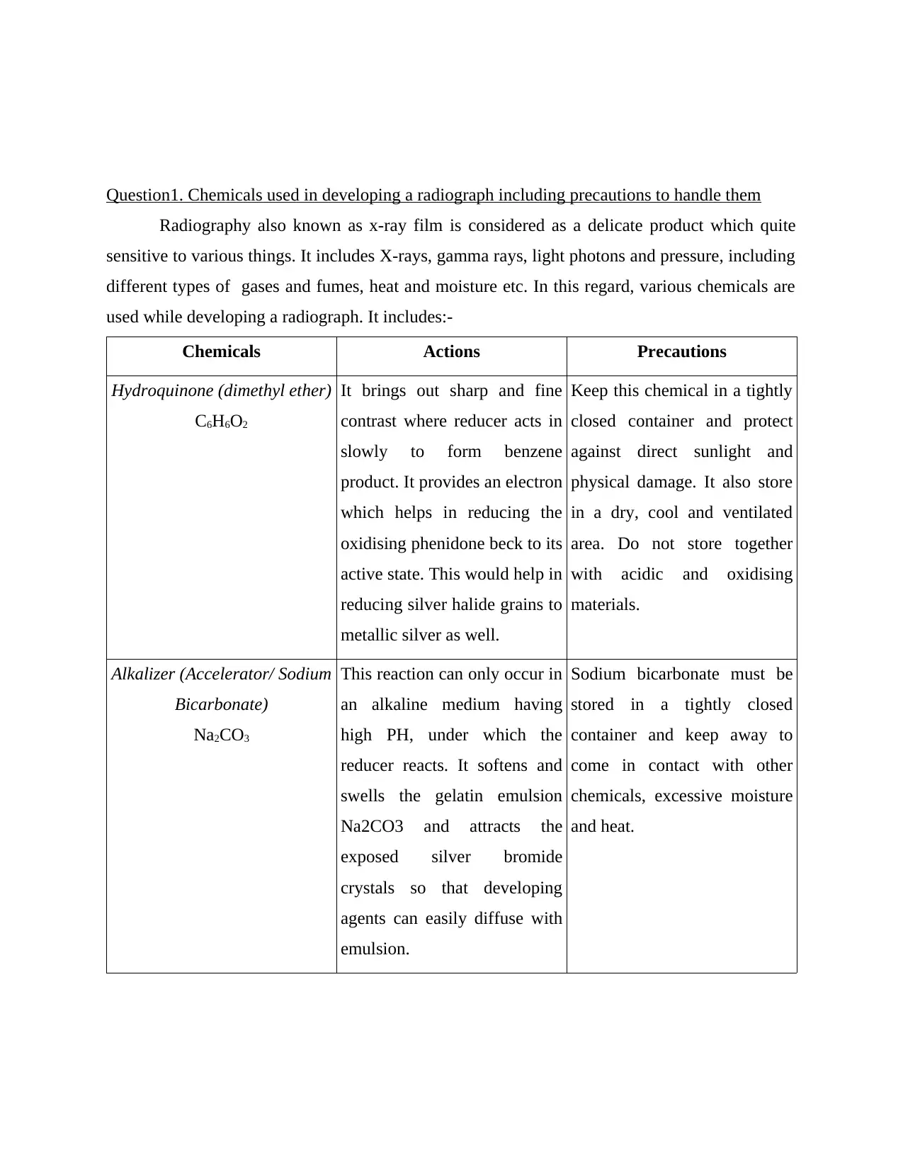
Question1. Chemicals used in developing a radiograph including precautions to handle them
Radiography also known as x-ray film is considered as a delicate product which quite
sensitive to various things. It includes X-rays, gamma rays, light photons and pressure, including
different types of gases and fumes, heat and moisture etc. In this regard, various chemicals are
used while developing a radiograph. It includes:-
Chemicals Actions Precautions
Hydroquinone (dimethyl ether)
C6H6O2
It brings out sharp and fine
contrast where reducer acts in
slowly to form benzene
product. It provides an electron
which helps in reducing the
oxidising phenidone beck to its
active state. This would help in
reducing silver halide grains to
metallic silver as well.
Keep this chemical in a tightly
closed container and protect
against direct sunlight and
physical damage. It also store
in a dry, cool and ventilated
area. Do not store together
with acidic and oxidising
materials.
Alkalizer (Accelerator/ Sodium
Bicarbonate)
Na2CO3
This reaction can only occur in
an alkaline medium having
high PH, under which the
reducer reacts. It softens and
swells the gelatin emulsion
Na2CO3 and attracts the
exposed silver bromide
crystals so that developing
agents can easily diffuse with
emulsion.
Sodium bicarbonate must be
stored in a tightly closed
container and keep away to
come in contact with other
chemicals, excessive moisture
and heat.
Document Page
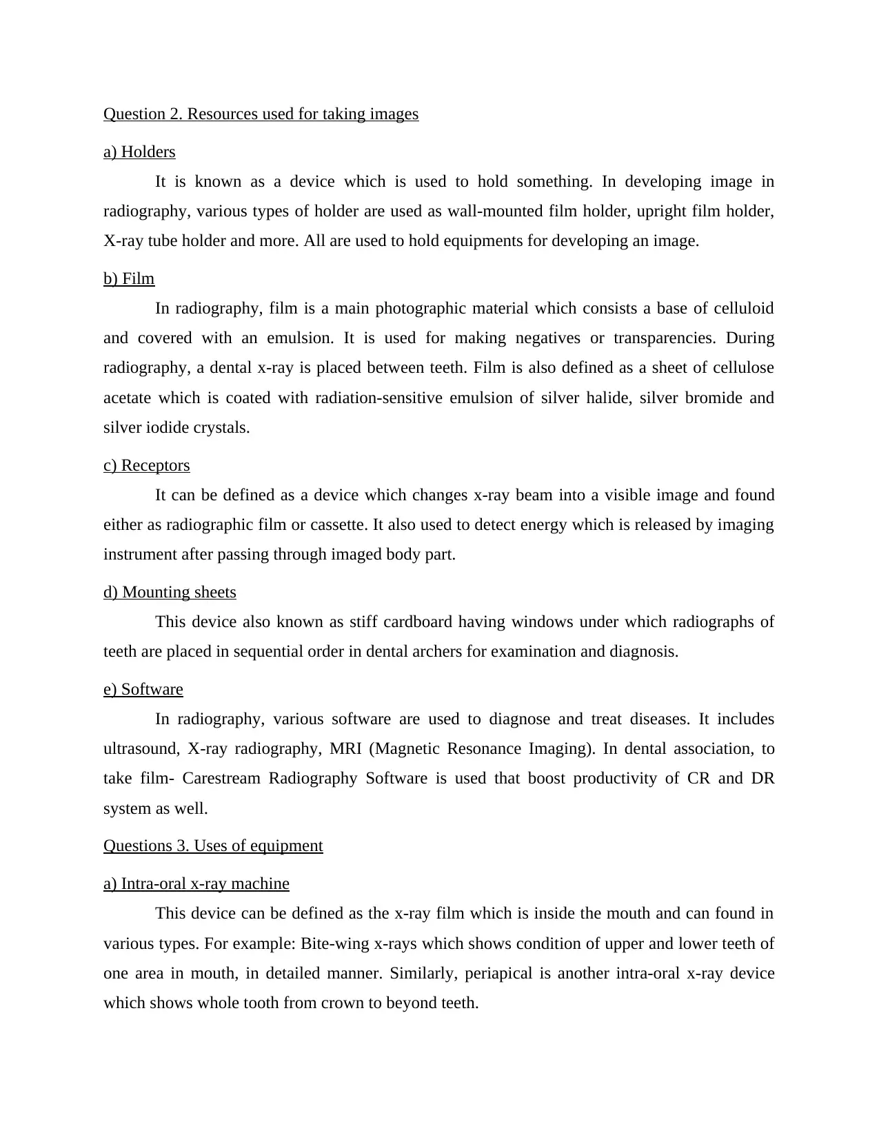
Question 2. Resources used for taking images
a) Holders
It is known as a device which is used to hold something. In developing image in
radiography, various types of holder are used as wall-mounted film holder, upright film holder,
X-ray tube holder and more. All are used to hold equipments for developing an image.
b) Film
In radiography, film is a main photographic material which consists a base of celluloid
and covered with an emulsion. It is used for making negatives or transparencies. During
radiography, a dental x-ray is placed between teeth. Film is also defined as a sheet of cellulose
acetate which is coated with radiation-sensitive emulsion of silver halide, silver bromide and
silver iodide crystals.
c) Receptors
It can be defined as a device which changes x-ray beam into a visible image and found
either as radiographic film or cassette. It also used to detect energy which is released by imaging
instrument after passing through imaged body part.
d) Mounting sheets
This device also known as stiff cardboard having windows under which radiographs of
teeth are placed in sequential order in dental archers for examination and diagnosis.
e) Software
In radiography, various software are used to diagnose and treat diseases. It includes
ultrasound, X-ray radiography, MRI (Magnetic Resonance Imaging). In dental association, to
take film- Carestream Radiography Software is used that boost productivity of CR and DR
system as well.
Questions 3. Uses of equipment
a) Intra-oral x-ray machine
This device can be defined as the x-ray film which is inside the mouth and can found in
various types. For example: Bite-wing x-rays which shows condition of upper and lower teeth of
one area in mouth, in detailed manner. Similarly, periapical is another intra-oral x-ray device
which shows whole tooth from crown to beyond teeth.
Document Page
b) Extra-oral x-ray machine
This device is used to detect dental issues within jaw and skull. It is also found in
different types like Panormic x-rays, Tomograms, sialogram and more. Panormic x-rays is used
to show entire area of mouth while cephalometric for head.
c) Computer Programme
It includes CDR (Computed Dental Radiography) technology used for capturing a
computerised image of teeth and gums. It intakes near about 90% less radiations as compared to
conventional x-rays film.
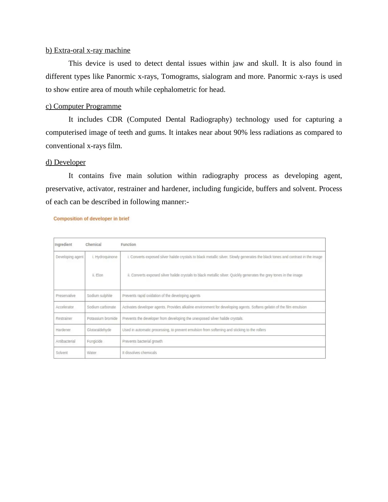
d) Developer
It contains five main solution within radiography process as developing agent,
preservative, activator, restrainer and hardener, including fungicide, buffers and solvent. Process
of each can be described in following manner:-
tabler-icon-diamond-filled.svg

Paraphrase This Document

Need a fresh take? Get an instant paraphrase of this document with our AI Paraphraser
Document Page
e) Image receptors
It is described as a medium which covers X-rays beams into a visible image. In dental
association, image receptors includes film, electronic sensors used in computed tomography and
digital imaging, film-screen combinations, etc.
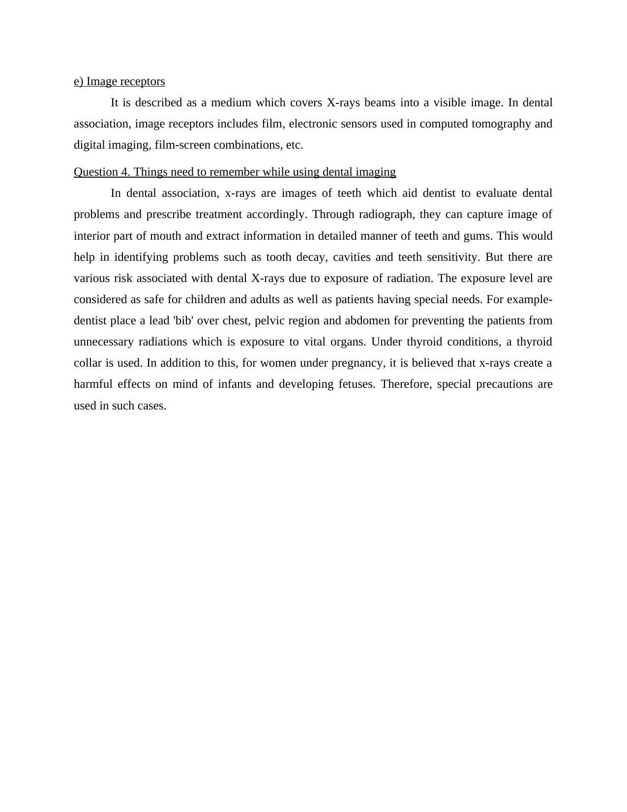
Question 4. Things need to remember while using dental imaging
In dental association, x-rays are images of teeth which aid dentist to evaluate dental
problems and prescribe treatment accordingly. Through radiograph, they can capture image of
interior part of mouth and extract information in detailed manner of teeth and gums. This would
help in identifying problems such as tooth decay, cavities and teeth sensitivity. But there are
various risk associated with dental X-rays due to exposure of radiation. The exposure level are
considered as safe for children and adults as well as patients having special needs. For example-
dentist place a lead 'bib' over chest, pelvic region and abdomen for preventing the patients from
unnecessary radiations which is exposure to vital organs. Under thyroid conditions, a thyroid
collar is used. In addition to this, for women under pregnancy, it is believed that x-rays create a
harmful effects on mind of infants and developing fetuses. Therefore, special precautions are
used in such cases.
chevron_up_icon
1 out of 5
circle_padding
hide_on_mobile
zoom_out_icon
[object Object]