Radiology Report: Analysis of Radiological Tests for Mr. Tomy's Injury

Verified

Added on  2023/01/18

|5
|1154
|24
Report
AI Summary
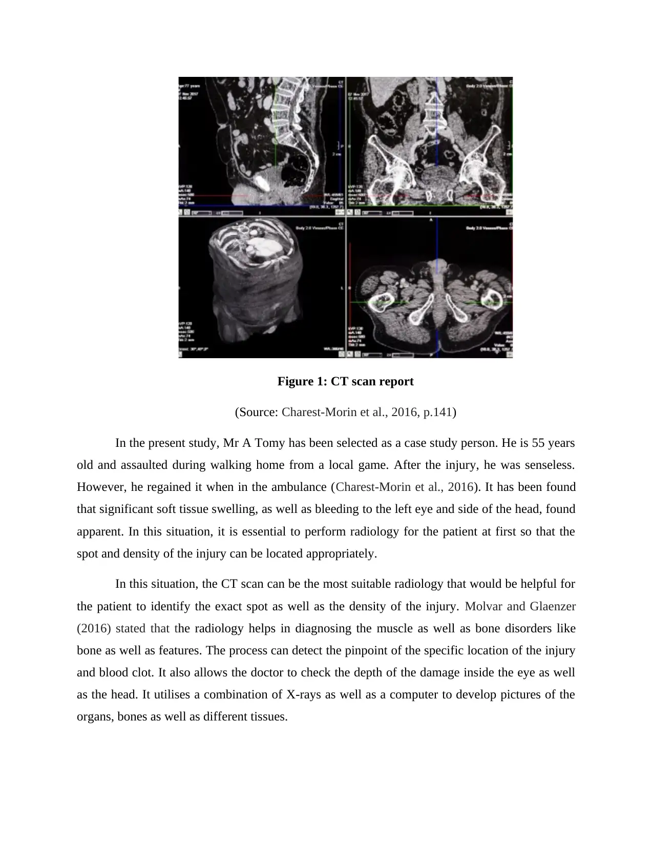
This report presents a radiology case study of Mr. A. Tomy, a 55-year-old male who sustained a head injury following an assault. The report details the clinical indications, which include significant soft tissue swelling and bleeding to the left eye and side of the head, along with a suspicion of a closed head injury with coup and contrecoup injuries. The study focuses on the suitability and methodology of a CT scan, explaining how the radiological test was conducted and discussing the main radiological findings using appropriate terminology. It highlights the diagnostic utility of CT scans in identifying the extent and location of injuries, including bone injuries and blood clots, and its role in guiding treatment plans. The report references relevant literature, including works by Charest-Morin et al. (2016) and Molvar & Glaenzer (2016), to support the analysis. The report emphasizes the importance of appropriate radiology selection for effective patient treatment and recovery. It also includes a discussion of the imaging process and how it is utilized to develop pictures of organs, bones and different tissues.
Document Page
RADIOLOGY REQUEST AND REPORT
Student Name
University Name
Author note
tabler-icon-diamond-filled.svg

Secure Best Marks with AI Grader

Need help grading? Try our AI Grader for instant feedback on your assignments.
Document Page
Radiology reports immediately after the exam of CBCT, which is before surgery. It helps
to reduce liability that may present to the implanting clinician. On the other hand, formal
radiology reports accountability may be gained from several sources. It is preferably from the
proper qualified as well as board certified maxillofacial radiologist. There are several types of
radiology services that are present in the current day's healthcare plan (de Baere, 2015). CT scan
or computed axial tomography is one of the types that deal with a non-invasive medical test. It
utilises special x-ray equipment with different types as well as sophisticated systems to generate
several images or pictures of the inside of the body.
On the other hand, CT lung cancer screening deals with low dose CT scan of the lungs
that are used to identify lung cancer. Also, fluoroscopy deals with a medical test where a
constant x-ray beam is passed across the body. However, magnetic resonance imagining scan is
essential for the procedure where it is required to use a non-invasive medical test or examination
that utilises a large magnet as well as a computer taking an image of the inside body. Ultrasound
named as sonography we considered as a medical test that uses high-frequency sound waves. A
computer creates images of the blood vessels, tissues as well as organs (Anzidei et al., 2015).
The virtual imaging process utilises CT scanning as well as advanced computer hardware to
generate 2d as well as 3d images of the colon, which can be viewed on the video screen. X-rays
is one of the most popular techniques that uses invisible electromagnetic energy beams to
generate the images of internal tissues, bones as well as organs on a film.
Document Page
Figure 1: CT scan report
(Source: Charest-Morin et al., 2016, p.141)
In the present study, Mr A Tomy has been selected as a case study person. He is 55 years
old and assaulted during walking home from a local game. After the injury, he was senseless.
However, he regained it when in the ambulance (Charest-Morin et al., 2016). It has been found
that significant soft tissue swelling, as well as bleeding to the left eye and side of the head, found
apparent. In this situation, it is essential to perform radiology for the patient at first so that the
spot and density of the injury can be located appropriately.
In this situation, the CT scan can be the most suitable radiology that would be helpful for
the patient to identify the exact spot as well as the density of the injury. Molvar and Glaenzer
(2016) stated that the radiology helps in diagnosing the muscle as well as bone disorders like
bone as well as features. The process can detect the pinpoint of the specific location of the injury
and blood clot. It also allows the doctor to check the depth of the damage inside the eye as well
as the head. It utilises a combination of X-rays as well as a computer to develop pictures of the
organs, bones as well as different tissues.
Document Page
The CT scan process utilises a narrow x-ray beam that can circle the parts of the body. It
also provides a series of images of the body. The procedure is generally developed to generate a
detailed picture of the organs and blood vessels (Cheshire et al., 2016). For instance, the surgeon
may utilise the scan for preparing an operation. CT scan can be useful to detect the bone injury
as well as joint problems such as complicated bone features as well as tumours. In addition, they
can assist in locating the injuries as well as blooding like it is caused by accident. Doctors use for
guiding the treatment plans as well as processes like biopsies and surgeries as well as radiation
therapy (Cheshire et al., 2018). In addition, it helps to compare finding out whether specific
treatments are working correctly. The scans of the injury places over time can show if the
responding chemotherapy or radiation.
With the help of CT scan, the solid substances such as bones injury of Mr Tomy can be
easily seen. However, the soft tissues do not show up. To assist in the process, it is required for a
special dye named as contrast material (Molvar & Glaenzer, 2016). The x-rays can be blocked
as well as appear on the scan. It helps to highlight the blood vessel, organs as well as different
structures. Hence, it is essential to select the best possible and suitable radiology for the patient
so that proper treatment can be performed and the patient recovers quickly.
tabler-icon-diamond-filled.svg

Secure Best Marks with AI Grader

Need help grading? Try our AI Grader for instant feedback on your assignments.
Document Page
References
Anzidei, M., Lucatelli, P., Napoli, A., Jens, S., Saba, L., Cartocci, G., Sedati, P., d'Adamo, A. &
Catalano, C., 2015. CT angiography & magnetic resonance angiography findings after
surgical & interventional radiology treatment of peripheral arterial obstructive
disease. Journal of cardiovascular computed tomography, 9(3), pp.165-182.
Charest-Morin, R., Boriani, S., Fisher, C.G., Patel, S.R., Kawahara, N., Mendel, E., Bettegowda,
C. & Rhines, L.D., 2016. Benign tumours of the spine: has new chemotherapy &
interventional radiology changed the treatment paradigm?. Spine, 41, pp.S178-S185.
Cheshire, S.C., Board, R.E., Lewis, A.R., Gudur, L.D. & Dobson, M.J., 2018. Pembrolizumab-
induced sarcoid-like reactions during treatment of metastatic
melanoma. Radiology, 289(2), pp.564-567.
de Baere, T., Deschamps, F., Tselikas, L., Ducreux, M., Planchard, D., Pearson, E., Berdelou, A.,
Leboulleux, S., Elias, D. & Baudin, E., 2015. GEP-NETS update: Interventional
radiology: role in the treatment of liver metastases from GEP-NETs. European journal of
endocrinology, 172(4), pp.R151-R166.
Molvar, C. & Glaenzer, B., 2016, December. Choledocholithiasis: evaluation, treatment, &
outcomes. In Seminars in interventional radiology (Vol. 33, No. 04, pp. 268-276).
Thieme Medical Publishers.
chevron_up_icon
1 out of 5
circle_padding
hide_on_mobile
zoom_out_icon
logo.png

Your All-in-One AI-Powered Toolkit for Academic Success.

Available 24*7 on WhatsApp / Email

[object Object]