Access to HE (Nursing): Reproduction and Inheritance Assignment
VerifiedAdded on 2022/08/22
|23
|4977
|22
Homework Assignment
AI Summary
This assignment, designed for an Access to HE (Nursing) course, covers the fundamental concepts of human reproduction and inheritance. Task 1 focuses on the human reproductive system, including the structure and function of both male and female reproductive systems, gamete formation, fertilization, embryonic and foetal development, and the birthing process. Students are required to label diagrams, complete tables detailing the function of each component, explain the stages of fertilization, provide an overview of embryonic and foetal development, and create a poster describing the birthing process. The assignment also includes questions related to hormones and the menstrual cycle. The student demonstrates understanding through detailed explanations, diagrams, and a poster, addressing learning outcomes related to understanding the human reproductive system and its processes. The assignment also references sources like Kent (2000), Jones and Lopez (2006), and Mackean (2004) to support the answers provided.
Student name:
Access
Course:
Access to HE (Nursing)
Unit Title: Reproduction and
Inheritance WJG347
Assignment title and number
(link with whole unit assessment,
e.g. 1 of 2)
Assignment:
Task 1 Questions and a poster
Task 2- questions and punnet squares
Task 3 – Short answer questions
Conditions for completion supervised / monitored / independent
Name of assessor Rajeshwari Thammineni/Gabriella Rowley
Internal Verification (name, month)
Mapping to unit
Learning Outcomes/ Assessment
Criteria to be covered in this
assignment (e.g. 1.1, 1.2, 2.1, etc)
LO1: Criteria 1.1, 1.2, 1.3,1.4;
LO2: Criteria 2.1, 2.2, 2.3;
LO3: Criteria 3.1
Grade descriptors to be indicated
in this assignment GD 1,2,5,7
Declaration: I confirm that this assignment is all my own work and that it conforms to the
course policy on plagiarism as stated in the course handbook.
Print name: Learner signature: Date:
Further assessment and grading guidance
Presentation Follow task book instructions. It is recommended that you
reference your sources where appropriate.
Completion of task and
achievement of learning
outcomes
What you have to do: See tasks below
SUBMISSION DEADLINE:
1
Paraphrase This Document
Need a fresh take? Get an instant paraphrase of this document with our AI Paraphraser
Learning Outcome Assessment Criteria
1. Understand the human
reproductive
system.
1.1. Describe the structure and
function of
the male and female reproductive
systems, including gamete formation.
1.2. Explain fertilization.
1.3. Describe human embryonic and
foetal
development.
1.4. Describe the birthing process.
1.5. Outline the menstrual cycle.
Learning Outcome Assessment Criteria
2. Understand meiosis and Mendelian
genetics.
2.1. State Mendel’s laws.
2.2. Describe chromosome behaviour in the
meiotic divisions.
2.3. Predict outcomes of genetic crosses
with Punnett squares for dominant-recessive,
co-dominant, sex-linked and
multi-allelic inheritance patterns.
Learning Outcome Assessment Criteria
3. Understand human inherited
conditions.
3.1. Identify classes of genetic mutations in
humans.
3.2. Explain how an allele associated with a
human genetic condition might confer a
selective advantage.
3.3. Interpret human karyotypes in terms of
the sex of the individual and any
abnormalities in the chromosome pairs.
What this means for this assignment
(Please give guidance to the student to explain what the grade descriptor components
mean within the context of the task in order that s/he may be able to achieve Merits or a
Distinctions – see previous page)
All tasks
To gain a merit or distinction for this assignment, the work should be factually correct (GD2) and
written in full sentences with appropriate use of relevant scientific keywords and referencing (GD5).
The written work should contain evidence of additional reading and research, outside of content
provided in class (GD2).
All drawings should be neat and coloured and/or annotated appropriately and any tables or other
means of presentation completed accurately and neatly (GD 1, 7).
Any pictures drawn or inserted from an external source should be appropriately referenced with a
figure legend (GD5).
2
All seven tasks should be presented in a neat and orderly way with all of the
tasks completed to the best of your ability (GD7).
All three tasks should be presented in a neat and orderly way with all
of the tasks completed to the best of your ability
Grading information Grade Descriptors for this assignment, with components & guidance.
Grade descriptor: 1. Understanding of the subject
Component(s): 1b
For a pass you should: Meet the assessment criteria to achieve the learning outcomes.
For a merit you
should:
Produce work that is generally informed by the major conventions and
practices of human anatomy & physiology
For a distinction you
should:
Produce work that is consistently informed by the major conventions
and practices of human anatomy & physiology.
Additional Guidance
notes
We want to see evidence that you have understood the basic rules of
human anatomy & physiology correctly.
Grade descriptor: 2. Application of knowledge
Component(s): 2ac
For a pass you should: Meet the assessment criteria to achieve the learning outcomes.
For a merit you
should:
Produce work that makes use of relevant facts and ideas with very
good levels of accuracy.
For a distinction you
should:
Produce work that makes use of relevant facts and ideas with excellent
levels of accuracy.
Additional Guidance
notes
We are looking for appropriate reasoning to show that you can make
use of your knowledge.
Grade descriptor: 5. Communication and presentation
Component(s): 5a
For a pass you should: Meet the assessment criteria to achieve the learning outcomes.
For a merit you
should:
Produce work that shows very good command of use of images.
For a distinction you
should:
Produce work that shows excellent command of use of images.
Additional Guidance
notes
We want to see that your diagrams and drawings support your
arguments well and tie in with your written text.
Grade descriptor: 7. Quality
Component(s): 7b
For a pass you should: Meet the assessment criteria to achieve the learning outcomes.
For a merit you
should:
Produce work where arguments and ideas are generally unambiguous
and cogent.
For a distinction you
should:
Produce work where arguments and ideas are consistently
unambiguous and cogent.
Additional Guidance We want to see that you make your answers as clear and unequivocal
3
⊘ This is a preview!⊘
Do you want full access?
Subscribe today to unlock all pages.
Trusted by 1+ million students worldwide
notes as possible.
Task 1
Course Title: Access to HE Diploma:
Unit Title: Reproduction and Inheritance
Task(s) Short answer questions and a poster to address learning outcome 1
Task Title:
1. Labelling structures and completing a table
2. Labelling structures and completing a table
3. Explain stages of fertilisation
4. Write an overview of stages of foetal development
5. A4 poster on the stages of birth
6. Hormones
7. Outline of the menstrual cycle
Date of Internal
Moderation:
Description of Assessment Task (mapped to ACs) Assessmen
t criteria
Task 1: In completing this task, students are required to undertake the following seven
questions:
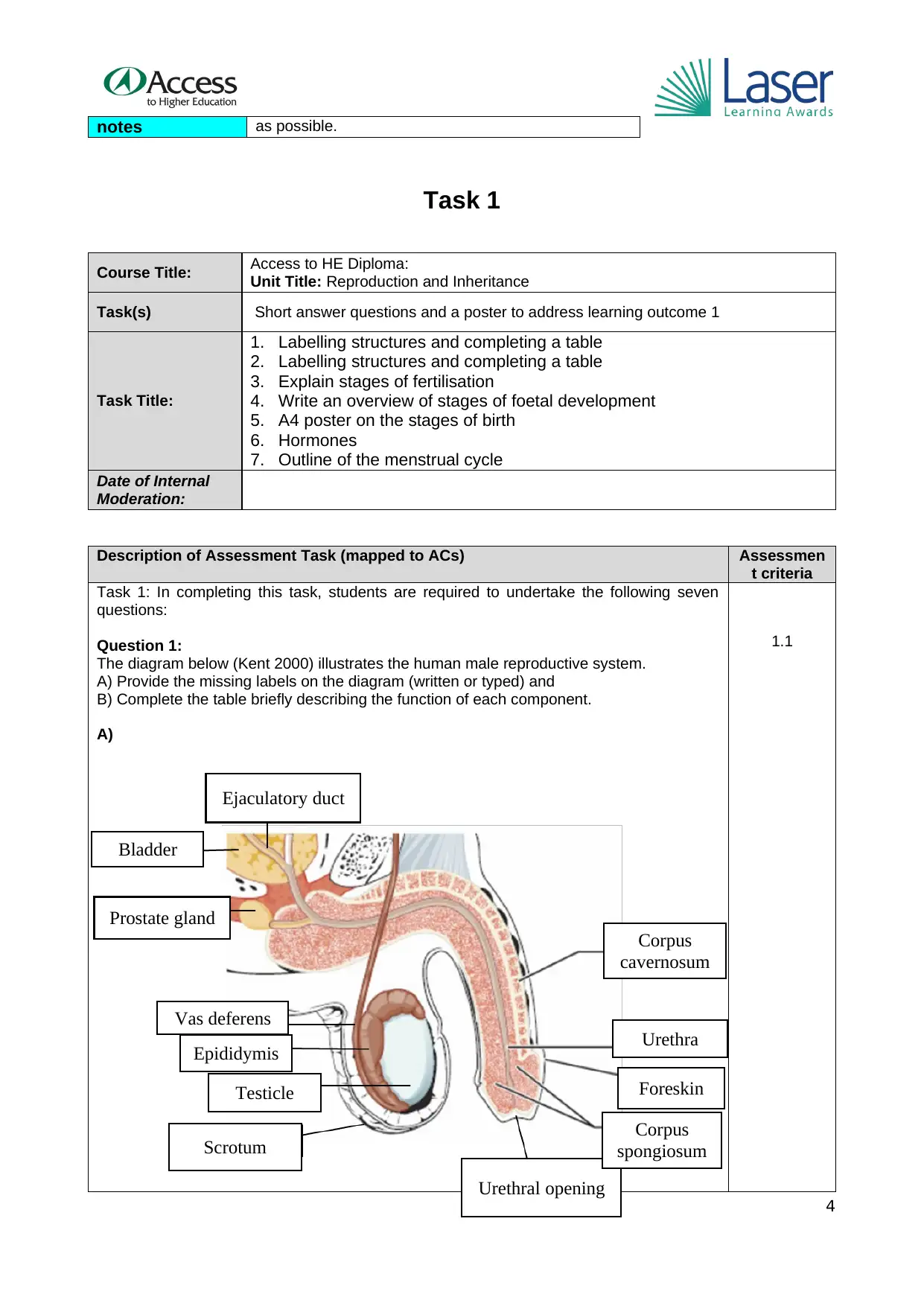
Question 1:
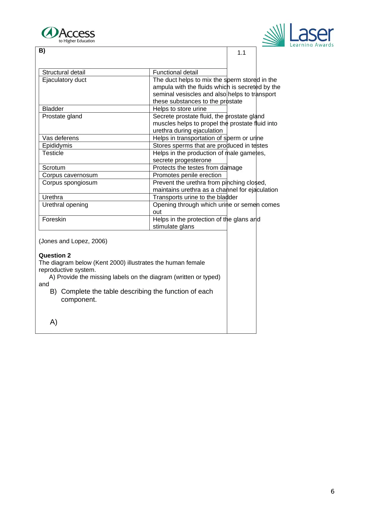
The diagram below (Kent 2000) illustrates the human male reproductive system.
A) Provide the missing labels on the diagram (written or typed) and
B) Complete the table briefly describing the function of each component.
A)
1.1
4
Ejaculatory duct
Urethral opening
Testicle
Epididymis
Vas deferens
Bladder
Prostate gland
Scrotum
Corpus
cavernosum
Urethra
Foreskin
Corpus
spongiosum
Task 1
Course Title: Access to HE Diploma:
Unit Title: Reproduction and Inheritance
Task(s) Short answer questions and a poster to address learning outcome 1
Task Title:
1. Labelling structures and completing a table
2. Labelling structures and completing a table
3. Explain stages of fertilisation
4. Write an overview of stages of foetal development
5. A4 poster on the stages of birth
6. Hormones
7. Outline of the menstrual cycle
Date of Internal
Moderation:
Description of Assessment Task (mapped to ACs) Assessmen
t criteria
Task 1: In completing this task, students are required to undertake the following seven
questions:
Question 1:
The diagram below (Kent 2000) illustrates the human male reproductive system.
A) Provide the missing labels on the diagram (written or typed) and
B) Complete the table briefly describing the function of each component.
A)
1.1
4
Ejaculatory duct
Urethral opening
Testicle
Epididymis
Vas deferens
Bladder
Prostate gland
Scrotum
Corpus
cavernosum
Urethra
Foreskin
Corpus
spongiosum
Paraphrase This Document
Need a fresh take? Get an instant paraphrase of this document with our AI Paraphraser

5
B)
Structural detail Functional detail
Ejaculatory duct The duct helps to mix the sperm stored in the
ampula with the fluids which is secreted by the
seminal vesiscles and also helps to transport
these substances to the prostate
Bladder Helps to store urine
Prostate gland Secrete prostate fluid, the prostate gland
muscles helps to propel the prostate fluid into
urethra during ejaculation
Vas deferens Helps in transportation of sperm or urine
Epididymis Stores sperms that are produced in testes
Testicle Helps in the production of male gametes,
secrete progesterone
Scrotum Protects the testes from damage
Corpus cavernosum Promotes penile erection
Corpus spongiosum Prevent the urethra from pinching closed,
maintains urethra as a channel for ejaculation
Urethra Transports urine to the bladder
Urethral opening Opening through which urine or semen comes
out
Foreskin Helps in the protection of the glans and
stimulate glans
(Jones and Lopez, 2006)
Question 2
The diagram below (Kent 2000) illustrates the human female
reproductive system.
A) Provide the missing labels on the diagram (written or typed)
and
B) Complete the table describing the function of each
component.
A)
1.1
6
⊘ This is a preview!⊘
Do you want full access?
Subscribe today to unlock all pages.
Trusted by 1+ million students worldwide
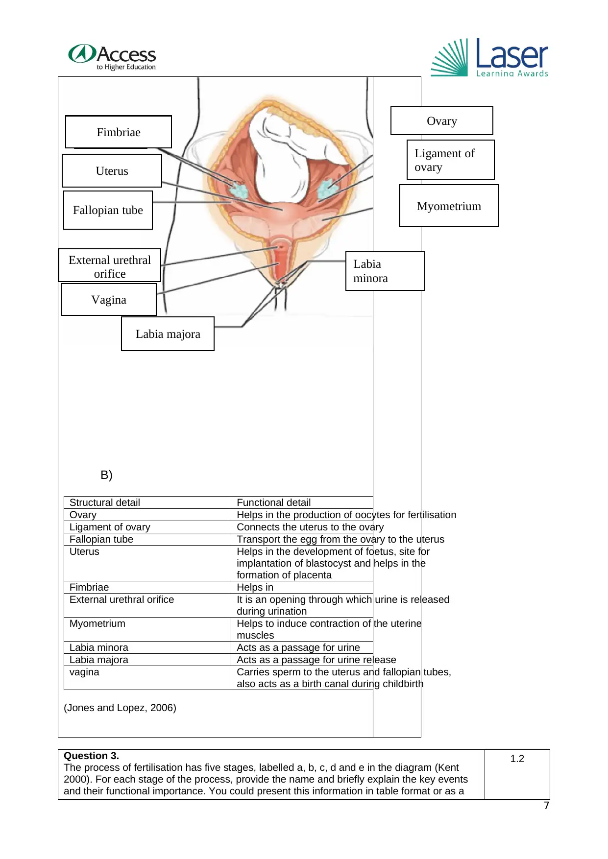
B)
Structural detail Functional detail
Ovary Helps in the production of oocytes for fertilisation
Ligament of ovary Connects the uterus to the ovary
Fallopian tube Transport the egg from the ovary to the uterus
Uterus Helps in the development of foetus, site for
implantation of blastocyst and helps in the
formation of placenta
Fimbriae Helps in
External urethral orifice It is an opening through which urine is released
during urination
Myometrium Helps to induce contraction of the uterine
muscles
Labia minora Acts as a passage for urine
Labia majora Acts as a passage for urine release
vagina Carries sperm to the uterus and fallopian tubes,
also acts as a birth canal during childbirth
(Jones and Lopez, 2006)
Question 3.
The process of fertilisation has five stages, labelled a, b, c, d and e in the diagram (Kent
2000). For each stage of the process, provide the name and briefly explain the key events
and their functional importance. You could present this information in table format or as a
1.2
7
Fimbriae
Uterus
Fallopian tube
Labia
minora
External urethral
orifice
Vagina
Ovary
Ligament of
ovary
Labia majora
Myometrium
Paraphrase This Document
Need a fresh take? Get an instant paraphrase of this document with our AI Paraphraser
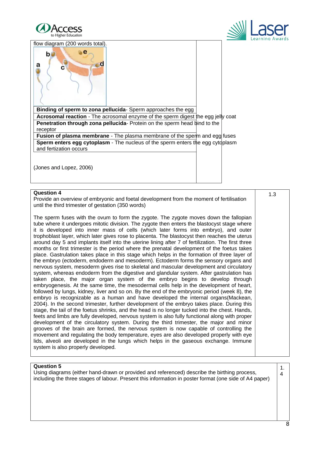
flow diagram (200 words total).
Binding of sperm to zona pellucida- Sperm approaches the egg
Acrosomal reaction - The acrosomal enzyme of the sperm digest the egg jelly coat
Penetration through zona pellucida- Protein on the sperm head bind to the
receptor
Fusion of plasma membrane - The plasma membrane of the sperm and egg fuses
Sperm enters egg cytoplasm - The nucleus of the sperm enters the egg cytoplasm
and fertization occurs
(Jones and Lopez, 2006)
Question 4
Provide an overview of embryonic and foetal development from the moment of fertilisation
until the third trimester of gestation (350 words)
The sperm fuses with the ovum to form the zygote. The zygote moves down the fallopian
tube where it undergoes mitotic division. The zygote then enters the blastocyst stage where
it is developed into inner mass of cells (which later forms into embryo), and outer
trophoblast layer, which later gives rose to placenta. The blastocyst then reaches the uterus
around day 5 and implants itself into the uterine lining after 7 of fertilization. The first three
months or first trimester is the period where the prenatal development of the foetus takes
place. Gastrulation takes place in this stage which helps in the formation of three layer of
the embryo (ectoderm, endoderm and mesoderm). Ectoderm forms the sensory organs and
nervous system, mesoderm gives rise to skeletal and mascular development and circulatory
system, whereas endoderm from the digestive and glandular system. After gastrulation has
taken place, the major organ system of the embryo begins to develop through
embryogenesis. At the same time, the mesodermal cells help in the development of heart,
followed by lungs, kidney, liver and so on. By the end of the embryonic period (week 8), the
embryo is recognizable as a human and have developed the internal organs(Mackean,
2004). In the second trimester, further development of the embryo takes place. During this
stage, the tail of the foetus shrinks, and the head is no longer tucked into the chest. Hands,
feets and limbs are fully developed, nervous system is also fully functional along with proper
development of the circulatory system. During the third trimester, the major and minor
grooves of the brain are formed, the nervous system is now capable of controlling the
movement and regulating the body temperature, eyes are also developed properly with eye
lids, alveoli are developed in the lungs which helps in the gaseous exchange. Immune
system is also properly developed.
1.3
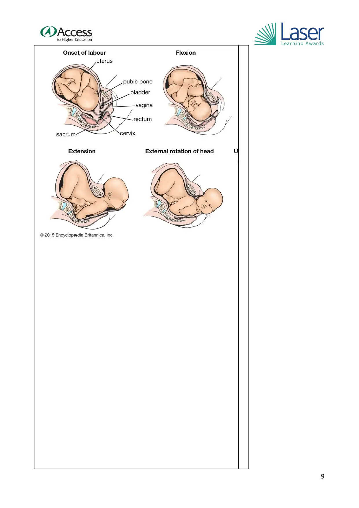
Question 5
Using diagrams (either hand-drawn or provided and referenced) describe the birthing process,
including the three stages of labour. Present this information in poster format (one side of A4 paper)
1.
4
8
Binding of sperm to zona pellucida- Sperm approaches the egg
Acrosomal reaction - The acrosomal enzyme of the sperm digest the egg jelly coat
Penetration through zona pellucida- Protein on the sperm head bind to the
receptor
Fusion of plasma membrane - The plasma membrane of the sperm and egg fuses
Sperm enters egg cytoplasm - The nucleus of the sperm enters the egg cytoplasm
and fertization occurs
(Jones and Lopez, 2006)
Question 4
Provide an overview of embryonic and foetal development from the moment of fertilisation
until the third trimester of gestation (350 words)
The sperm fuses with the ovum to form the zygote. The zygote moves down the fallopian
tube where it undergoes mitotic division. The zygote then enters the blastocyst stage where
it is developed into inner mass of cells (which later forms into embryo), and outer
trophoblast layer, which later gives rose to placenta. The blastocyst then reaches the uterus
around day 5 and implants itself into the uterine lining after 7 of fertilization. The first three
months or first trimester is the period where the prenatal development of the foetus takes
place. Gastrulation takes place in this stage which helps in the formation of three layer of
the embryo (ectoderm, endoderm and mesoderm). Ectoderm forms the sensory organs and
nervous system, mesoderm gives rise to skeletal and mascular development and circulatory
system, whereas endoderm from the digestive and glandular system. After gastrulation has
taken place, the major organ system of the embryo begins to develop through
embryogenesis. At the same time, the mesodermal cells help in the development of heart,
followed by lungs, kidney, liver and so on. By the end of the embryonic period (week 8), the
embryo is recognizable as a human and have developed the internal organs(Mackean,
2004). In the second trimester, further development of the embryo takes place. During this
stage, the tail of the foetus shrinks, and the head is no longer tucked into the chest. Hands,
feets and limbs are fully developed, nervous system is also fully functional along with proper
development of the circulatory system. During the third trimester, the major and minor
grooves of the brain are formed, the nervous system is now capable of controlling the
movement and regulating the body temperature, eyes are also developed properly with eye
lids, alveoli are developed in the lungs which helps in the gaseous exchange. Immune
system is also properly developed.
1.3
Question 5
Using diagrams (either hand-drawn or provided and referenced) describe the birthing process,
including the three stages of labour. Present this information in poster format (one side of A4 paper)
1.
4
8
9
⊘ This is a preview!⊘
Do you want full access?
Subscribe today to unlock all pages.
Trusted by 1+ million students worldwide
(Mackean, 2004)
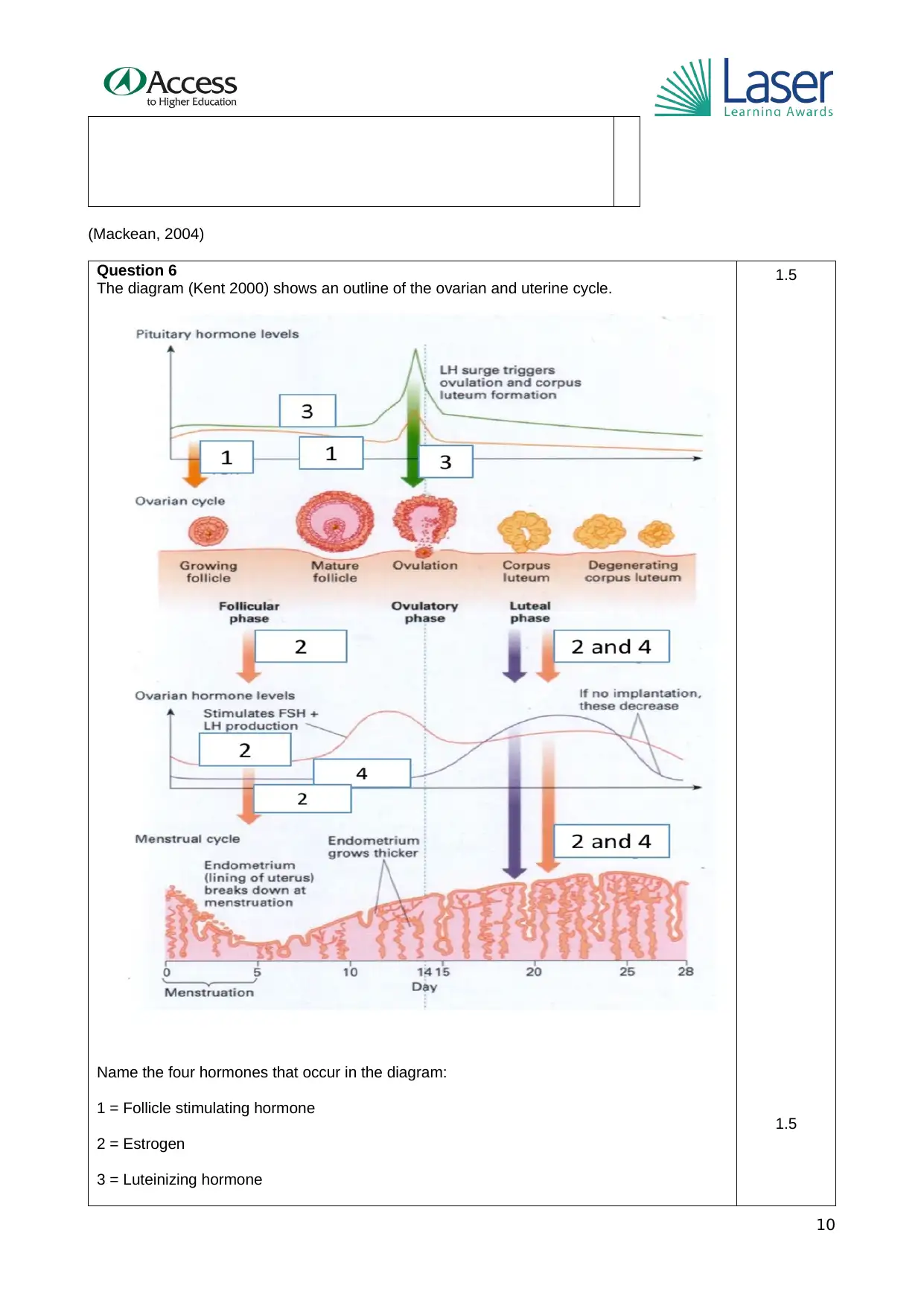
Question 6
The diagram (Kent 2000) shows an outline of the ovarian and uterine cycle.
Name the four hormones that occur in the diagram:
1 = Follicle stimulating hormone
2 = Estrogen
3 = Luteinizing hormone
1.5
1.5
10
Question 6
The diagram (Kent 2000) shows an outline of the ovarian and uterine cycle.
Name the four hormones that occur in the diagram:
1 = Follicle stimulating hormone
2 = Estrogen
3 = Luteinizing hormone
1.5
1.5
10
Paraphrase This Document
Need a fresh take? Get an instant paraphrase of this document with our AI Paraphraser
4 = Progesterone
Question 7
With reference to the diagram, explain briefly how the ovarian cycle
(changes in the ovary) and the menstrual cycle are regulated by
blood hormonal levels (250 words).
The ovarian and menstrual cycle in women is contolled by
hormones. Gonadotrophin releasing hormone secreted from
hypothalamus, stimulates the release of Follicle stimulating
hormone(FSH) and Luteinizing hormone(LH) from the anterior
pituitary gland. The FSH helps in follicular growth and secretion of
estrogen from them. LH helps in the development of ovarian
follicles, promotes formation of corpus luteum(Toole & Toole,
2014). The menstrual cycle has three parts: Follicular, Ovulatory
and Luteal. During the onset of the follicular phase, the level of
estrogen and progestrerone are low, which leads to the shedding
of the uteruas lining or endometrium, thus menstrual bleeding
occurs. At this time, the level of FSH increases, stimulating the
development of follicles in the ovaries. In the later phase, the FSH
level decreases. The ovulatory phase begins with a surge in the LH
and FSH level. LH helps in ovulation. The estrogen level remains
low during this stage and progesterone level rises. During the
luteal phase, LH and FSH level decreases. The ruptured follicles
fuses with the eggs to form corpus luteum, which produces
progesterone. During this stage, estrogen level remain high, both
progesterone and estrogen helps in the thickening of the uterine
wall preparing for fertilisation. If the eggs are not fertilized, the
corpus luteum degenerates and the progesterone and estrogen
release is stopped., their level decreases. This leads to the
shedding of the uterine wall and thus the menstrual bleeding
happens. And if egg are fertilized, the corpus luteum continues to
function properly during early pregnancy and helps in the
maintainance of pregnancy(Maloy, Hughes and Brenner, 2013).
Task 2
Course Title: Access to HE Diploma:
Unit Title: Reproduction and Inheritance
Student Name:
Task(s) Short answer questions
Task Title:
8. Mendel’s laws
9. Stages of meiosis
10. Punnett squares
Date of Internal
Moderation:
Description of Assessment Task (mapped to ACs) As
11
s
e
s
s
m
e
n
t
c
ri
te
ri
a
In completing this assignment, students are required to undertake the
following three tasks:
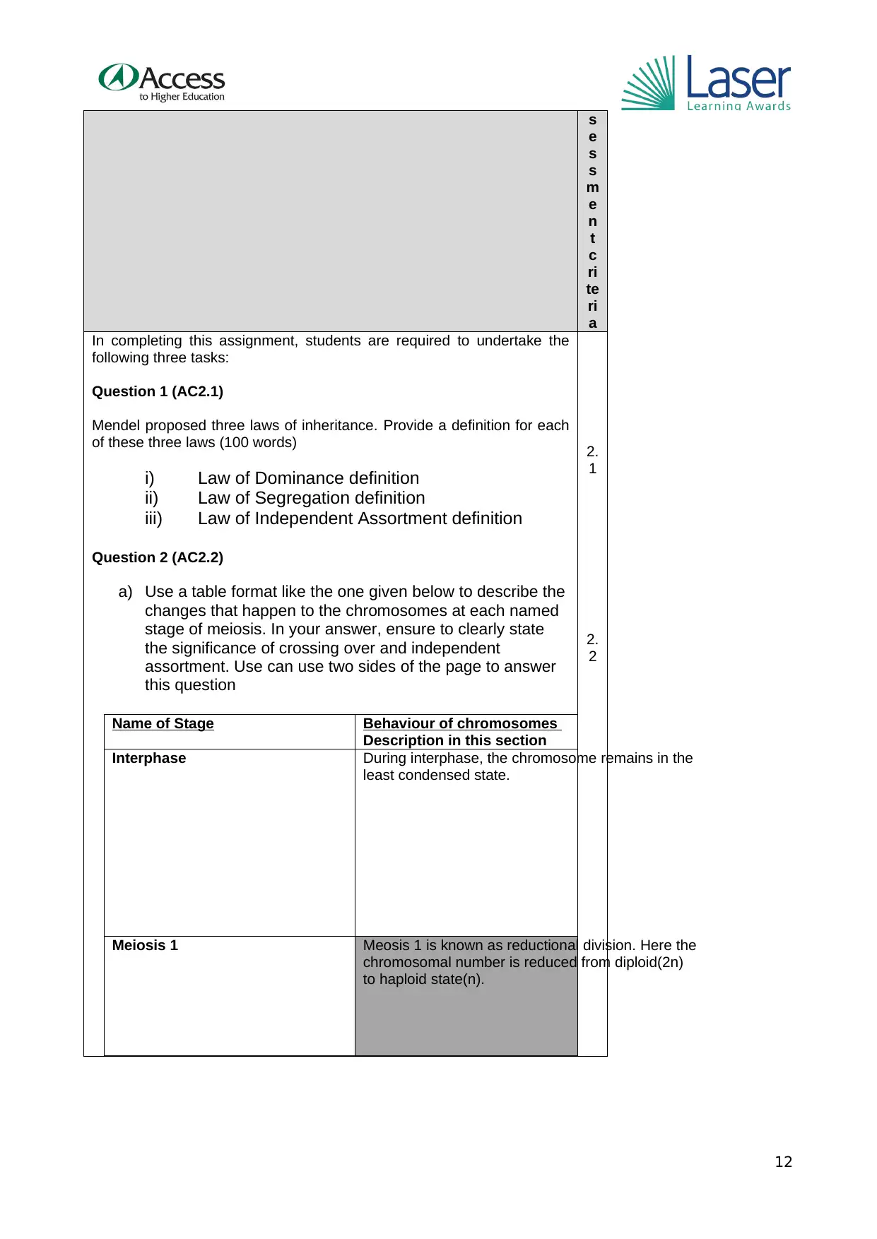
Question 1 (AC2.1)
Mendel proposed three laws of inheritance. Provide a definition for each
of these three laws (100 words)
i) Law of Dominance definition
ii) Law of Segregation definition
iii) Law of Independent Assortment definition
Question 2 (AC2.2)
a) Use a table format like the one given below to describe the
changes that happen to the chromosomes at each named
stage of meiosis. In your answer, ensure to clearly state
the significance of crossing over and independent
assortment. Use can use two sides of the page to answer
this question
Name of Stage Behaviour of chromosomes
Description in this section
Interphase During interphase, the chromosome remains in the
least condensed state.
Meiosis 1 Meosis 1 is known as reductional division. Here the
chromosomal number is reduced from diploid(2n)
to haploid state(n).
2.
1
2.
2
12
e
s
s
m
e
n
t
c
ri
te
ri
a
In completing this assignment, students are required to undertake the
following three tasks:
Question 1 (AC2.1)
Mendel proposed three laws of inheritance. Provide a definition for each
of these three laws (100 words)
i) Law of Dominance definition
ii) Law of Segregation definition
iii) Law of Independent Assortment definition
Question 2 (AC2.2)
a) Use a table format like the one given below to describe the
changes that happen to the chromosomes at each named
stage of meiosis. In your answer, ensure to clearly state
the significance of crossing over and independent
assortment. Use can use two sides of the page to answer
this question
Name of Stage Behaviour of chromosomes
Description in this section
Interphase During interphase, the chromosome remains in the
least condensed state.
Meiosis 1 Meosis 1 is known as reductional division. Here the
chromosomal number is reduced from diploid(2n)
to haploid state(n).
2.
1
2.
2
12
⊘ This is a preview!⊘
Do you want full access?
Subscribe today to unlock all pages.
Trusted by 1+ million students worldwide
1 out of 23
Related Documents


Your All-in-One AI-Powered Toolkit for Academic Success.
+13062052269
info@desklib.com
Available 24*7 on WhatsApp / Email
![[object Object]](/_next/static/media/star-bottom.7253800d.svg)
Unlock your academic potential
Copyright © 2020–2026 A2Z Services. All Rights Reserved. Developed and managed by ZUCOL.