Structural Biology Project: SilE Protein Folding and Silver Resistance
VerifiedAdded on 2023/04/24
|9
|2230
|293
Project
AI Summary
This project investigates the mechanism of bacterial resistance to silver by focusing on the SilE protein. The project aims to overexpress SilE in E. coli, purify the protein, and characterize its silver-induced folding using NMR spectroscopy. The study seeks to obtain structural information on the folded, silver-bound form of the protein, potentially through X-ray crystallography. The research begins with constructing a plasmid for SilE expression, followed by protein purification using chromatography techniques. NMR spectroscopy will be employed to analyze the protein's folding in response to silver, and either NMR or X-ray crystallography will be used to determine the structure of the silver-bound protein. The findings are expected to enhance the understanding of bacterial silver resistance, potentially leading to strategies to prevent or circumvent antibiotic resistance in the future. The project also discusses the background of silver's antimicrobial properties, the role of efflux pumps, and previous research on SilE's structure and silver binding capabilities, including the importance of methionine and histidine residues in Ag+ binding.
Abstract
The increase in bacterial resistance to antibiotics has encouraged augmented usage of
antimicrobials like silver in dressing wounds. This in turn had resulted in the subsequent
isolation of bacteria that are silver resistant and generate two periplasmic silver binding
proteins and an efflux pump, which are thought to act in a way that allows expulsion of silver
from periplasm, thus stopping silver from disturbing the cytoplasmic membrane. The
particulars of silver resistance mechanism have not been completely explored yet. SilE is a
major silver binding protein, which is considered to display native unfolded configuration.
The protein generates tertiary fold upon binding with silver and has also been found to confer
resistance. The purpose of this assignment is to characterize silver-associated SilE folding
and retrieve adequate structural information related to the silver bound and folded form. The
assignment will also involve SilE overexpression in E. coli cells and subsequent protein
purification. It will then involve NMR spectroscopy for determining the protein folding, in
relation to silver. The assembly of the folded, silver-associated protein will be then assessed
with the use of NMR spectroscopy and/or X-ray crystallography. The findings of the research
will significantly contribute gaining a sound understanding of bacterial resistance to silver.
By gaining an awareness of the resistance mechanisms, antibiotic resistance can be prevented
or circumvented in future.
Key words: Structural biology, NMR spectroscopy, protein purification
Hypothesis and Aims
It is hypothesised that bacterial resistance to antibiotics has prompted increased This
has led to the isolation of silver resistant bacteria that produce an efflux pump and two
periplasmic silver binding proteins that are thought to act to expel silver from the periplasm,
preventing silver from perturbing the cytoplasmic membrane.
Paraphrase This Document
Need a fresh take? Get an instant paraphrase of this document with our AI Paraphraser
Therefore, the main aims of this project are to:
• overexpression of SilE in E. coli and protein purification
• Characterise silver induced folding
• Obtain structural information on the folded, silver bound form
Background
Internationally, the wide spread of bacteria resistance made a significant impact on
the efficacy of the antibiotic which is saved many lives from this organism. The antibiotic
resistance situation happened due to the overuse of this drug and the lack of developing new
medication by the pharmaceutical companies (Ventola, 2015). Silver is considered an
antimicrobial agent a long time before the antibiotic was discovered, as well as silver
consider the most effective antibacterial and less toxicity out of all metals that have
antimicrobial properties. The usage of silver decreased by the time the antibiotic discovered.
However, due to the antibiotic resistance bacteria endanger our lives, an interest of silver as
an antimicrobial agent begun to re-use. Silver resistance in Gram-negative bacteria was first
encountered in a strain of Salmonella typhimurium that caused an outbreak on a burns ward
in 1975 and resulted in the death of three people (WHO. int, 2019). Silver resistance bacteria
develop a technique to expel the silver from the cell membrane using an efflux pump and two
periplasmic silver binding proteins. There have been studies that show the significant role of
SilE in the occurrence of bacteria silver resistance. However, there is a poor understanding of
the genetic mechanism that underlies the susceptibility of the resistance gene that contributes
to the resistance. Owing to the high antimicrobial activity of ionic silver against a plethora of
microorganisms, silver has been recognized as a key component for numerous commercially
accessible healthcare products (Zheng et al., 2018).
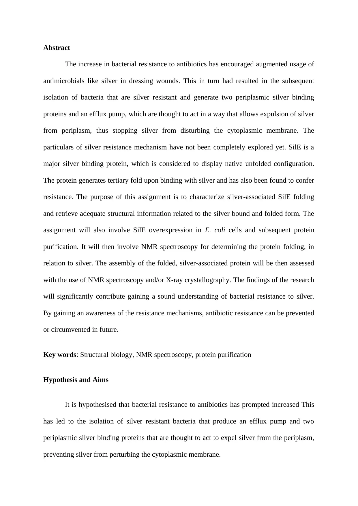
Figure 1- Silver resistance operon
Source- (Asiani et al., 2016)
The usage of silver is snowballing speedily in the arena of wound care, and a
widespread diversity of silver-impregnated wound dressings are currently commonplace such
as, polyurethane foams, hydrofiber dressing, and gauzes. The primary benefit of silver-
containing wound dressings can be accredited to the fact that they provide a moist wound
environment, thus hastening the process of wound recovery, with proven effectiveness in
local infection or wounds having bioburden (Wu et al., 2014). In addition, efflux pumps
provides the opportunity to microorganisms for regulating the internal environment, which in
turn is facilitates removal of toxic substances, together with metabolites, antimicrobial agents,
and quorum sensing signal molecules (Chacón et al., 2014).
⊘ This is a preview!⊘
Do you want full access?
Subscribe today to unlock all pages.
Trusted by 1+ million students worldwide
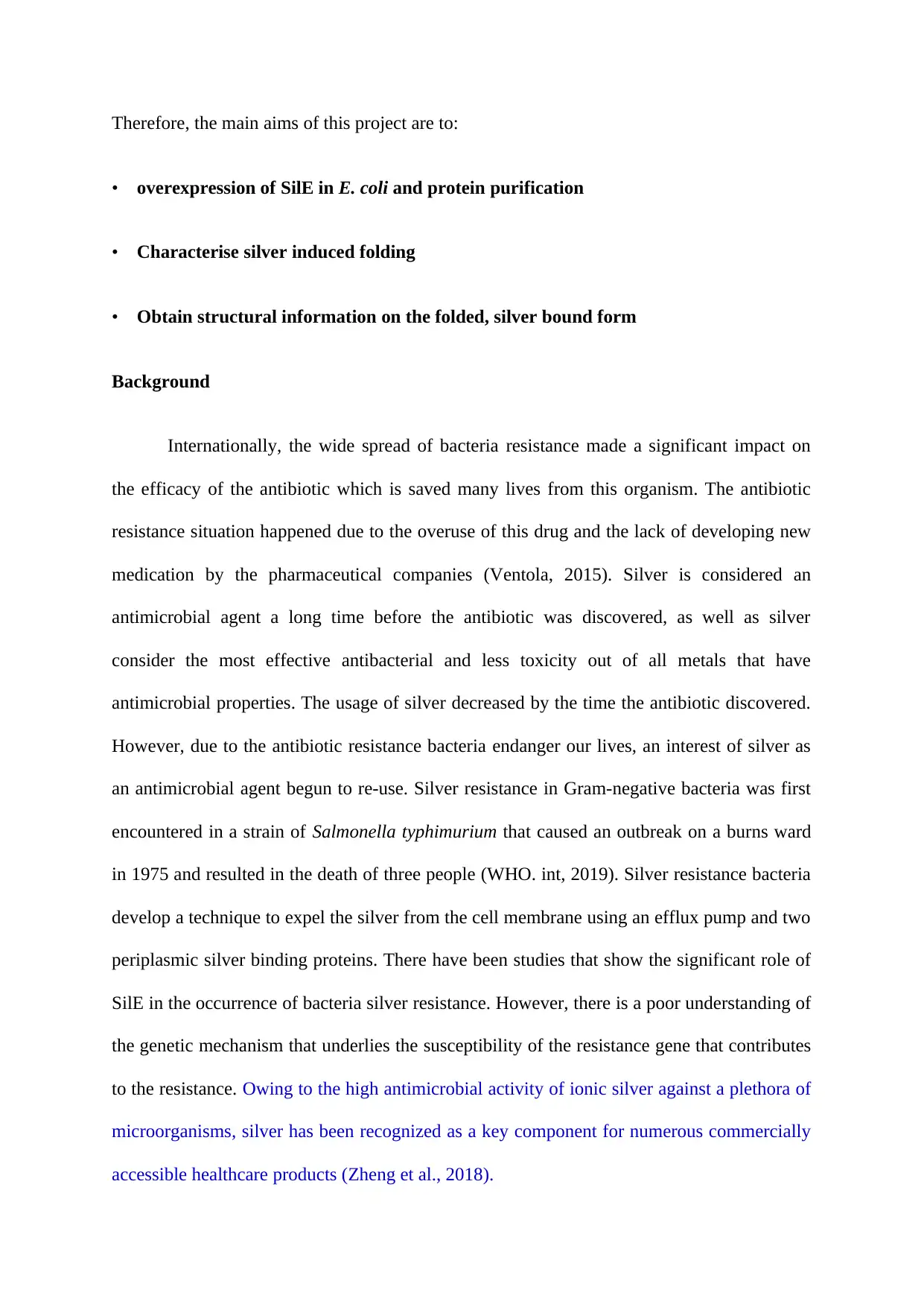
Figure 2- Silver wound dressings
Source- (Medline.com., 2019)
Previous studies
Asiani et al. (2016) tested the hypothesis that was based on putative model of Ag+
resistance and found that SilE is a protein that is intrinsically disordered in the free apo‐form.
However, the protein gets folded to a condensed arrangement, upon achieving optimum
binding to six major Ag+ ions in the holo‐configuration. Furthermore, conduction of site‐
directed mutagenesis and sequence analyses helped in establishing the prominence of
methionine and histidine encompassing motifs for binding of Ag+, besides assisting in the
identification of a nucleation core that inducts Ag+‐facilitated SilE folding. Findings from
another study suggests that the physico-chemical attributes of nanoparticles containing silver
and their contact with the living cells varies considerably from the attributes of silver ions.
Furthermore, the diversity of the characteristics and forms of countless silver nanoparticles
are also accountable for modifications in the antibacterial mechanism of action and most
likely in the bacterial mechanism of resistance as well (Kędziora et al., 2018). Chabert et al.
(2017) also provided evidence for the buffering role of Ag+ for SilE, in relation to high
Paraphrase This Document
Need a fresh take? Get an instant paraphrase of this document with our AI Paraphraser
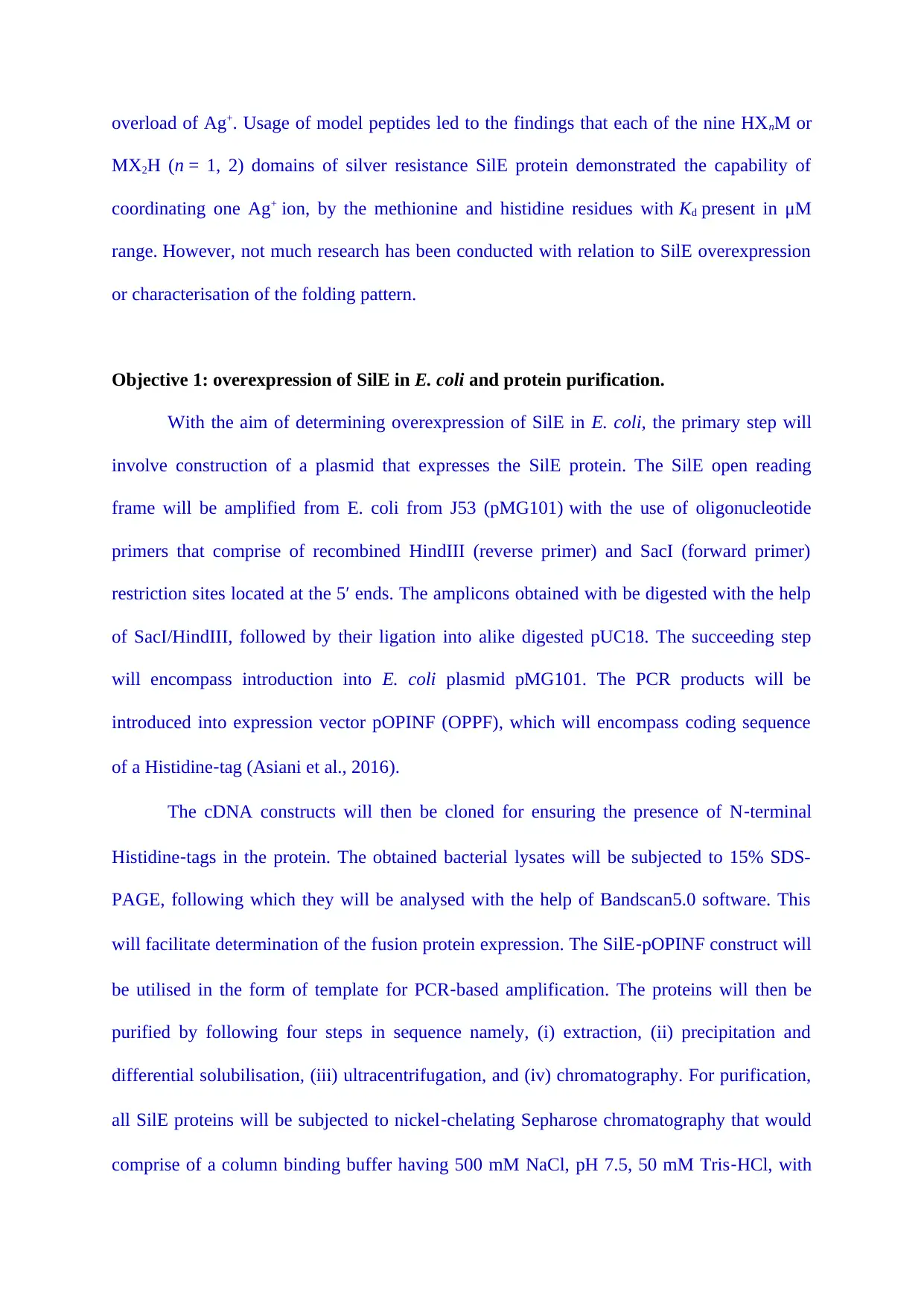
overload of Ag+. Usage of model peptides led to the findings that each of the nine HXnM or
MX2H (n = 1, 2) domains of silver resistance SilE protein demonstrated the capability of
coordinating one Ag+ ion, by the methionine and histidine residues with Kd present in μM
range. However, not much research has been conducted with relation to SilE overexpression
or characterisation of the folding pattern.
Objective 1: overexpression of SilE in E. coli and protein purification.
With the aim of determining overexpression of SilE in E. coli, the primary step will
involve construction of a plasmid that expresses the SilE protein. The SilE open reading
frame will be amplified from E. coli from J53 (pMG101) with the use of oligonucleotide
primers that comprise of recombined HindIII (reverse primer) and SacI (forward primer)
restriction sites located at the 5′ ends. The amplicons obtained with be digested with the help
of SacI/HindIII, followed by their ligation into alike digested pUC18. The succeeding step
will encompass introduction into E. coli plasmid pMG101. The PCR products will be
introduced into expression vector pOPINF (OPPF), which will encompass coding sequence
of a Histidine‐tag (Asiani et al., 2016).
The cDNA constructs will then be cloned for ensuring the presence of N‐terminal
Histidine‐tags in the protein. The obtained bacterial lysates will be subjected to 15% SDS-
PAGE, following which they will be analysed with the help of Bandscan5.0 software. This
will facilitate determination of the fusion protein expression. The SilE‐pOPINF construct will
be utilised in the form of template for PCR‐based amplification. The proteins will then be
purified by following four steps in sequence namely, (i) extraction, (ii) precipitation and
differential solubilisation, (iii) ultracentrifugation, and (iv) chromatography. For purification,
all SilE proteins will be subjected to nickel‐chelating Sepharose chromatography that would
comprise of a column binding buffer having 500 mM NaCl, pH 7.5, 50 mM Tris‐HCl, with
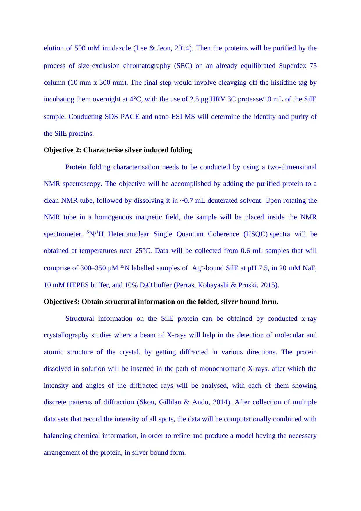
elution of 500 mM imidazole (Lee & Jeon, 2014). Then the proteins will be purified by the
process of size‐exclusion chromatography (SEC) on an already equilibrated Superdex 75
column (10 mm x 300 mm). The final step would involve cleavging off the histidine tag by
incubating them overnight at 4°C, with the use of 2.5 μg HRV 3C protease/10 mL of the SilE
sample. Conducting SDS‐PAGE and nano‐ESI MS will determine the identity and purity of
the SilE proteins.
Objective 2: Characterise silver induced folding
Protein folding characterisation needs to be conducted by using a two-dimensional
NMR spectroscopy. The objective will be accomplished by adding the purified protein to a
clean NMR tube, followed by dissolving it in ~0.7 mL deuterated solvent. Upon rotating the
NMR tube in a homogenous magnetic field, the sample will be placed inside the NMR
spectrometer. 15N/1H Heteronuclear Single Quantum Coherence (HSQC) spectra will be
obtained at temperatures near 25°C. Data will be collected from 0.6 mL samples that will
comprise of 300–350 μM 15N labelled samples of Ag+‐bound SilE at pH 7.5, in 20 mM NaF,
10 mM HEPES buffer, and 10% D2O buffer (Perras, Kobayashi & Pruski, 2015).
Objective3: Obtain structural information on the folded, silver bound form.
Structural information on the SilE protein can be obtained by conducted x-ray
crystallography studies where a beam of X-rays will help in the detection of molecular and
atomic structure of the crystal, by getting diffracted in various directions. The protein
dissolved in solution will be inserted in the path of monochromatic X-rays, after which the
intensity and angles of the diffracted rays will be analysed, with each of them showing
discrete patterns of diffraction (Skou, Gillilan & Ando, 2014). After collection of multiple
data sets that record the intensity of all spots, the data will be computationally combined with
balancing chemical information, in order to refine and produce a model having the necessary
arrangement of the protein, in silver bound form.
⊘ This is a preview!⊘
Do you want full access?
Subscribe today to unlock all pages.
Trusted by 1+ million students worldwide
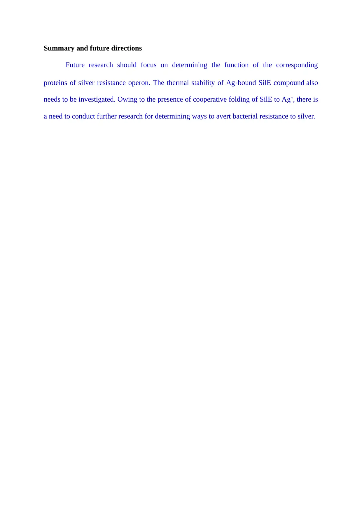
Summary and future directions
Future research should focus on determining the function of the corresponding
proteins of silver resistance operon. The thermal stability of Ag‐bound SilE compound also
needs to be investigated. Owing to the presence of cooperative folding of SilE to Ag+, there is
a need to conduct further research for determining ways to avert bacterial resistance to silver.
Paraphrase This Document
Need a fresh take? Get an instant paraphrase of this document with our AI Paraphraser
References
Asiani, K. R., Williams, H., Bird, L., Jenner, M., Searle, M. S., Hobman, J. L., ... &
Soultanas, P. (2016). SilE is an intrinsically disordered periplasmic “molecular
sponge” involved in bacterial silver resistance. Molecular microbiology, 101(5), 731-
742.
Chabert, V., Hologne, M., Sénèque, O., Crochet, A., Walker, O., & Fromm, K. M. (2017).
Model peptide studies of Ag+ binding sites from the silver resistance protein
SilE. Chemical Communications, 53(45), 6105-6108.
Chacón, K. N., Mealman, T. D., McEvoy, M. M., & Blackburn, N. J. (2014). Tracking metal
ions through a Cu/Ag efflux pump assigns the functional roles of the periplasmic
proteins. Proceedings of the National Academy of Sciences, 111(43), 15373-15378.
Kędziora, A., Speruda, M., Krzyżewska, E., Rybka, J., Łukowiak, A., & Bugla-Płoskońska,
G. (2018). Similarities and differences between silver ions and silver in nanoforms as
antibacterial agents. International journal of molecular sciences, 19(2), 444.
Lee, J. Y., & Jeon, S. J. (2014). Characterization and immobilization on nickel-chelated
Sepharose of a glutamate decarboxylase A from Lactobacillus brevis BH2 and its
application for production of GABA. Bioscience, biotechnology, and
biochemistry, 78(10), 1656-1661.
Perras, F. A., Kobayashi, T., & Pruski, M. (2015). Natural abundance 17O DNP two-
dimensional and surface-enhanced NMR spectroscopy. Journal of the American
Chemical Society, 137(26), 8336-8339.
Skou, S., Gillilan, R. E., & Ando, N. (2014). Synchrotron-based small-angle X-ray scattering
of proteins in solution. Nature protocols, 9(7), 1727.
Wu, J., Zheng, Y., Song, W., Luan, J., Wen, X., Wu, Z., ... & Guo, S. (2014). In situ
synthesis of silver-nanoparticles/bacterial cellulose composites for slow-released
antimicrobial wound dressing. Carbohydrate polymers, 102, 762-771.
Zheng, K., Setyawati, M. I., Leong, D. T., & Xie, J. (2018). Antimicrobial silver
nanomaterials. Coordination Chemistry Reviews, 357, 1-17.
Medline.com. (2019). SilvaSorb Silver Antimicrobial Wound Dressing. Retrieved from
https://www.medline.com/product/SilvaSorb-Silver-Antimicrobial-Wound-
Dressing/Z05-PF00207
⊘ This is a preview!⊘
Do you want full access?
Subscribe today to unlock all pages.
Trusted by 1+ million students worldwide
1 out of 9

Your All-in-One AI-Powered Toolkit for Academic Success.
+13062052269
info@desklib.com
Available 24*7 on WhatsApp / Email
![[object Object]](/_next/static/media/star-bottom.7253800d.svg)
Unlock your academic potential
Copyright © 2020–2026 A2Z Services. All Rights Reserved. Developed and managed by ZUCOL.