Detailed Report on Skin Rashes and Lesions: Diagnosis Methods

Verified

Added on  2023/06/15

|12
|1766
|342
Report
AI Summary
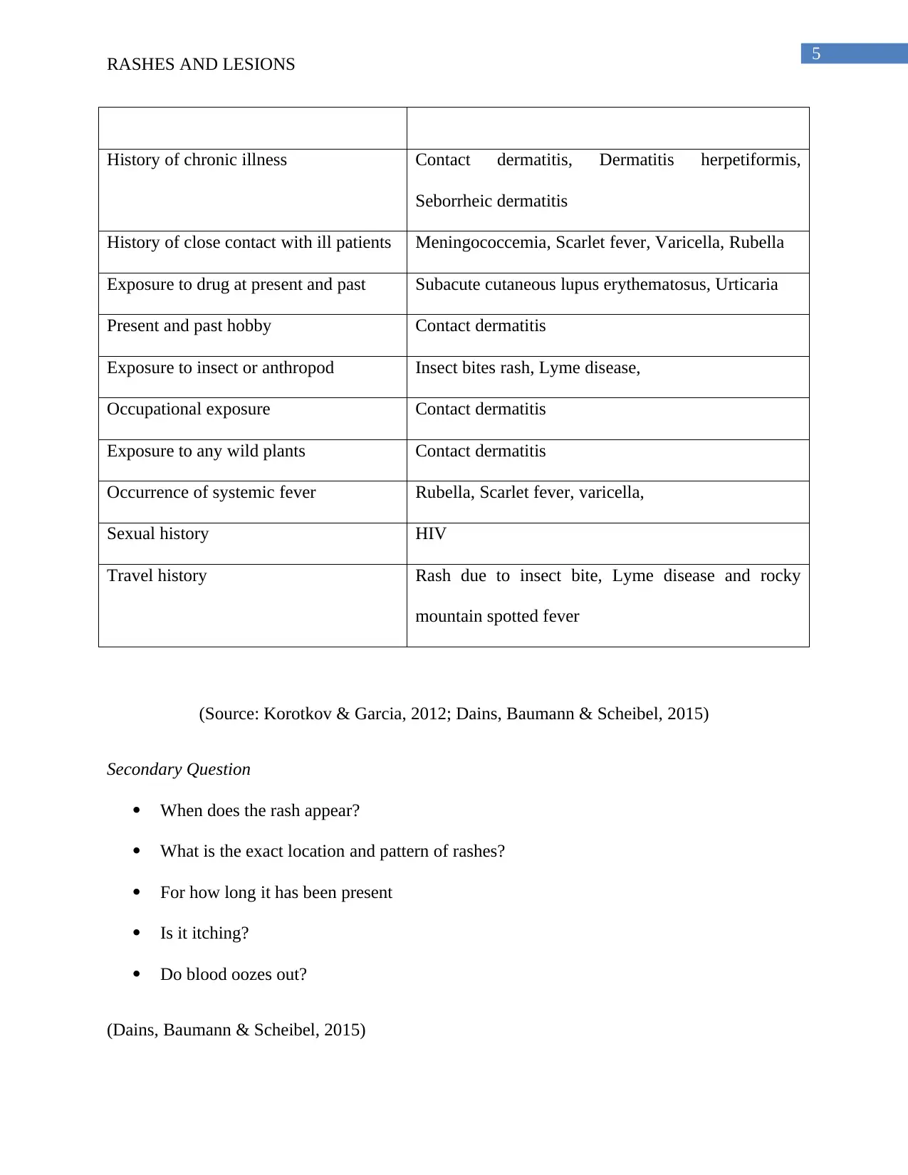
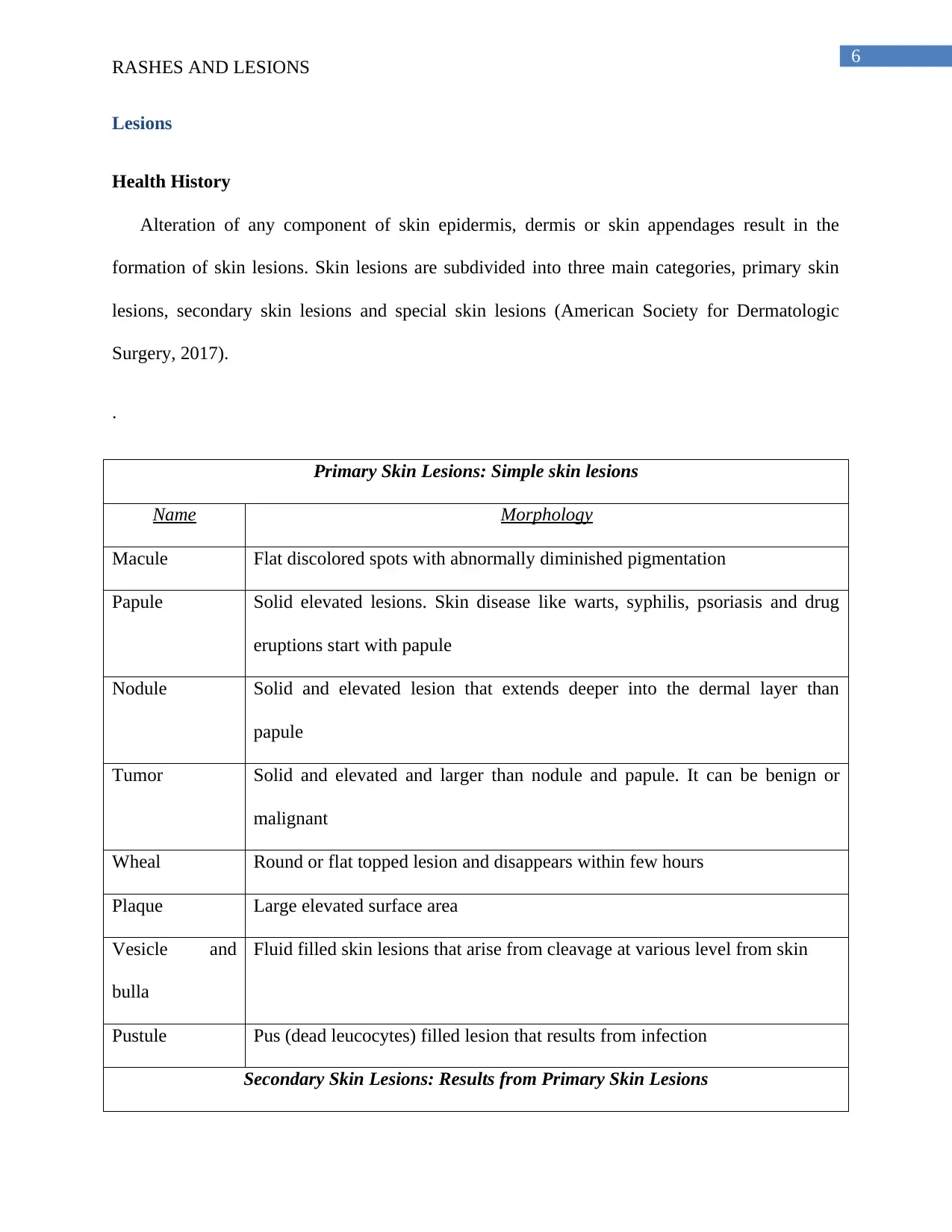
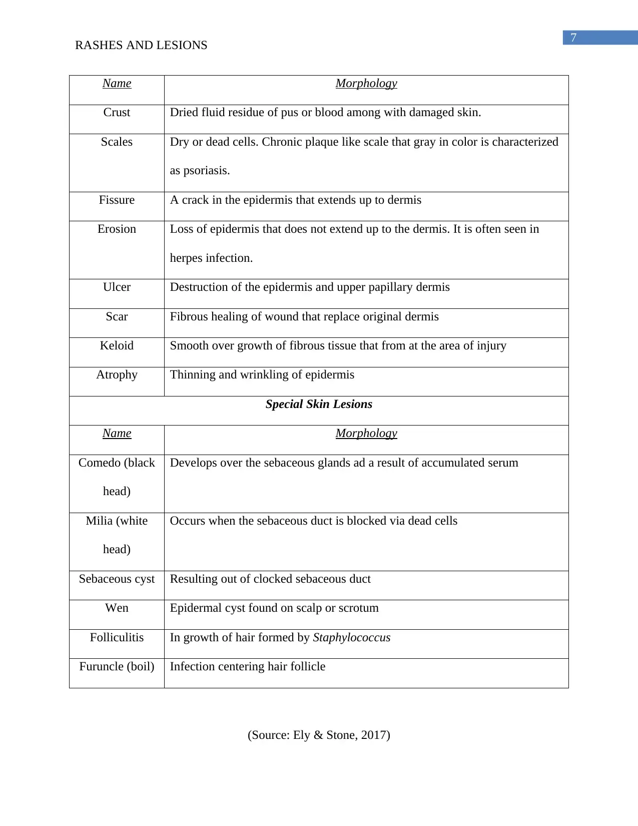
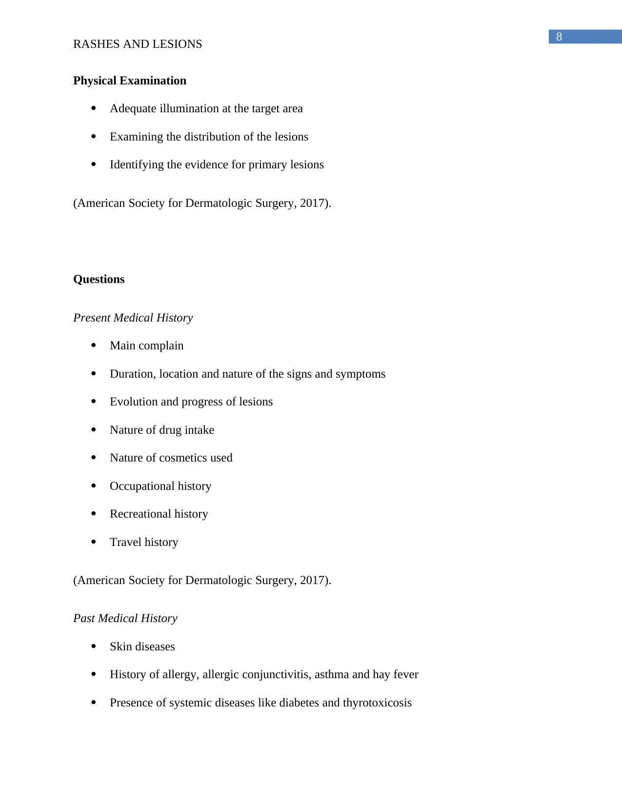
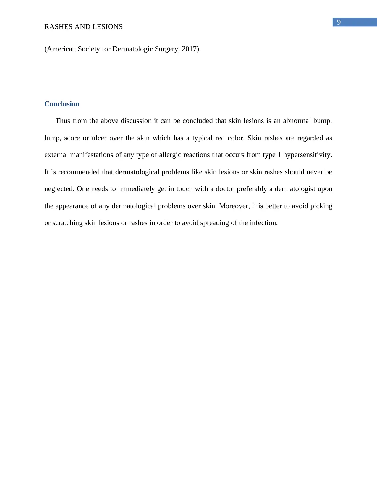
This report provides a detailed analysis of skin rashes and skin lesions, differentiating them based on health history, morphology, and physical examination methods. It discusses the importance of considering factors such as travel history, insect bites, drug reactions, and exposure to infectious agents when diagnosing skin rashes. The report also outlines various types of skin lesions, including primary, secondary, and special lesions, along with their respective characteristics and morphologies. Furthermore, it emphasizes the significance of a thorough medical history, including present and past conditions, to accurately diagnose and manage dermatological problems. The report concludes by recommending prompt consultation with a dermatologist for any skin-related issues and advises against picking or scratching lesions or rashes to prevent the spread of infection. Desklib provides a platform for students to access this and other solved assignments.
Document Page
0Running head: RASHES AND LESIONS
Rashes and Lesions
Name of the Student
Name of University
Author’s note
tabler-icon-diamond-filled.svg

Paraphrase This Document

Need a fresh take? Get an instant paraphrase of this document with our AI Paraphraser
Document Page
1
RASHES AND LESIONS
Table of Contents
Introduction......................................................................................................................................2
Rashes..............................................................................................................................................2
Health History..............................................................................................................................2
Physical Examination..................................................................................................................3
Morphological criteria.................................................................................................................3
Questions.....................................................................................................................................3
Primary Questions...................................................................................................................3
Secondary Question.................................................................................................................4
Lesions.............................................................................................................................................4
Health History..............................................................................................................................4
Physical Examination..................................................................................................................6
Questions.....................................................................................................................................7
Present Medical History..........................................................................................................7
Past Medical History................................................................................................................7
Conclusion.......................................................................................................................................8
References........................................................................................................................................9
Document Page
2
RASHES AND LESIONS
Document Page
3
RASHES AND LESIONS
Introduction
Skin lesions for example acnes are disorder of the skin and skin rashes are defined as
temporary eruptions or discolorations of the skin which arise out of inflammation (Dains,
Baumann & Scheibel, 2015). Both rash and lesions are skin problems or dermatological
disorders however; there exists a thin line of difference between them (Iaccarino et al., 2014).
The following report aims to highlight the differential classification of skin rash and skin lesions
based on their health history, morphology and the methods employed for physical examination.
Rashes
Health History
When the diagnosis of generalized manifestation of skin rash is not obvious then the
patients should be enquired about their recent travel history, past issues of insect bites, exposure
towards wild plants, drug reactions and contact with other infectious agents (Bhole et al., 2012).
Other history which is associated with the development of skin rash includes adverse chemical
reactions over skin due to the exposure from cosmetics, sexual history and recent systemic
symptoms like fever. Patients’ health history in relation to skin rash also deals with painful
lesions, personal or family history of atopy like allergic rhinitis, childhood eczema and asthma.
Moreover, people who are genetically predisposed with high concentration of IgE in blood or
eosinophil are more prone of getting affected with allergic syndromes and most important
manifestation of allergic syndrome is skin rashes (Bhole et al., 2012).
tabler-icon-diamond-filled.svg

Paraphrase This Document

Need a fresh take? Get an instant paraphrase of this document with our AI Paraphraser
Document Page
4
RASHES AND LESIONS
The age of the patient may also help to make the possible diagnosis. Like acute
maculopapular rashes in children are formed as a result of viral infection where as in adult, it it’s
a manifestation of adverse drug reaction (Bhole et al., 2012).
Physical Examination
Characteristic of rashes help to streamline the differential diagnosis. Other factors that
must be taken into consideration during physical examination include the affected area, size, and
the severity of redness and the shape of the rashes. The size of rashes can vary from pinpoint to
total body redness as in case of erythroderma. The shape of the individual rashes and they
tendency to remain cluster together can also provide numerous effective clue towards physical
identification. For example liner pattern of erythema or vesicles of erythema are regarded as
poison ivy (Romano & Caubet, 2014). Oval shape of the rashes determines the pituriasis rosea.
Round shaped rashes are mummular eczema and annular rashes are tinea corporis. The majority
of generalized rashes are pink in color however; there are also brown or deep red rashes too
(Romano & Caubet, 2014).
Morphological criteria
Morphology of rash varies from pinpoint rash to whole body rash. Muscular rashes are
flat spots and are generally red in color. Papular rashes are small solid bumps and they tend to
enlarge (Flett, et al., 2012). Populosquammous rashes are characterized by papules which is
surrounded by scaly areas. Vesicular rashes form blisters (Flett, et al., 2012)
Questions
Primary Questions
Questions Outcomes associated with it
Document Page
5
RASHES AND LESIONS
History of chronic illness Contact dermatitis, Dermatitis herpetiformis,
Seborrheic dermatitis
History of close contact with ill patients Meningococcemia, Scarlet fever, Varicella, Rubella
Exposure to drug at present and past Subacute cutaneous lupus erythematosus, Urticaria
Present and past hobby Contact dermatitis
Exposure to insect or anthropod Insect bites rash, Lyme disease,
Occupational exposure Contact dermatitis
Exposure to any wild plants Contact dermatitis
Occurrence of systemic fever Rubella, Scarlet fever, varicella,
Sexual history HIV
Travel history Rash due to insect bite, Lyme disease and rocky
mountain spotted fever
(Source: Korotkov & Garcia, 2012; Dains, Baumann & Scheibel, 2015)
Secondary Question
When does the rash appear?
What is the exact location and pattern of rashes?
For how long it has been present
Is it itching?
Do blood oozes out?
(Dains, Baumann & Scheibel, 2015)
Document Page
6
RASHES AND LESIONS
Lesions
Health History
Alteration of any component of skin epidermis, dermis or skin appendages result in the
formation of skin lesions. Skin lesions are subdivided into three main categories, primary skin
lesions, secondary skin lesions and special skin lesions (American Society for Dermatologic
Surgery, 2017).
.
Primary Skin Lesions: Simple skin lesions
Name Morphology
Macule Flat discolored spots with abnormally diminished pigmentation
Papule Solid elevated lesions. Skin disease like warts, syphilis, psoriasis and drug
eruptions start with papule
Nodule Solid and elevated lesion that extends deeper into the dermal layer than
papule
Tumor Solid and elevated and larger than nodule and papule. It can be benign or
malignant
Wheal Round or flat topped lesion and disappears within few hours
Plaque Large elevated surface area
Vesicle and
bulla
Fluid filled skin lesions that arise from cleavage at various level from skin
Pustule Pus (dead leucocytes) filled lesion that results from infection
Secondary Skin Lesions: Results from Primary Skin Lesions
tabler-icon-diamond-filled.svg

Paraphrase This Document

Need a fresh take? Get an instant paraphrase of this document with our AI Paraphraser
Document Page
7
RASHES AND LESIONS
Name Morphology
Crust Dried fluid residue of pus or blood among with damaged skin.
Scales Dry or dead cells. Chronic plaque like scale that gray in color is characterized
as psoriasis.
Fissure A crack in the epidermis that extends up to dermis
Erosion Loss of epidermis that does not extend up to the dermis. It is often seen in
herpes infection.
Ulcer Destruction of the epidermis and upper papillary dermis
Scar Fibrous healing of wound that replace original dermis
Keloid Smooth over growth of fibrous tissue that from at the area of injury
Atrophy Thinning and wrinkling of epidermis
Special Skin Lesions
Name Morphology
Comedo (black
head)
Develops over the sebaceous glands ad a result of accumulated serum
Milia (white
head)
Occurs when the sebaceous duct is blocked via dead cells
Sebaceous cyst Resulting out of clocked sebaceous duct
Wen Epidermal cyst found on scalp or scrotum
Folliculitis In growth of hair formed by Staphylococcus
Furuncle (boil) Infection centering hair follicle
(Source: Ely & Stone, 2017)
Document Page
8
RASHES AND LESIONS
Physical Examination
Adequate illumination at the target area
Examining the distribution of the lesions
Identifying the evidence for primary lesions
(American Society for Dermatologic Surgery, 2017).
Questions
Present Medical History
Main complain
Duration, location and nature of the signs and symptoms
Evolution and progress of lesions
Nature of drug intake
Nature of cosmetics used
Occupational history
Recreational history
Travel history
(American Society for Dermatologic Surgery, 2017).
Past Medical History
Skin diseases
History of allergy, allergic conjunctivitis, asthma and hay fever
Presence of systemic diseases like diabetes and thyrotoxicosis
Document Page
9
RASHES AND LESIONS
(American Society for Dermatologic Surgery, 2017).
Conclusion
Thus from the above discussion it can be concluded that skin lesions is an abnormal bump,
lump, score or ulcer over the skin which has a typical red color. Skin rashes are regarded as
external manifestations of any type of allergic reactions that occurs from type 1 hypersensitivity.
It is recommended that dermatological problems like skin lesions or skin rashes should never be
neglected. One needs to immediately get in touch with a doctor preferably a dermatologist upon
the appearance of any dermatological problems over skin. Moreover, it is better to avoid picking
or scratching skin lesions or rashes in order to avoid spreading of the infection.
tabler-icon-diamond-filled.svg

Paraphrase This Document

Need a fresh take? Get an instant paraphrase of this document with our AI Paraphraser
Document Page
10
RASHES AND LESIONS
References
American Society for Dermatologic Surgery . Asds.net. (2017). Skin Lesions. [online] Available
at: https://www.asds.net/Skin-Lesions/ [Accessed 16 Feb. 2018].
Bhole, M. V., Manson, A. L., Seneviratne, S. L., & Misbah, S. A. (2012). IgE-mediated allergy
to local anaesthetics: separating fact from perception: a UK perspective. British journal
of anaesthesia, 108(6), 903-911.
Dains, J. E., Baumann, L. C., & Scheibel, P. (2015). Advanced Health Assessment & Clinical
Diagnosis in Primary Care-E-Book. Elsevier Health Sciences.
Ely, J., & Stone, M. (2017). The Generalized Rash: Part II. Diagnostic Approach. Aafp.org.
Retrieved 16 February 2018, from https://www.aafp.org/afp/2010/0315/p735.html
Flett, K., Youngster, I., Huang, J., McAdam, A., Sandora, T. J., Rennick, M., ... & Gellis, S.
(2012). Hand, foot, and mouth disease caused by coxsackievirus A6. Emerging infectious
diseases, 18(10), 1702.
Iaccarino, L., Ghirardello, A., Bettio, S., Zen, M., Gatto, M., Punzi, L., & Doria, A. (2014). The
clinical features, diagnosis and classification of dermatomyositis. Journal of
autoimmunity, 48, 122-127.
Korotkov, K., & Garcia, R. (2012). Computerized analysis of pigmented skin lesions: a
review. Artificial intelligence in medicine, 56(2), 69-90.
Document Page
11
RASHES AND LESIONS
Romano, A., & Caubet, J. C. (2014). Antibiotic allergies in children and adults: from clinical
symptoms to skin testing diagnosis. The Journal of Allergy and Clinical Immunology: In
Practice, 2(1), 3-12.
chevron_up_icon
1 out of 12
circle_padding
hide_on_mobile
zoom_out_icon
[object Object]