Engineering Management Report: Cost-Effective Spinal Biomaterials
VerifiedAdded on 2023/06/12
|33
|8912
|466
Report
AI Summary
This report provides a comprehensive analysis of the cost-effectiveness of various biomaterials used in spinal fusion surgery. It begins with an introduction to biomaterials and their applications, followed by a literature review covering autografts, allografts, bioactive glass, bone morphogenetic protein, demineralized bone matrix, hydroxyapatite, and calcium phosphate/calcium triphosphate. The report details the advantages and disadvantages of each material, along with considerations for their use. It outlines the data collection methodology used for the cost comparison, presents the results of the cost analysis between autografts and allografts, and discusses surgeons' opinions on different materials and their usage. The report concludes by identifying the most cost-effective material based on the evidence presented, utilizing charts and figures to support the findings. The report also includes a PowerPoint presentation summarizing the key findings, including a price comparison chart, and adheres to APA referencing style. Desklib provides this and other solved assignments to aid students in their studies.
ENGINEERING MANAGEMENT
[Author Name(s), First M. Last, Omit Titles and Degrees]
[Institutional Affiliation(s)]
[Author Name(s), First M. Last, Omit Titles and Degrees]
[Institutional Affiliation(s)]
Paraphrase This Document
Need a fresh take? Get an instant paraphrase of this document with our AI Paraphraser
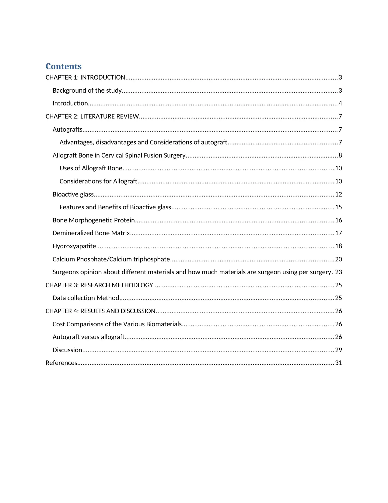
Contents
CHAPTER 1: INTRODUCTION........................................................................................................................3
Background of the study..........................................................................................................................3
Introduction.............................................................................................................................................4
CHAPTER 2: LITERATURE REVIEW................................................................................................................7
Autografts................................................................................................................................................7
Advantages, disadvantages and Considerations of autograft..............................................................7
Allograft Bone in Cervical Spinal Fusion Surgery......................................................................................8
Uses of Allograft Bone.......................................................................................................................10
Considerations for Allograft...............................................................................................................10
Bioactive glass.......................................................................................................................................12
Features and Benefits of Bioactive glass............................................................................................15
Bone Morphogenetic Protein................................................................................................................16
Demineralized Bone Matrix...................................................................................................................17
Hydroxyapatite......................................................................................................................................18
Calcium Phosphate/Calcium triphosphate............................................................................................20
Surgeons opinion about different materials and how much materials are surgeon using per surgery. 23
CHAPTER 3: RESEARCH METHODLOGY......................................................................................................25
Data collection Method.........................................................................................................................25
CHAPTER 4: RESULTS AND DISCUSSION.....................................................................................................26
Cost Comparisons of the Various Biomaterials......................................................................................26
Autograft versus allograft......................................................................................................................26
Discussion..............................................................................................................................................29
References.................................................................................................................................................31
CHAPTER 1: INTRODUCTION
Background of the study
A surgical repair or replacement is necessitated by trauma, diseases or degeneration.
When an individual experiences pain in the joints, the main focus tends to relief of the pain
restoration of the health of the individual to a not only healthy but functional lifestyle. This
normally comes with the replacement of some of the skeletal parts including hips, finger joints,
elbows, teeth, knees, vertebrae or even repair of the mandibles. The value of biomedical
materials globally currently stands at approximately $24,000M with dental and orthopedic
applications accounting for about 55% of the total market for biomaterials. In the year 2000, the
value of orthopedic products in the market was more than $13 billion and an expansion in such
trends expected to continue citing a number of factors among them the ageing population,
advancements and improvements in technology and lifestyle, an increased taste and preference
by the younger to middle aged population to undertake surgery, desire for better function,
enhanced aesthetics as well as better comprehension of the functionality of the body.
The use and requirements for biomaterials in tissue engineering is strictly defined with
biocompatibility being the top of the agenda for the choice of any material for use for their
orthopedic or diagnostic purposes. For the case of biocompatibility, the scaffolds and bioreactors
have to attain qualities among them tissue friendly so as not to invoke an immune-response. At
best, biomaterials should be supportive of the cellular and tissue functions among the adhesion,
proliferation and differentiation through its special surface in chemistry. Another important
requirement for scaffolds is porosity which is supposed to be at least 90% so as to allow cells to
seed evenly and promote vascular growth after implantation. Controlled biodegradation gains
concern especially under circumstance that implanted materials are replaced by healthy tissues
⊘ This is a preview!⊘
Do you want full access?
Subscribe today to unlock all pages.

Trusted by 1+ million students worldwide
and then biomaterial slowly degrades in the body of the host. Biomaterials are either natural or
synthetic.
Introduction
The science of biomaterials was in place more than a century ago when artificial devices
and objects were enhanced to the point where they could replace different components of the
human body. A biomaterial is a substance that can has been tailored to interact with the systems
of biology for the purposes of medical practice. These composites have the ability to be in
contact with the fluids of the body and tissues from relatively long periods of time without
invoking any significant if any adverse reactions with the body systems. Biomedical materials
are used either of therapeutic i.e. treatment, replacement or repair of a tissue that functions in the
body or they can be used for diagnostic purposes (Bandyopadhyay, 2013). They are non-viable
materials that have the capability to be implanted to replace or repair a tissue that is missing or
worn out. Biomaterial can either be synthesized in the laboratory or be of natural origin.
Biomaterials traces its roots as far back as the ancient Phoenicia during which gold wires
were used in binding together loose teeth by tying artificial teeth to the neighboring natural teeth.
Implementation of bone plates was successful in stabilization of bone fractures and thus
accelerating the process of healing in the 1900s. Artificial hip joints and heart valves were under
development and replacement of blood vessels was undergoing clinical trials in the 1950s and
60’s (Black, 2014).
Biomaterials are categorized into metals, ceramics, polymers and natural biomaterials.
Metals are the group of biomaterials that is most commonly used due to the ability to bear loads.
This load bearing ability makes metals ideal for such practices as knee implants and total hip
replacements. These metallic implants comes in various ranges of shapes and sizes and do vary
Paraphrase This Document
Need a fresh take? Get an instant paraphrase of this document with our AI Paraphraser
from modest wires to screw to plates for fixation of fractures and to as complicated as total joint
prostheses. artificial joint implantation have been successful on joints including ankles, shoulder,
hops and knees with different metals currently being used in various medical applications to
meet different surgical needs. Cobalt-base alloys, stainless steel, titanium alloys and pure
titanium are among the most commonly used metals as biomaterials (Dobke, 2013).
A good biomaterial of metallic nature should have the same values of Young’s modulus
as the bone or the tissues for which it is replacing. The surrounding tissues may degenerate as a
result of lack of mechanical loading in cases where the biomaterial is carrying too much loading.
On the other hand, a biomaterial that cannot carry adequate load cannot make an adequate
replacement (DiMasi, 2014).
Ceramics and composites: these biomaterials are mostly used on dental applications
including fillings, crowns and dentures. The application and utilization of ceramics is limited to
the cats that they have poor toughness to fracture due to their brittle property. Ceramics cannot
be applied in implantations that required high tolerance of loads. Despite the shortcomings,
calcium phosphates have been applied in augmentation and bone repair while alumina and
zirconia have formed fundamental materials in joint replacement (Black, 2014).
Composites widely find their applications in dentistry. PMMA-glass filter and BIS-
GMA-quartz-silica filler are the two most commonly used composites and are mostly used for
dental filling and restoration. Other applications of composts include prostheses which are
favored by heir light weight and high loading capability.
Polymers are grouped as one of the most versatile biomaterials and have the widest range
of applications from scaffolds used in tissue engineering to the PLGA that is used in drug
delivery. It is possible to mold polymers into any desirable shapes or size. Collagen or synthetic
fibre is used in making scaffolds that are used in tissue engineering (Bronzino, 2016). Scaffolds
are very important as they offer both the molecular and physical cues that control development
of the donor cells. The extra cellular matrix is normally used in the provision of these cues.
Nevertheless, the polymers also have the capability to provide protection to the donor cells from
the different immune systems that are available as a result of the transplant. Lastly, the three
dimensional space offers a convenient way of package to the cells thereby prompting growth
after transplantation has been done. It is possible to design the pores in the polymers in such a
way that they are large enough to allow the growth of endothelial cells and blood vessels into the
scaffold.
⊘ This is a preview!⊘
Do you want full access?
Subscribe today to unlock all pages.

Trusted by 1+ million students worldwide
CHAPTER 2: LITERATURE REVIEW
Autografts
Autograft, also known as autologous bone or autogenous bone graft is harvesting of the bone
from a patient and transferring it to the part of the spine that is to be fused. A separate spinal
fusion procedure is carried out during spinal fusion surgery to extract a bone from another part of
the body of the patent and place it in the region of the spine that is to undergo fusion. This is a
surgical process and is termed as harvesting of the bone graft (Bronzino, 2016). The procedure is
normally carried out through a separate incision on anterior fusions and through a same incision
in the posterior fusions.
Harvesting of the bone is normally done from one of the bones of the patient in the iliac
crest also known as the pelvis. Under other circumstances, this can also be extracted from the rib
or another region of the spine. Very few spine procedures include autograft harvesting. This is
attributed to the morbid nature of the bone graft harvest procedure as well as an ever increasing
number of better and more reasonable alternatives to autograft.
Advantages, disadvantages and Considerations of autograft
As a result of bearing all the features that are required for a solid bridge to grown, autograft is
perceived to be the gold standard in the achievement of a successful and solid spine fusion.
Among those characteristics include:
It is composed of osteophytes which are the bone growing cells as well as bone-growing
portions also called bone morphogenic proteins which are all used in facilitating the
growth of new bone in the patient.
It offers the spinal fusion with calcium scaffolding which forms a base on to which the
new bone grows (Williams, 2014)
Paraphrase This Document
Need a fresh take? Get an instant paraphrase of this document with our AI Paraphraser
There are two main benefits or advantages that are associated with autograft:
There is no risk of transmission of disease
There are higher chances of success of fusion versus allograft (cadaver bone) as well as
some types of substitutes for bone graft
Risks and chances of complications that are associated with carrying out any surgical
procedure is the main disadvantage of autograft. Among such risks include bleeding, injury to
the nerves as well as surgical wound problems for example infection (Guda, 2017).
Still, there is a risk of chronic pain associated with autograft procedures at the site of harvest
of the bone. The incidence of ongoing pain is a factor of the technique adopted in the harvesting
for the case of posterior incisions. The incidences of such pain are usually very low in actual
practice. Research has established that site pain of ongoing graft is relatively higher in separate
incision rather than using the same incision. The pain is worse in cases where the graft requires
three cortical surfaces for example a structural bone graft that is to be use on an interbody fusion
(Gunzburg, 2012).
Limitation in the supply of this type of bone graft is as well a disadvantage. Under some
circumstances, bone graft may need to be supplemented with some bone graft substitute form in
order to meet the required expectations.
Allograft Bone in Cervical Spinal Fusion Surgery
Allograft is a bone that has been extracted from a cadaver by a tissue bank for the purposes of
medical procedures. Allograft can be prepared in a number of various forms among them chips
for use in a spine fusion. In as much as allograft bone only offers a calcium scaffolding, has not
any bone growing cells or proteins that grow bones needed to ignite growth of a new bone and
thus has lower chances of fusion in comparison to using the patient’s own bone, it has been
proved to be comparable in some studies to autograft when it comes to generating successful
fusions (Ramalingam, 2012).
Allograft bone also known as donor bone or bank bone from a cadaver does away with the need
to harvest the bone of the patient. Instead, the donor graft serves as bone scaffolding that offers
the platform for the growth of the patient’s bone and finally replaces it over years. Chances of
rejection of the graft are very minimal as there are no living cells in the bone graft just contrary
to the case of organ transplant (Patel, 2013).
This notwithstanding, healing of bone graft still remains an issue of concern as there is higher
likelihood that the bone graft may fail with the allograft bone in comparison with autograft. This
has then generated studies that have been able to make comparison between allograft and
autograft with regard to production of successful fusions (Peterson, 2012). The rate of healing in
allografts is relatively slower as compared to a fusion involving an autograft bone. Still,
Allograft produces almost equivalent rates of fusion as autograft bone in one level spinal
fusion
More significance is continuously being attached to the differences in the rates of fusion
between autograft and allograft as a result of the ever increasing number of levels that are
to be fused or grafted (Nakai, 2015)
To increase the rates of fusion, anterior cervical instrumentation such as plates and
screws are used.
⊘ This is a preview!⊘
Do you want full access?
Subscribe today to unlock all pages.

Trusted by 1+ million students worldwide
A theoretical risk of transmission of disease from a donor exists and such a risk has been
established to be between 1 in 200,000 and 1 in 1 million for the cases if HIV and hepatitis. This
risk has been reduced even further with modern procurement and sterilization methods.
Uses of Allograft Bone
Allograft bone is usable either on its own or as a supplement to the patient’s own bone. It
is used on its own in the lumbar spine in which its use is restricted to PLIF or ALIF procedures.
In either of these procures, the bone graft is put in compression between the vertebrae and as the
compression continues, a better healing process is achieved for the bone. The use of allograft
bone by itself in spinal fusion of a posterolateral gutter where the bone is put in tension may not
yield a solid fusion (Kaur, 2017). In the case of using allograft bone as a supplement to the
patient’s own bone, the chips of allograft are used in augmenting the bone of the patient in a
posterolateral gutter fusion. This is also applicable in cases where more bone graft is required in
fusion procedures that are more extensive for example spin fusion for adolescent scoliosis.
Considerations for Allograft
Any surgical risk associated with harvesting of the patient’s own bone is eliminated when
allograft bone is used. The main drawbacks of allograft include:
Risks of disease transmission: In as much as there are strict rules and regulations with
regard to processing and procedures of human tissue for tissue banks, a small potential
risk for disease transmission still exists from using cadaver bone (Manbachi, 2016)
Low chances of fusion: This is probable due to the fact that allograft bone is not
composed of living bone cells which reduces the effectiveness of stimulation of fusion as
the own bone of the patient.
Paraphrase This Document
Need a fresh take? Get an instant paraphrase of this document with our AI Paraphraser
Bone banks also called tissue banks are the main source of allograft bone that is used in spine
fusion procedures. The tissue bank is controlled and regulated by the US Food and Drug
Administration so as to reduce the risk of contamination of tissue as well as transmission of
diseases. Demineralized bone matrix, (DBM) is one of the forms of allograft that is commonly
used. Demineralized bone matrix is a commercialized form of the bone morphogenic protein that
is a derivative of processing cadaver bone from various donors (Nakai, 2015). The quantity of
BPM available in the material varies from one manufacturer to another but is far less available
through the use of synthetic recombinant BPM. The stem cell technology is a recent form of
cadaver-donated tissue that makes use of stem cells from a donor to aid in the creation of a bone.
Biomaterials are classified depending on the responses of tissues into three categories:
bioactive, bio inert and bioresorbable. Bio inert biomaterials are devices that do not have any
interactions with the surrounding body tissues once they are placed in the human body. Among
such materials include stainless steel, polyethene, alumina, stabilized zirconia, titanium among
other materials (Benzel, 2012). It is possible that a fibrous capsule forms a bio inert implant
making the functionality of the implant be a factor of tissue integration through the implant
made. Bioactive biomaterials on the other hand refer to materials that interact with the
surrounding bones of the bone upon as soon as they are placed with the human body. In some
cases, these materials react with the soft tissues as well. The reaction takes place through a
kinematic modification of the surface that depends on time and is invoked by the implantation
within the living tissue or bone.
The interaction results into a reaction of exchange of cations between the nearby body
fluids and the bioactive implant which leads to the formation of a carbonate apatite layer which
is biologically active. This layer is formed on the implant and is equivalent to the mineral phase
in the bone in terms of the chemical and crystallographic composition (Ong, 2017). The most
common examples of these materials include bio glass, hydroxyapatite as well as glass ceramic
A-W.
Bioresorbable materials on the other hand are biomaterials that dissolve and are gradually
replaced by advancing tissues such as bones when they are placed within the human body.
Tricalcium phosphate and polylactic-polyglycolic acid copolymers are the most commonly
examples of bioresorbable biomaterials.
Bioactive glass
These are inorganic materials that have clear bonding of bones and osteoinductive
properties. The characteristics of the surface of bio active glass have an impact in the facilitation
of rapid colonization and bonding of the cells that form bones. Cells that form bone differentiate
due to the release of ions when the material comes into contact with the body fluids. Among the
ions release includes zinc, copper and strontium (Kalaskar, 2016). Bioactive glass materials are
effective in the treatment of osteomyelitis in which they are associated with the production of
antigenic factors. They influence degradation and bioactivity through changing the chemical
composition and structure.
Clinical evidence in the application of bio active materials in bone replacement is limited
even though the mechanical and surface characteristic properties of the material are favorable for
the said application. A recent clinical trial and study was conducted in tibia plateau fractured and
the findings illustrated that the material is comparable to autologous bone.
A promising material has been identified in bioactive glass which is differentiated from
the ancient synthetics such as calcium phosphates since the material has the potential of
⊘ This is a preview!⊘
Do you want full access?
Subscribe today to unlock all pages.

Trusted by 1+ million students worldwide
1 out of 33
Your All-in-One AI-Powered Toolkit for Academic Success.
+13062052269
info@desklib.com
Available 24*7 on WhatsApp / Email
![[object Object]](/_next/static/media/star-bottom.7253800d.svg)
Unlock your academic potential
Copyright © 2020–2026 A2Z Services. All Rights Reserved. Developed and managed by ZUCOL.

