Detailed Technical Report: Blood Pumping in the Human Heart
VerifiedAdded on 2023/05/28
|6
|1149
|113
AI Summary
This technical report provides a detailed explanation of how the human heart pumps blood throughout the circulatory system. It begins with an introduction to the heart's role in the circulatory system, emphasizing its muscular structure and connection to major blood vessels like the aorta and vena cava. The report describes the heart's four chambers—the atria and ventricles—and their coordinated function in pumping blood. It also details the four heart valves (mitral, tricuspid, aortic, and pulmonic) and their role in preventing backflow. The report explains the blood flow through both the right and left sides of the heart, including the oxygenation process in the lungs. The right side receives deoxygenated blood from the vena cava, which then flows to the lungs via the pulmonary artery. The left side receives oxygenated blood from the pulmonary vein and pumps it to the rest of the body via the aorta, concluding with the importance of continuous blood circulation for sustaining life. Desklib provides this document along with other solved assignments for students.
Running head: ADVANCED TECHNICAL WRITING 1
Advanced technical Writing
Name
Institution Affiliation
Advanced technical Writing
Name
Institution Affiliation
Paraphrase This Document
Need a fresh take? Get an instant paraphrase of this document with our AI Paraphraser
ADVANCED TECHNICAL WRITING 2
Description of How the Heart Pumps Blood in the Human Body
Introduction
The heart has been place at the central place of the circulatory system of all human
beings. Accordingly, the circulatory system is a network of blood vessels which convey
blood to each part of the human body. Therefore, the blood carries oxygen as well as other
significant elements such as nutrients required by the rest of the human body organs for their
healthy stay and proper functioning. When the heart is viewed from outside one will notice
that it made of muscles. Therefore, the heart is a muscular organ whose primary job is to
pump blood throughout the circulatory system. Indeed, the heart has strong muscular walls to
allow for contraction in order to pump blood out of the heart through the arteries (Clayton, &
Hose, 2017). The arteries are muscular-walled tubes which form part of the circulatory
system through which blood that mainly contain oxygen is transported from the heart to all
part of the body.
Accordingly, the heart has main blood vessels which are connected to it such as the
aorta which is the chief artery of the body, the superior vena cava, the inferior vena cava and
the pulmonary artery. The superior vena cava is the primary vein which convey deoxygenated
blood coming from different body parts into the heart. Therefore there are two types of vena
cava in the human body the superior vena cava which carry blood from the head, and other
upper body parts while the inferior vena cava carries blood from the lower body parts. The
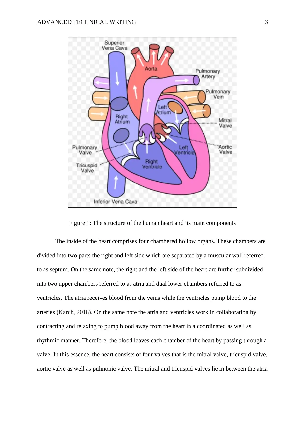
figure below is in an illustration of the structure of the human heart and its main components.
In this sense the arrows pointing away from the heart represents the flow of blood way from
the heart to other body parts while arrows pointing towards the heart is the flow of blood
from towards the heart from other body parts.
ADVANCED TECHNICAL WRITING 3
Figure 1: The structure of the human heart and its main components
The inside of the heart comprises four chambered hollow organs. These chambers are
divided into two parts the right and left side which are separated by a muscular wall referred
to as septum. On the same note, the right and the left side of the heart are further subdivided
into two upper chambers referred to as atria and dual lower chambers referred to as
ventricles. The atria receives blood from the veins while the ventricles pump blood to the
arteries (Karch, 2018). On the same note the atria and ventricles work in collaboration by
contracting and relaxing to pump blood away from the heart in a coordinated as well as
rhythmic manner. Therefore, the blood leaves each chamber of the heart by passing through a
valve. In this essence, the heart consists of four valves that is the mitral valve, tricuspid valve,
aortic valve as well as pulmonic valve. The mitral and tricuspid valves lie in between the atria
⊘ This is a preview!⊘
Do you want full access?
Subscribe today to unlock all pages.

Trusted by 1+ million students worldwide
ADVANCED TECHNICAL WRITING 4
and ventricles while the aortic and pulmonic valves are found in between the ventricles and
main blood vessels leaving the heart.
Consequently, the function of these valves is to prevent the blood from flowing in the
back direction. As a result, each valve consists of a set of flaps known as cusps. These valves
comprises three cusps each with the exception of the mitral valve which has only two cusps.
These cusps are ascribed to and sustained by a rings of fibrous and tough tissues referred to
as annulus which aid to maintaining the appropriate form of the valve. Similarly, the cusps
located on the tricuspid and mitral vales are supported by additional tough fibrous strings
known as chordae tendineae. The above figure demonstrates the location of different valves
on the heart.
How the blood flow through the heart
The heart has two side the right and left side which work together in the following
pattern in a repeated manner to enable the continuous flow of blood to the heart, the lungs
and all other parts of the body.
The right side of the heart
The bloods finds its way to the heart via two major veins the inferior and superior
vena cava containing blood with deprived oxygen from the right atrium. The atrium
diminishes to allow blood flow to the right ventricle through the tricuspid valve. When the
ventricles get full the tricuspid vale close preventing the back flow of blood to the right
atrium while the ventricles diminishes (Colli, et al., 2015). When the ventricle diminishes the
blood moves out of the heart through the pulmonary valve to the pulmonary artery to the
lungs for oxygenation. The blood rich in oxygen flow back to the heart via the pulmonary
vein.
Left side of the heart
Paraphrase This Document
Need a fresh take? Get an instant paraphrase of this document with our AI Paraphraser
ADVANCED TECHNICAL WRITING 5
The pulmonary vein is responsible for emptying blood rich in oxygen from the lungs
through the left atrium which diminishes to allow blood to flow to the left ventricles via the
mitral valve (Moazami et al., 2015). Once the ventricle is full of blood the mitral valve closes
to prevent the backflow of blood to the atrium while the ventricles contracts allowing the
blood to flow out of the heart via the aortic valve to the aorta for transport to different parts of
the body.
In conclusion, the blood circulation maintains a constant flow through the heart
constantly pumping throughout an individual life.
ADVANCED TECHNICAL WRITING 6
References
Clayton, R. H., & Hose, D. R. (2017). Excitation-Contraction in the Heart. In Cardiovascular
Biomechanics (pp. 107-126). Springer, Cham.
Colli, A., Zucchetta, F., Torregrossa, G., Manzan, E., Bizzotto, E., Besola, L., & Gerosa, G.
(2015). Transapical off-pump mitral valve repair with Neochord Implantation (TOP-
MINI): step-by-step guide. Annals of cardiothoracic surgery, 4(3), 295.
Karch, D. (2018). U.S. Patent Application No. 10/080,830.
Moazami, N., Dembitsky, W. P., Adamson, R., Steffen, R. J., Soltesz, E. G., Starling, R. C.,
& Fukamachi, K. (2015). Does pulsatility matter in the era of continuous-flow blood
pumps?
⊘ This is a preview!⊘
Do you want full access?
Subscribe today to unlock all pages.

Trusted by 1+ million students worldwide
1 out of 6
Related Documents
Your All-in-One AI-Powered Toolkit for Academic Success.
+13062052269
info@desklib.com
Available 24*7 on WhatsApp / Email
![[object Object]](/_next/static/media/star-bottom.7253800d.svg)
Unlock your academic potential
Copyright © 2020–2025 A2Z Services. All Rights Reserved. Developed and managed by ZUCOL.


