Rapid Review: MRI Effectiveness in Breast Cancer Diagnosis
VerifiedAdded on 2022/09/17
|14
|2920
|23
Report
AI Summary
This rapid review analyzes the effectiveness of MRI imaging in the early diagnosis of breast cancer, particularly among young, high-risk women. The study examines existing literature, focusing on systematic reviews to understand how MRI enhances the detection of breast cancer. The review considers the limitations of traditional screening methods like mammography, especially for younger women, and highlights MRI's potential for detecting small lesions. The methodology includes a detailed search strategy across databases like MEDLINE, PubMed, and EMBASE, using specific keywords and eligibility criteria (studies published between 2009-2019 in English). The methodological quality of the included studies will be assessed using AMSTAR 2. The data extracted will include study population, intervention (MRI imaging), and outcomes. The findings, presented narratively with tables and figures, aim to determine the significance of MRI in early breast cancer detection and address the increasing prevalence among young women.

A rapid review of the effectiveness of MRI Imaging in early diagnosis of breast cancer
in high-risk women
in high-risk women
Paraphrase This Document
Need a fresh take? Get an instant paraphrase of this document with our AI Paraphraser
Objectives
The main purpose of this rapid review is to analyse and summarise the most appropriate
evidences from the literature in order to understand the effectiveness of MRI Imaging in early
diagnosis of breast cancer in high-risk women. More specifically this rapid review will focus
on the following question:
How MRI imaging can help in enhancing the possibility of detection of breast cancer among
young and high risk women?
Keywords: MRI imaging, breast cancer, MRI in cancer screening, detecting breast cancer,
systematic reviews
Background
Breast cancer is the most common form of cancer occurring among the women. Women, who
have the strong family history of breast cancer, have undergone any kind of chest radiation
therapy before 30 years of age, or have the predisposing genetic mutation
(e.g., BRCA1 or BRCA2 mutation) are at the higher risk of developing breast cancer (20% to
65%) in comparison to general population (11%) (Wang, 2017). The effective diagnosis of
breast cancer at the early stage can significantly help in reducing the breast cancer related
mortality and morbidities. Most of the techniques used for breast cancer screening have some
limitations, such as they are expensive, time consuming and not suitable for younger women
(Roganovic et al, 2015). Magnetic resonance imaging is being considered as the most
effective screening modality for early detection of breast cancer. Therefore, this study will
conduct a critical review of the existing evidences from the literature in order to understand
the effectiveness of MRI Imaging in early diagnosis of breast cancer in high-risk women.
The aim of this research is to examine and analyse the effectiveness of MRI imaging for the
early diagnosis of breast cancer among the women who are at the higher risk. The main
objective is to understand the significance of early detection of the breast cancer, as the
prevalence and incidences of breast cancer is significantly increasing among young women.
Memon et al (2015) have informed that the family history of breast cancer and exposure to
radiations can result in increasing the risk of this cancer among young women and the late
diagnosis of the disease can increase the mortality rates (Paluch-Shimon et al, 2016).
Therefore, there is a substantial need of appropriate and effective screening tools for the early
detection of the breast cancer among young women.
Various forms of diagnostic approaches for breast cancer have been studied by the
investigators, such as computerised tomography, mammography, positron emission
tomography, biopsy, and ultrasound (Roganovic et al, 2015). Garber (2009) and Roganovic et
al (2015) identified that MRI imaging could be highly significant in early detection of breast
cancer. Mammography is the most popular and current standard used for the diagnosis and
detection of breast cancer. However, studies have identified that, this screening tool is not
effective for women under the age of 40 (Onega et al, 2016). Hassan & El-Shenawee (2011)
informed that MRI imaging could be more effective in detecting the small lesions among
young women that could not be detected through mammography. However, a gap has been
identified in the literature towards understanding the significance of MRI in early diagnosis
of breast cancer specifically in the case of young women who are at higher risk. This study
will help in filling this gap by informing about the effectiveness of MRI for early detection of
breast cancer among younger women.
Criteria for Considering Studies for this Review
Types of Studies
The systematic review studies will be evaluated in this study in order to identify the
effectiveness of MRI imaging in early detection of breast cancer among young high risk
women. Systematic reviews are considered to be most significant in developing evidence
based knowledge (Edwards, 2014). Studies have identified that breast cancer is also the most
common malignancy that is identified among 6.6% cases diagnosed among the young women
(Assi et al, 2013). According to Australian Government Cancer Statistics, in the year 2019
breast cancer accounted for 23% of all the new cancer cases among the women aged between
20 and 39. Breast cancer also resulted in 21% deaths among the women aged 20-39 (Cancer
Australia, 2019). Therefore, breast cancer is being considered as the most common form of
cancer resulting in higher rate of mortality among young women.
Types of Participants
This review will include the study that have the appropriate context and have included ‘young
women’ who are at high risk of breast cancer, or screening and imaging for early detection of
breast cancer. Diagnosis and treatment of breast cancer among young women require special
consideration, because of the various complexities associated with cancer diagnosis in young
age (Houssami & Cho, 2018). Therefore, the context of the research is to focus on wider
⊘ This is a preview!⊘
Do you want full access?
Subscribe today to unlock all pages.
Trusted by 1+ million students worldwide
literature and conduct a critical analysis of the studies that mainly discuss the significance of
early detection of breast cancer in younger women by using MRI imaging.
Types of Interventions
Rocha-Brischiliari et al (2017) have indicated the significance of high sensitivity and rapid
detection techniques for early diagnosis of breast cancer among young and high risk women.
Studies have also identified that detection of breast cancer among young women is more
challenging than in older women, as the cancer symptoms may present differently and may
be difficult to be detected (Houssami & Cho, 2018). Therefore, the breast cancer screening
tools and techniques will be considered in order to identify the significance and importance of
Magnetic resonance imaging for the rapid early detection of breast cancer in younger women
who are at higher risk of developing this disease (Cho et al, 2017). Stuckey & Onstad, (2015)
espoused that MRI is highly effective in detecting invasive carcinoma as well as carcinoma in
situ, which is not visible through other screening modalities and thus considered as highly
significant in high risk patients. Therefore, the main intervention included in this study will
be MRI imaging and how significant and effective it is for early detection of breast cancer.
Search Strategy
The main of the search strategy is to identify the peer reviewed systematic review studies. All
the studies that were searched through online search were assessed on the basis of their
relevance to the study problem provided in their title, abstract and the other description. The
search of the studies is also based on the appropriate PRISMA Flow (included in Appendix
A). Therefore, the main medical databases will be searched with appropriate use of keywords.
The five important data bases that will be used for conducting search are:
1. MEDLINE
2. PubMed
3. Cochrane
4. EMBASE
5. CINAHL
The eligibility criteria that would be applied for the inclusion of the studies are that all studies
that are published with the period of 2009 to 2019 will be included for the review. The time
period of 10 years is identified to be significant for this research, as it will help in obtaining
Paraphrase This Document
Need a fresh take? Get an instant paraphrase of this document with our AI Paraphraser
the recent evidences. Studies that are only published in English language will be included.
The detailed search strategy can be accessed in Appendix B.
Methods of Review
Assessment of methodological quality
The methodological validity and quality of the studies will be analysed on the basis of critical
appraisal instruments called as Assessment of Multiple Systematic Reviews (AMSTAR 2).
According to Shea et al (2017) that main aim of AMSTAR is to evaluate the systematic
review of the randomised control trials. AMSTAR 2 has been developed in order to improve
the process of reviewing systematic reviews (Lorenz et al, 2019). This tool contains 16
different items that help in assessing the validity and reliability of the systematic review
studies. The domain specific questions are answered in this tool under which ‘yes’ denotes
positive response and in case of some missing information ‘no’ is used (Shea et al, 2017). In
AMSTAR 2, the ‘partial yes’ has also been provided as an additional response for some
specific instances, where the researcher identifies any kind of partial response to the review
standard (Lorenz et al, 2019).
Data Extraction
The data collected for this review will be based on the appropriate information collection.
The data extracted will provide the specific details about the study population, intervention,
study methods and outcomes. MRI has been identified as a high sensitive technique that helps
in effective detection of malignant breast lesion in young women, who have dense breast
tissues in comparison to older women (Paluch-Shimon et al, 2016). For this study the data
extracted will inform if the study population are young women who are at high risk or not,
including the intervention of use of MRI imaging in early cancer detection, as well as the
study outcome that will inform if MRI imaging is effective in early detection of breast cancer
in young women or not.
Data Synthesis
The narrative approach will be used for providing the findings of this rapid review. The
findings will also be presented in the form of tables and figures where ever appropriate.
References
Assi, H. A., Khoury, K. E., Dbouk, H., Khalil, L. E., Mouhieddine, T. H., & El Saghir, N. S.
(2013). Epidemiology and prognosis of breast cancer in young women. Journal of
thoracic disease, 5(Suppl 1), S2.
Cancer Australia. (2019). Breast Cancer in Young Women. Australian Government.
Retrieved from:
https://breast-cancer-in-young-women.canceraustralia.gov.au/statistics
Cho, N., Han, W., Han, B. K., Bae, M. S., Ko, E. S., Nam, S. J., ... & Song, B. J. (2017).
Breast cancer screening with mammography plus ultrasonography or magnetic
resonance imaging in women 50 years or younger at diagnosis and treated with breast
conservation therapy. JAMA oncology, 3(11), 1495-1502.
Edwards, T. B. (2014). What is the value of a systematic review?. Journal of shoulder and
elbow surgery, 23(1), 1-2.
Garber, J. E. (2009). Identifying and assessing women at high risk for breast cancer. Breast
Cancer Research, 11(1), S1.
Hassan, A. M., & El-Shenawee, M. (2011). Review of electromagnetic techniques for breast
cancer detection. IEEE Reviews in Biomedical Engineering, 4, 103-118.
Houssami, N., & Cho, N. (2018). Screening women with a personal history of breast cancer:
overview of the evidence on breast imaging surveillance. Ultrasonography, 37(4),
277.
Lorenz, R. C., Matthias, K., Pieper, D., Wegewitz, U., Morche, J., Nocon, M., ... & Jacobs,
A. (2019). A psychometric study found AMSTAR 2 to be a valid and moderately
reliable appraisal tool. Journal of clinical epidemiology.
Memon, Z. A., Kanwal, N., Sami, M., Larik, P. A., & Farooq, M. Z. (2015). Risk of breast
cancer among young women and importance of early screening. Asian Pac J Cancer
Prev, 16(17), 7485-9.
Onega, T., Goldman, L. E., Walker, R. L., Miglioretti, D. L., Buist, D. S., Taplin, S., ... &
Smith-Bindman, R. (2016). Facility mammography volume in relation to breast
cancer screening outcomes. Journal of medical screening, 23(1), 31-37.
⊘ This is a preview!⊘
Do you want full access?
Subscribe today to unlock all pages.
Trusted by 1+ million students worldwide
Paluch-Shimon, S., Pagani, O., Partridge, A. H., Bar-Meir, E., Fallowfield, L., Fenlon, D., ...
& Harbeck, N. (2016). Second international consensus guidelines for breast cancer in
young women (BCY2). The Breast, 26, 87-99.
Rocha-Brischiliari, S. C., de Oliveira, R. R., Andrade, L., Brischiliari, A., Gravena, A. A. F.,
de Barros Carvalho, M. D., & Pelloso, S. M. (2017). The rise in mortality from breast
cancer in young women: trend analysis in Brazil. PloS one, 12(1), e0168950.
Roganovic, D., Djilas, D., Vujnovic, S., Pavic, D., & Stojanov, D. (2015). Breast MRI, digital
mammography and breast tomosynthesis: comparison of three methods for early
detection of breast cancer. Bosnian journal of basic medical sciences, 15(4), 64.
Shea, B. J., Reeves, B. C., Wells, G., Thuku, M., Hamel, C., Moran, J., ... & Henry, D. A.
(2017). AMSTAR 2: a critical appraisal tool for systematic reviews that include
randomised or non-randomised studies of healthcare interventions, or both, Bmj, 358,
j4008.
Wang, L. (2017). Early diagnosis of breast cancer. sensors, 17(7), 1572.
Paraphrase This Document
Need a fresh take? Get an instant paraphrase of this document with our AI Paraphraser
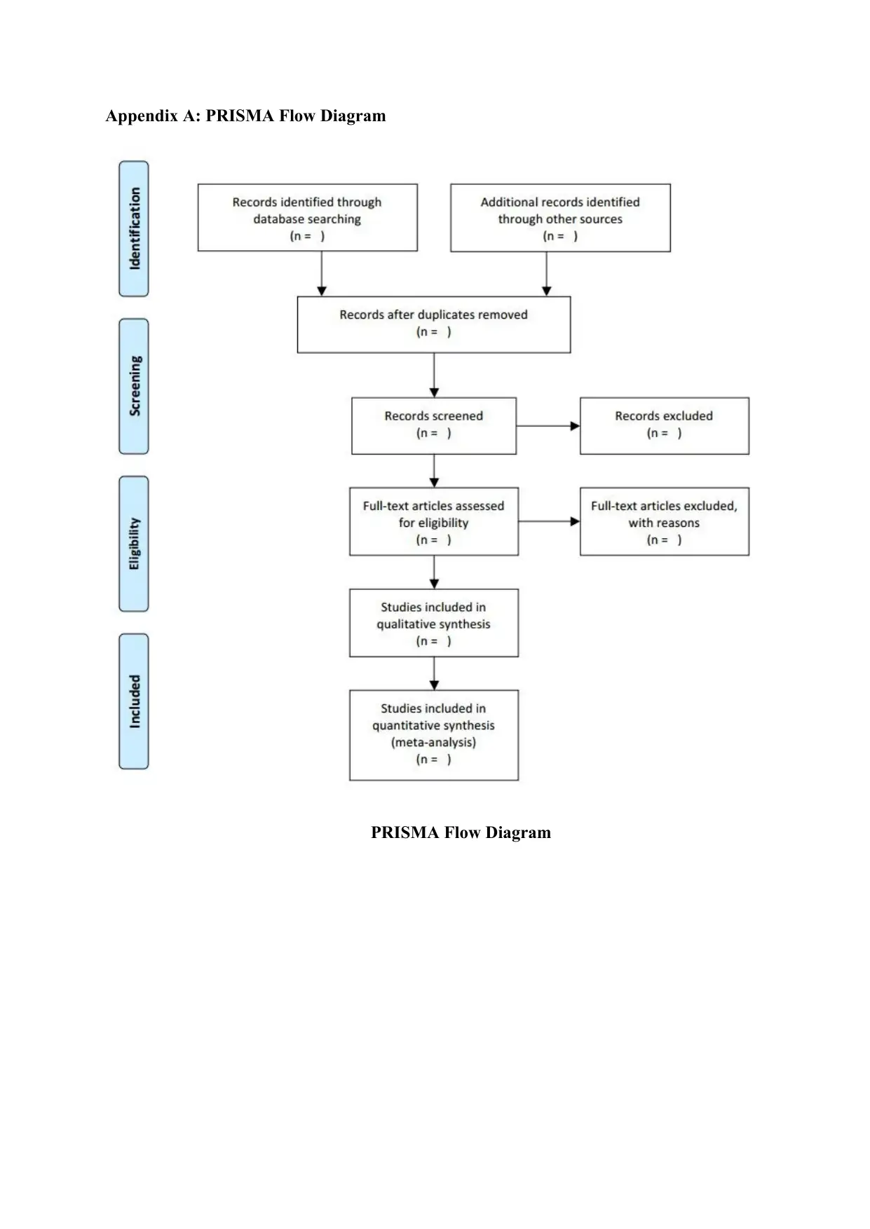
Appendix A: PRISMA Flow Diagram
PRISMA Flow Diagram
PRISMA Flow Diagram

Appendix B: Search Strategy for PubMed
1. ‘Breast cancer’
2. ‘Breast cancer and young women’ or ‘breast cancer prevalence’
3. ‘Cancer Screening’
4. ‘MRI imaging and cancer screening’
5. ‘MRI’ or ‘mammography’
6. ‘cancer in women’
7. ‘High risk women and breast cancer’
8. ‘MRI imaging in high risk women’
9. ‘Cancer detection among women’
10. ‘Breast cancer detection and young women’
11. ‘systematic review’
12. ‘meta-analysis’ and ‘breast cancer’
13. ‘Language – English
14. Year 2009- current
⊘ This is a preview!⊘
Do you want full access?
Subscribe today to unlock all pages.
Trusted by 1+ million students worldwide
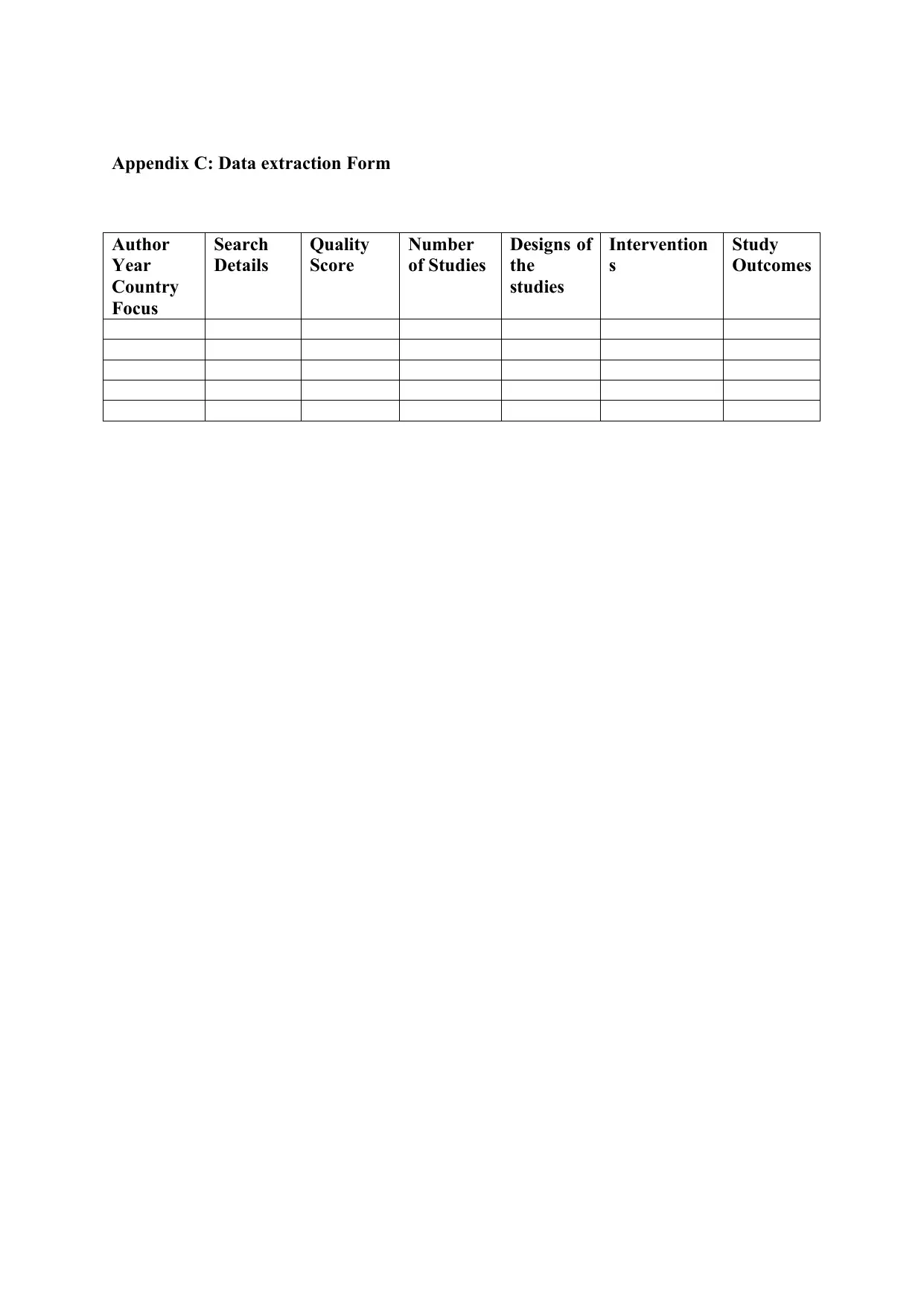
Appendix C: Data extraction Form
Author
Year
Country
Focus
Search
Details
Quality
Score
Number
of Studies
Designs of
the
studies
Intervention
s
Study
Outcomes
Author
Year
Country
Focus
Search
Details
Quality
Score
Number
of Studies
Designs of
the
studies
Intervention
s
Study
Outcomes
Paraphrase This Document
Need a fresh take? Get an instant paraphrase of this document with our AI Paraphraser
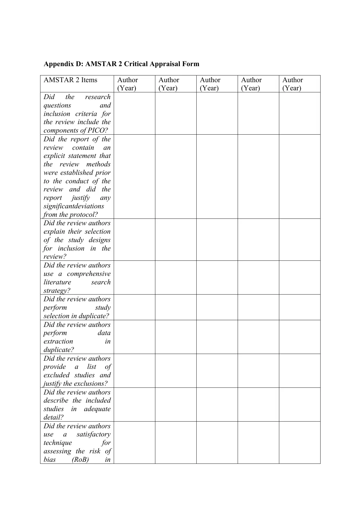
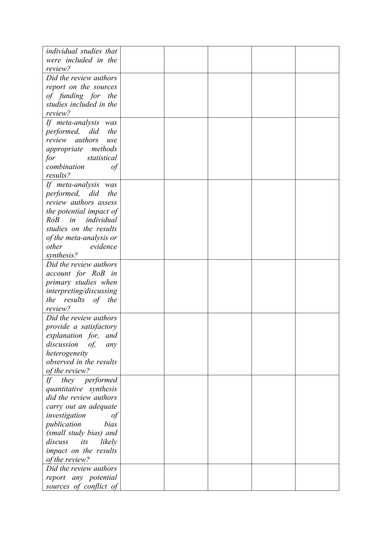
Appendix D: AMSTAR 2 Critical Appraisal Form
AMSTAR 2 Items Author
(Year)
Author
(Year)
Author
(Year)
Author
(Year)
Author
(Year)
Did the research
questions and
inclusion criteria for
the review include the
components of PICO?
Did the report of the
review contain an
explicit statement that
the review methods
were established prior
to the conduct of the
review and did the
report justify any
significantdeviations
from the protocol?
Did the review authors
explain their selection
of the study designs
for inclusion in the
review?
Did the review authors
use a comprehensive
literature search
strategy?
Did the review authors
perform study
selection in duplicate?
Did the review authors
perform data
extraction in
duplicate?
Did the review authors
provide a list of
excluded studies and
justify the exclusions?
Did the review authors
describe the included
studies in adequate
detail?
Did the review authors
use a satisfactory
technique for
assessing the risk of
bias (RoB) in
individual studies that
were included in the
review?
Did the review authors
report on the sources
of funding for the
studies included in the
review?
If meta-analysis was
performed, did the
review authors use
appropriate methods
for statistical
combination of
results?
If meta-analysis was
performed, did the
review authors assess
the potential impact of
RoB in individual
studies on the results
of the meta-analysis or
other evidence
synthesis?
Did the review authors
account for RoB in
primary studies when
interpreting/discussing
the results of the
review?
Did the review authors
provide a satisfactory
explanation for, and
discussion of, any
heterogeneity
observed in the results
of the review?
If they performed
quantitative synthesis
did the review authors
carry out an adequate
investigation of
publication bias
(small study bias) and
discuss its likely
impact on the results
of the review?
Did the review authors
report any potential
sources of conflict of
⊘ This is a preview!⊘
Do you want full access?
Subscribe today to unlock all pages.
Trusted by 1+ million students worldwide
1 out of 14
Related Documents


Your All-in-One AI-Powered Toolkit for Academic Success.
+13062052269
info@desklib.com
Available 24*7 on WhatsApp / Email
![[object Object]](/_next/static/media/star-bottom.7253800d.svg)
Unlock your academic potential
Copyright © 2020–2026 A2Z Services. All Rights Reserved. Developed and managed by ZUCOL.