Acute Tubular Necrosis: Risk Factors, Aetiology, Pathophysiology, Clinical Manifestations, Diagnostic Tests, and Treatment
VerifiedAdded on 2023/06/09
|3
|1927
|234
AI Summary
Acute tubular necrosis encompasses a medical condition where there occurs death of the tubular epithelial cells present in the renal tubules. This condition is concomitant with acute kidney injury. Some of the risk factor that contribute to the onset of kidney injury include advanced age, diabetes, radiocontrast, trauma, haemorrhage, and sepsis, overdose of drugs and disease of the connective tissue. Following suspicion of acute tubular necrosis, a range of diagnostic tests were performed in the patient that are given below: Urinalysis, CT scan, Biopsy, Creatinine urine test. Gentamycin antibiotic is commonly used for the prevention of bacterial infections, in combination with other antibiotics. Higher dose of the drug have been found directly responsible for nephrotoxicity.
Contribute Materials
Your contribution can guide someone’s learning journey. Share your
documents today.
Compression of muscular
tissue
Altered renal function
Immunoregula
tion
Blood clot
Pathophysiology
Risk Factors
Aetiology
Clinical
manifestations
Diagnostic tests
Treatment
Dehydratio
n
Trauma
Kidney stones
Weakness
Internal
bleeding and
haemorrhage
Drowsiness
Stop
nephrotoxins
Reduced fluid intake
Reduced blood flow to
kidneys
Reduced urine output
Accumulation of wastes
Clogging of blood vessels
Fatigue
Black coloured urine
Gentamycin
Diuretics
Potassium controlling
medications
Limiting intake of
sodium and
potassium
Dialysis
Fluid retention
Tubular necrosis
Urinalysis
Key
Haemodynamic
measurements
Lethargy and being
physically drained
Secure Best Marks with AI Grader
Need help grading? Try our AI Grader for instant feedback on your assignments.
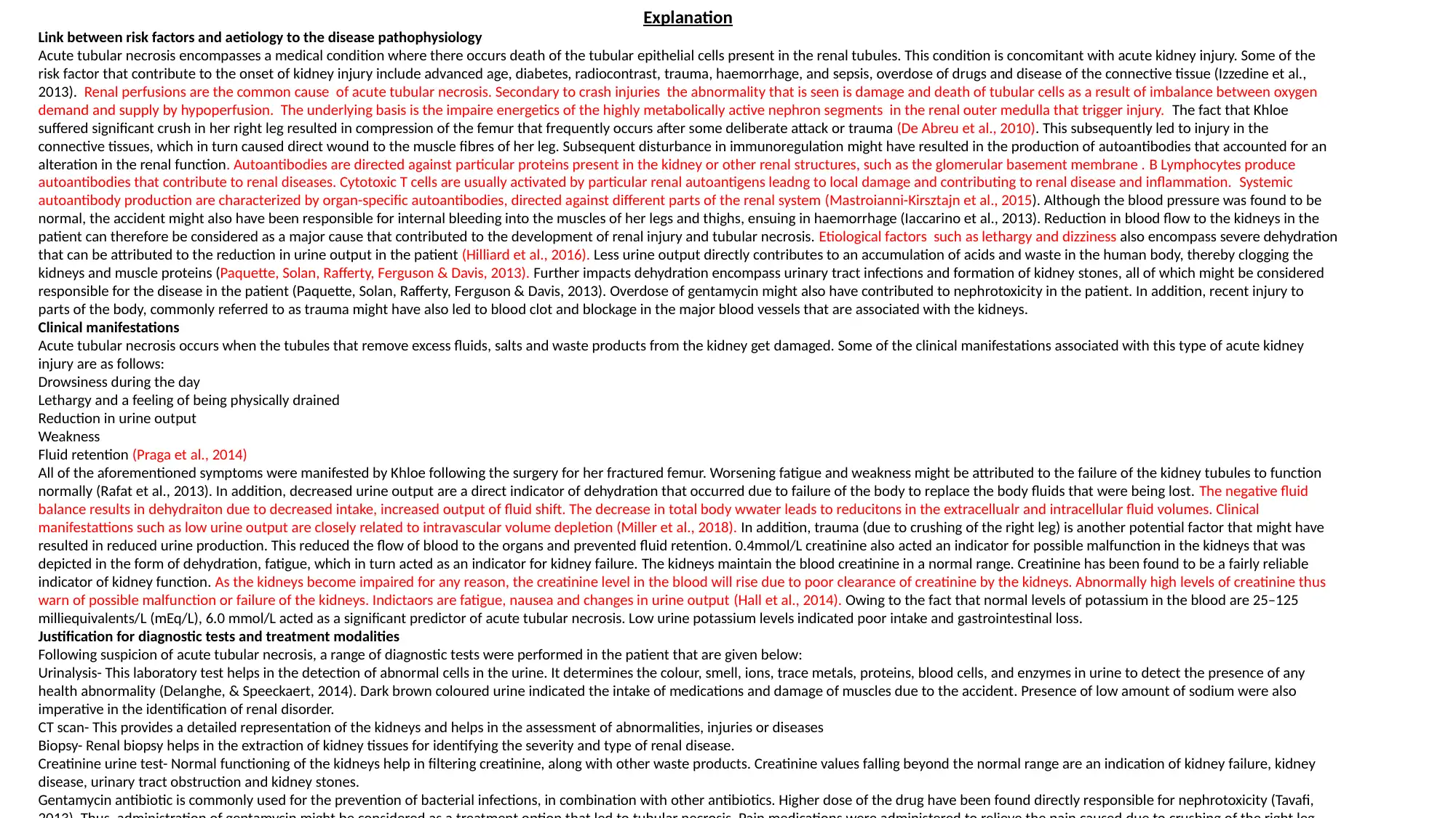
Explanation
Link between risk factors and aetiology to the disease pathophysiology
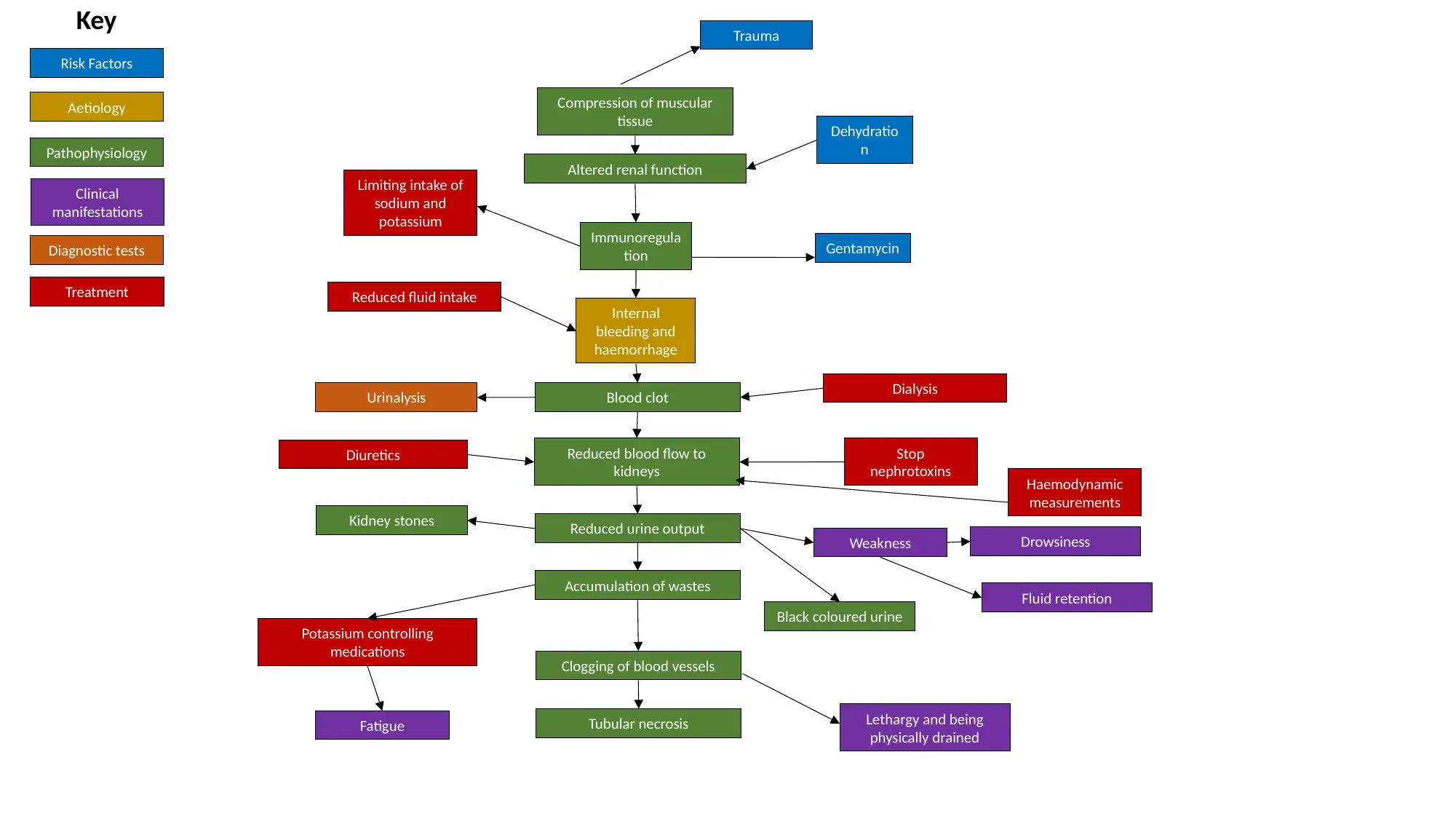
Acute tubular necrosis encompasses a medical condition where there occurs death of the tubular epithelial cells present in the renal tubules. This condition is concomitant with acute kidney injury. Some of the
risk factor that contribute to the onset of kidney injury include advanced age, diabetes, radiocontrast, trauma, haemorrhage, and sepsis, overdose of drugs and disease of the connective tissue (Izzedine et al.,
2013). Renal perfusions are the common cause of acute tubular necrosis. Secondary to crash injuries the abnormality that is seen is damage and death of tubular cells as a result of imbalance between oxygen
demand and supply by hypoperfusion. The underlying basis is the impaire energetics of the highly metabolically active nephron segments in the renal outer medulla that trigger injury. The fact that Khloe
suffered significant crush in her right leg resulted in compression of the femur that frequently occurs after some deliberate attack or trauma (De Abreu et al., 2010). This subsequently led to injury in the
connective tissues, which in turn caused direct wound to the muscle fibres of her leg. Subsequent disturbance in immunoregulation might have resulted in the production of autoantibodies that accounted for an
alteration in the renal function. Autoantibodies are directed against particular proteins present in the kidney or other renal structures, such as the glomerular basement membrane . B Lymphocytes produce
autoantibodies that contribute to renal diseases. Cytotoxic T cells are usually activated by particular renal autoantigens leadng to local damage and contributing to renal disease and inflammation. Systemic
autoantibody production are characterized by organ-specific autoantibodies, directed against different parts of the renal system (Mastroianni-Kirsztajn et al., 2015). Although the blood pressure was found to be
normal, the accident might also have been responsible for internal bleeding into the muscles of her legs and thighs, ensuing in haemorrhage (Iaccarino et al., 2013). Reduction in blood flow to the kidneys in the
patient can therefore be considered as a major cause that contributed to the development of renal injury and tubular necrosis. Etiological factors such as lethargy and dizziness also encompass severe dehydration
that can be attributed to the reduction in urine output in the patient (Hilliard et al., 2016). Less urine output directly contributes to an accumulation of acids and waste in the human body, thereby clogging the
kidneys and muscle proteins (Paquette, Solan, Rafferty, Ferguson & Davis, 2013). Further impacts dehydration encompass urinary tract infections and formation of kidney stones, all of which might be considered
responsible for the disease in the patient (Paquette, Solan, Rafferty, Ferguson & Davis, 2013). Overdose of gentamycin might also have contributed to nephrotoxicity in the patient. In addition, recent injury to
parts of the body, commonly referred to as trauma might have also led to blood clot and blockage in the major blood vessels that are associated with the kidneys.
Clinical manifestations
Acute tubular necrosis occurs when the tubules that remove excess fluids, salts and waste products from the kidney get damaged. Some of the clinical manifestations associated with this type of acute kidney
injury are as follows:
Drowsiness during the day
Lethargy and a feeling of being physically drained
Reduction in urine output
Weakness
Fluid retention (Praga et al., 2014)
All of the aforementioned symptoms were manifested by Khloe following the surgery for her fractured femur. Worsening fatigue and weakness might be attributed to the failure of the kidney tubules to function
normally (Rafat et al., 2013). In addition, decreased urine output are a direct indicator of dehydration that occurred due to failure of the body to replace the body fluids that were being lost. The negative fluid
balance results in dehydraiton due to decreased intake, increased output of fluid shift. The decrease in total body wwater leads to reducitons in the extracellualr and intracellular fluid volumes. Clinical
manifestattions such as low urine output are closely related to intravascular volume depletion (Miller et al., 2018). In addition, trauma (due to crushing of the right leg) is another potential factor that might have
resulted in reduced urine production. This reduced the flow of blood to the organs and prevented fluid retention. 0.4mmol/L creatinine also acted an indicator for possible malfunction in the kidneys that was
depicted in the form of dehydration, fatigue, which in turn acted as an indicator for kidney failure. The kidneys maintain the blood creatinine in a normal range. Creatinine has been found to be a fairly reliable
indicator of kidney function. As the kidneys become impaired for any reason, the creatinine level in the blood will rise due to poor clearance of creatinine by the kidneys. Abnormally high levels of creatinine thus
warn of possible malfunction or failure of the kidneys. Indictaors are fatigue, nausea and changes in urine output (Hall et al., 2014). Owing to the fact that normal levels of potassium in the blood are 25–125
milliequivalents/L (mEq/L), 6.0 mmol/L acted as a significant predictor of acute tubular necrosis. Low urine potassium levels indicated poor intake and gastrointestinal loss.
Justification for diagnostic tests and treatment modalities
Following suspicion of acute tubular necrosis, a range of diagnostic tests were performed in the patient that are given below:
Urinalysis- This laboratory test helps in the detection of abnormal cells in the urine. It determines the colour, smell, ions, trace metals, proteins, blood cells, and enzymes in urine to detect the presence of any
health abnormality (Delanghe, & Speeckaert, 2014). Dark brown coloured urine indicated the intake of medications and damage of muscles due to the accident. Presence of low amount of sodium were also
imperative in the identification of renal disorder.
CT scan- This provides a detailed representation of the kidneys and helps in the assessment of abnormalities, injuries or diseases
Biopsy- Renal biopsy helps in the extraction of kidney tissues for identifying the severity and type of renal disease.
Creatinine urine test- Normal functioning of the kidneys help in filtering creatinine, along with other waste products. Creatinine values falling beyond the normal range are an indication of kidney failure, kidney
disease, urinary tract obstruction and kidney stones.
Gentamycin antibiotic is commonly used for the prevention of bacterial infections, in combination with other antibiotics. Higher dose of the drug have been found directly responsible for nephrotoxicity (Tavafi,
Reference list
Delanghe, J., & Speeckaert, M. (2014). Preanalytical requirements of urinalysis. Biochemia medica: Biochemia medica, 24(1), 89- 104.
https://doi.org/10.11613/BM.2014.011
De Abreu, K. L. S., Silva Júnior, G. B., Barreto, A. G. C., Melo, F. M., Oliveira, B. B., Mota, R. M. S., … Daher, E. F. (2010). Acute kidney injury
after trauma: Prevalence, clinical characteristics and RIFLE classification. Indian Journal of Critical Care Medicine : Peer-
Reviewed, Official Publication of Indian Society of Critical Care Medicine, 14(3), 121–128. http://doi.org/10.4103/0972-5229.74170
Hall, J. A., Yerramilli, M., Obare, E., Yerramilli, M., & Jewell, D. E. (2014). Comparison of serum concentrations of symmetric dimethylarginine and creatinine as
kidney function biomarkers in cats with chronic kidney disease. Journal of veterinary internal medicine, 28(6), 1676-1683. DOI: https://
doi.org/10.1111/jvim.12445
Hilliard, L. M., Colafella, K. M. M., Bulmer, L. L., Puelles, V. G., Singh, R. R., Ow, C. P., ... & Denton, K. M. (2016). Chronic recurrent dehydration associated
with periodic water intake exacerbates hypertension and promotes renal damage in male spontaneously hypertensive
rats. Scientific reports, 6, 33855. Retrieved from https://www.nature.com/articles/srep33855
Iaccarino, L., Gatto, M., Bettio, S., Caso, F., Rampudda, M., Zen, M., ... & Doria, A. (2013). Overlap connective tissue disease
syndromes. Autoimmunity Reviews, 12(3), 363-373. https://doi.org/10.1016/j.autrev.2012.06.004
Izzedine, H., Escudier, B., Rouvier, P., Gueutin, V., Varga, A., Bahleda, R., & Soria, J. C. (2013). Acute tubular necrosis associated with mTOR inhibitor
therapy: a real entity biopsy-proven. Annals of oncology, 24(9), 2421-2425. https://doi.org/10.1093/annonc/mdt233
Mastroianni-Kirsztajn, G., Hornig, N., & Schlumberger, W. (2015). Autoantibodies in renal diseases–clinical significance and recent developments in
serological detection. Frontiers in immunology, 6, 221. DOI 10.3389/fimmu.2015.00221
Miller, J. B., Lee, A., Suszanski, J. P., Tustian, M., Corcoran, J. L., Moore, S., ... & Lewandowski, C. A. (2018). Challenge of intravascular volume assessment
in acute ischemic stroke. The American journal of emergency medicine, 36(6), 1018-1021. DOI https://doi.org/10.1016/j.ajem.2018.01.071
Paquette, I. M., Solan, P., Rafferty, J. F., Ferguson, M. A., & Davis, B. R. (2013). Readmission for dehydration or renal failure after ileostomy
creation. Diseases of the Colon & Rectum, 56(8), 974-979. doi: 10.1097/DCR.0b013e31828d02ba
Praga, M., Sevillano, A., Auñón, P., & González, E. (2014). Changes in the aetiology, clinical presentation and management of acute interstitial nephritis,
an increasingly common cause of acute kidney injury. Nephrology Dialysis Transplantation, 30(9), 1472-1479.
Rafat, C., Burbach, M., Brochériou, I., Zafrani, L., Callard, P., Rondeau, E., & Hertig, A. (2013). Bilirubin- associated acute tubular necrosis in a kidney
transplant recipient. American Journal of Kidney Diseases, 61(5), 782-785. https://doi.org/10.1053/j.ajkd.2012.11.046
Tavafi, M. (2013). Protection of renal tubules against gentamicin induced nephrotoxicity. Journal of renal injury prevention, 2(1), 5. DOI:
10.12861/jrip.2013.03
1 out of 3
Your All-in-One AI-Powered Toolkit for Academic Success.
+13062052269
info@desklib.com
Available 24*7 on WhatsApp / Email
Unlock your academic potential
© 2024 | Zucol Services PVT LTD | All rights reserved.