In-Depth Study: Respiratory System and Blood Component Functions
VerifiedAdded on 2023/04/21
|19
|3797
|442
Report
AI Summary
This report provides an in-depth analysis of the respiratory system and blood, detailing their structures, functions, and the critical processes they facilitate. It begins by exploring the anatomy of the respiratory system, including the nose, pharynx, trachea, bronchi, and lungs, emphasizing the role of alveoli in gas exchange. The report then discusses the mechanics of breathing, including pulmonary ventilation, external and internal respiration, and phonation. Conditions necessary for effective gaseous exchange, such as temperature, ventilation, cell wall permeability, surface area to volume ratio, and concentration gradients, are also examined. Furthermore, the report delves into the components of blood, focusing on plasma and red blood cells (erythrocytes). It elucidates the functions of plasma proteins like albumin, immunoglobulins, and fibrinogen, and describes the structure and function of red blood cells in oxygen and carbon dioxide transport. The process of oxygen and carbon dioxide transport in the blood is explained, along with the structural details and functions of arteries, veins, and capillaries in blood circulation. Finally, the report outlines the structure of the heart, providing a comprehensive understanding of these essential biological systems. Desklib offers a wealth of similar solved assignments and study resources for students.

Running head: BIOLOGY
BIOLOGY
Name of the student:
Name of the university:
Author note:
BIOLOGY
Name of the student:
Name of the university:
Author note:
Paraphrase This Document
Need a fresh take? Get an instant paraphrase of this document with our AI Paraphraser
1
BIOLOGY
Respiratory system:
Structure of respiratory system:
The respiratory system comprises of all organs involved in breathing procedures.
These are seen to include nose, pharynx, trachea, bronchi and the lungs. The first four
component mentioned all work like a system of pipes and through this pipeline like
system, air ultimately funnels down to the lungs (Rizzo 2015)
Very small air sacs that are called alveoli are present in the lungs mainly in the top
region of the bronchioles ( division of the bronchi). This helps in exchange of oxygen
and carbon-dioxide between the lungs and blood systems.
The lungs are two lobed paired cone shaped organs with right lung having three lobes
and left lung having two lobes. Lungs are broken into smaller portions called
bronchio-pulmonary segments. Air enters into the lungs through system of pipes
called bronchi. These mainly start at the bottom region of trachea as left and right
bronchi and branch many times throughout lungs until they form alveoli.
Lungs remain covered by smooth membranes called pleurae having two layers called
visceral and parietal layers helping the systems to breathe without any friction
(Beachy 2015)
For respiration, muscles are important. Diaphragm is a large sheet like muscle
stretching across the chest under the region of ribcage. This help in expanding the
space in chest allowing more air to enter. Other muscles are muscles of ribs called
intercostals muscles and help in moving the ribcage in and out.
2
BIOLOGY
Fig: structure of respiratory system: Source: (Beachy 2015)
Fig: structure of respiratory system: diaphragm; Source: (Protti et al. 2015)
BIOLOGY
Fig: structure of respiratory system: Source: (Beachy 2015)
Fig: structure of respiratory system: diaphragm; Source: (Protti et al. 2015)
⊘ This is a preview!⊘
Do you want full access?
Subscribe today to unlock all pages.

Trusted by 1+ million students worldwide
3
BIOLOGY
Function of respiratory system:
The respiratory system helps in breathing which is also called pulmonary ventilation.
In this process, air is inhaled through the nasal cavities as well as the oral cavities.
This air is then seen to move through pharynx, larynx and then trachea to finally the
lungs. Then air is exhaled flowing back through the same pathway. In this oxygen and
carbon-dioxide are taken in and exhaled out respectively by the respiratory system
(Protti et al. 2015).
Inside the lungs, oxygen gets exchanged for carbon dioxide waste through the
procedure called external respiration. This respiration tales place through millions of
alveoli which are microscopic sac like structure aiding the process
The bloodstream helps in delivering the oxygen towards the cells and thereby
removes carbon-dioxide through internal respiration. From the capillary system,
oxygen is realised in the tissues and then into cells. Glucose breaks down in presence
of oxygen to produce energy and carbon-dioxide is released. That is carried back by
the blood again to lungs to be exhaled out (Hansen 2017).
During exhalation, air passes from the lungs through the voice box or larynx. Air
vibrates the vocal cords to produce sound. Phonation is the creation of sound by the
different structures that are present in the region of the upper respiratory tract.
It also plays role in olfaction or smelling which is a chemical sensation (Scanlon and
Sanders 2018)
Conditions necessary for effective gaseous exchange:
A number of conditions need to be present so that gas exchange can take place successfully.
These are as follows:
Paraphrase This Document
Need a fresh take? Get an instant paraphrase of this document with our AI Paraphraser
4
BIOLOGY
Temperature: the warmer the conditions, the faster will be the rate of diffusion
between blood and alveoli and hence gaseous exchange will take place more
effectively
There is requirement of constant ventilation for maintaining a concentration gradient
and thereby stopping reaching the equilibrium points
Thin and permeable cell walls would be helping in rapid diffusion as well as in faster
gas exchange (Amin-Hanjani et al. 2015)
The surface area to that of the volume ratio should be large. If there are more and
more alveoli stacked together in the lungs, there would be more gaseous exchange
taking place.
There should be concentration difference between the gases from one side to that of
the other. Gases will always flow from higher concentration to that if lower
concentration constantly striving to reach equilibrium. This gradients needs to be
maintained by blood flow on one side that would be carrying oxygen quickly and by
airflow on other side.
Fig: conditions required for gas exchange Source: (Scanlon and Sanders 2018)
5
BIOLOGY
Blood:
Components of plasma and their function:
Plasma can be described as the the liquid constituent of blood where the RBC or
thered blood cells, platelets and the white blood cells remain in the suspended state. It
mainly comprises of more than half portions of that of the blood volume and
constitutes mainly of water containing proteins as well as dissolved salts.
Major protein is albumin. This prevents fluid from the action of that of leaking out
from the blood vessels and into the tissue systems. It helps by that of binding to and
thereby carrying materials such as the hormones and certain drugs (Ghersi-Egea et al.
2018).
Others proteins in the plasma are seen to include antibodies like that of immuno-
globulins. These actively help in defending the body against bacteria, virus, fungi and
the different cancer cells. By the procedures if electrophoresis, plasma globulins are
also found to be separated into α1, α2, β and ¥-globulins that are synthesized in liver.
¥-globulins are developed in the cells of reticulo-endothelial system and are mainly
called the immunoglobulins (Serlin et al. 2015).
Fibrinogen is a fibrous protein present in the plasma; it plays significant function in
the clotting of blood. It takes part in conversion to fibrin by thrombin. Pro-thrombin
present in α2-globulin fraction. At the same time, fibrinogen are seen to participate in
blood clotting procedures.
Prothrombin -> Thrombin (Presence – Thromboplastin)
Fibrinogen -> Fibrin (Clot) (Presence –Thrombin)
⊘ This is a preview!⊘
Do you want full access?
Subscribe today to unlock all pages.

Trusted by 1+ million students worldwide
6
BIOLOGY
Structure of red blood cells and function:
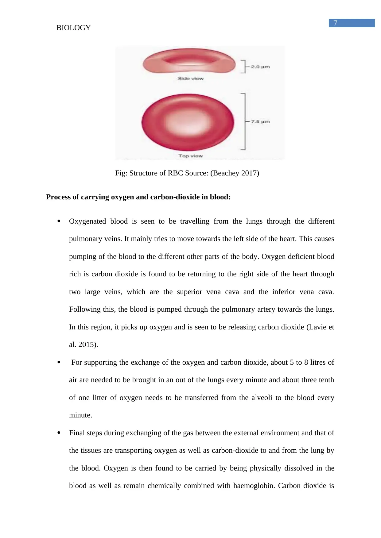
Red blood cells also called erythrocytes are specialised cells which circulate
throughout the body and help by delivering oxygen to the different tissues. In humans,
red blood cells also called RBC are small and biconcave structures that are thinnest in
the central region about only 7 – 8 μm in size. They do not contain mitochondria or
nucleus when they get matured (Verschakelen and De Wever 2018)
The above mentioned characteristics allow the red blood cells for performing their
task for oxygen transport effectively. Smaller size and biconcave shapes help in
increasing the surface area-to-volume ratio and thereby improve the process of gas
exchange. Lack of nucleus helps in making additional space for haemoglobin. This is
a key protein that is used for oxygen transport. Absence of the cell organelles of
mitochondria helps by preventing the RBC from utilising any portion of that of the
oxygen that they carry and thereby maximises the quantity delivered to the tissues of
the body.
In the lungs, red blood cells are seen to take up oxygen. They then circulate
throughout the rest of the body and release the oxygen to the different surrounding
tissues. These cells also play a significant role in carrying carbon-dioxide which is the
waste product from the tissues to the lungs. Some of the CO2 is seen to bind with
haemoglobin directly and are carried by RBC after an enzyme converts CO2 into
bicarbonate. This bicarbonate is seen to dissolve in the plasma and is thereby
transported into the lungs. Here, they are gain converted onto carbon dioxide and are
then released (Beachey 2017).
Paraphrase This Document
Need a fresh take? Get an instant paraphrase of this document with our AI Paraphraser
7
BIOLOGY
Fig: Structure of RBC Source: (Beachey 2017)
Process of carrying oxygen and carbon-dioxide in blood:
Oxygenated blood is seen to be travelling from the lungs through the different
pulmonary veins. It mainly tries to move towards the left side of the heart. This causes
pumping of the blood to the different other parts of the body. Oxygen deficient blood
rich is carbon dioxide is found to be returning to the right side of the heart through
two large veins, which are the superior vena cava and the inferior vena cava.
Following this, the blood is pumped through the pulmonary artery towards the lungs.
In this region, it picks up oxygen and is seen to be releasing carbon dioxide (Lavie et
al. 2015).
For supporting the exchange of the oxygen and carbon dioxide, about 5 to 8 litres of
air are needed to be brought in an out of the lungs every minute and about three tenth
of one litter of oxygen needs to be transferred from the alveoli to the blood every
minute.
Final steps during exchanging of the gas between the external environment and that of
the tissues are transporting oxygen as well as carbon-dioxide to and from the lung by
the blood. Oxygen is then found to be carried by being physically dissolved in the
blood as well as remain chemically combined with haemoglobin. Carbon dioxide is
8
BIOLOGY
also carried being physically dissolved in the blood as well as being chemically
combined to that of blood proteins as carbamino compounds and also as bicarbonate
(Isgaard et al. 2015)
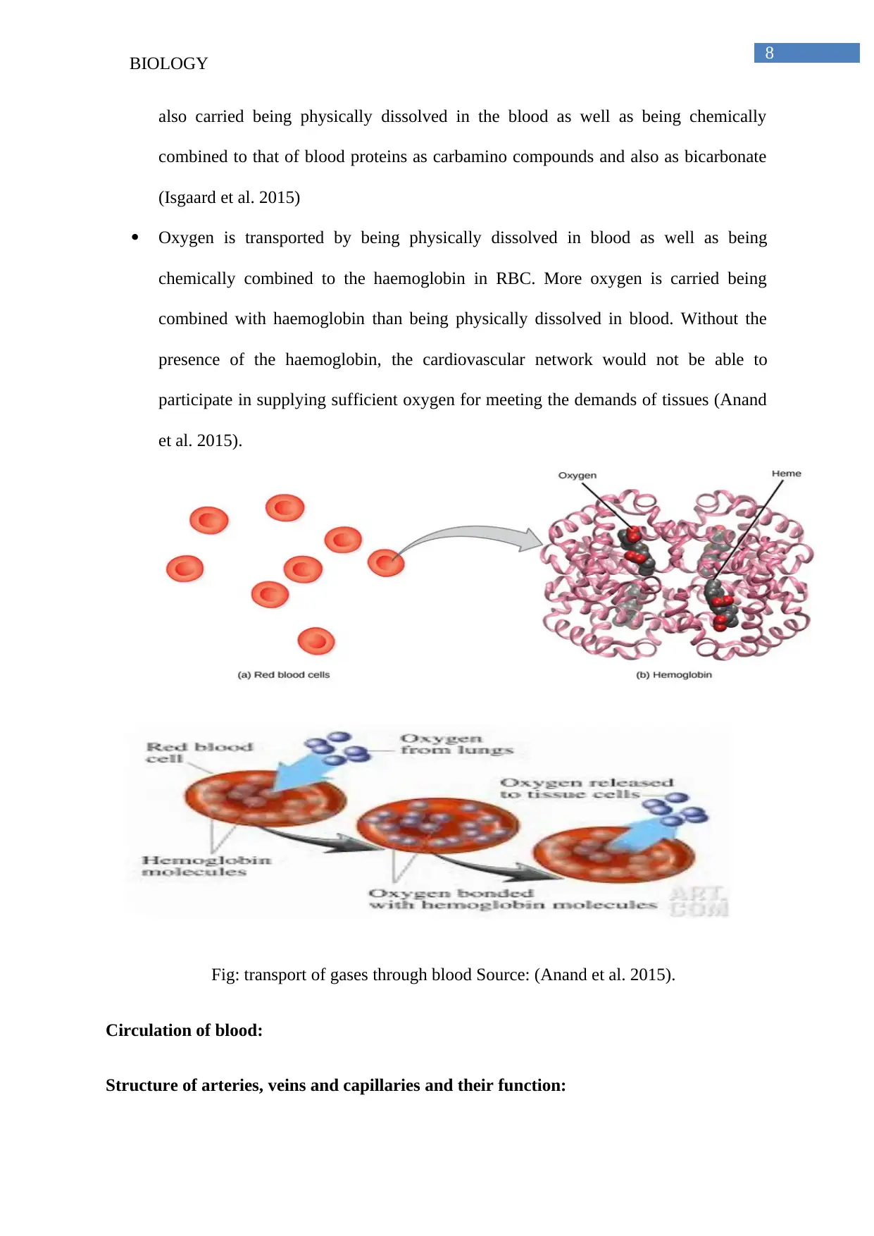
Oxygen is transported by being physically dissolved in blood as well as being
chemically combined to the haemoglobin in RBC. More oxygen is carried being
combined with haemoglobin than being physically dissolved in blood. Without the
presence of the haemoglobin, the cardiovascular network would not be able to
participate in supplying sufficient oxygen for meeting the demands of tissues (Anand
et al. 2015).
Fig: transport of gases through blood Source: (Anand et al. 2015).
Circulation of blood:
Structure of arteries, veins and capillaries and their function:
⊘ This is a preview!⊘
Do you want full access?
Subscribe today to unlock all pages.

Trusted by 1+ million students worldwide
9
BIOLOGY
Artery:
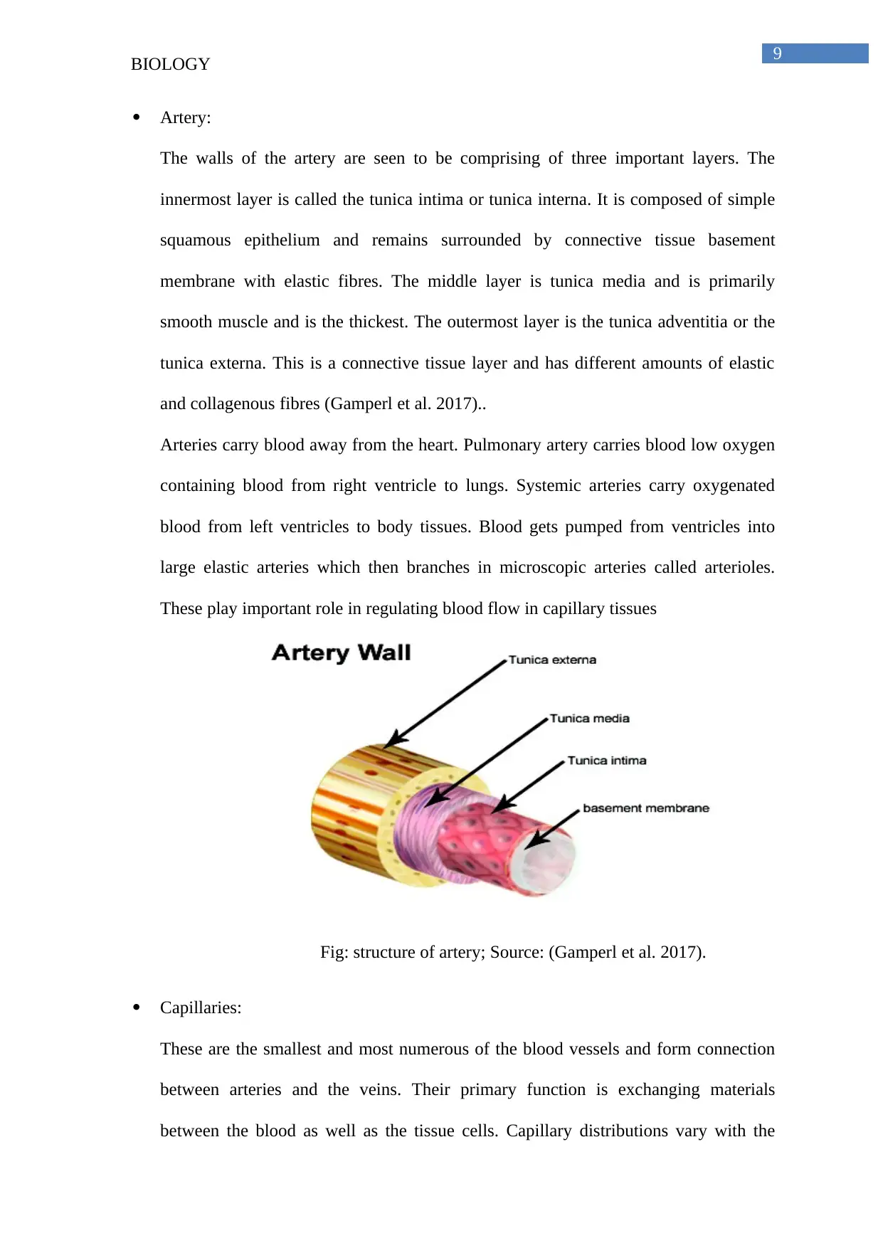
The walls of the artery are seen to be comprising of three important layers. The
innermost layer is called the tunica intima or tunica interna. It is composed of simple
squamous epithelium and remains surrounded by connective tissue basement
membrane with elastic fibres. The middle layer is tunica media and is primarily
smooth muscle and is the thickest. The outermost layer is the tunica adventitia or the
tunica externa. This is a connective tissue layer and has different amounts of elastic
and collagenous fibres (Gamperl et al. 2017)..
Arteries carry blood away from the heart. Pulmonary artery carries blood low oxygen
containing blood from right ventricle to lungs. Systemic arteries carry oxygenated
blood from left ventricles to body tissues. Blood gets pumped from ventricles into
large elastic arteries which then branches in microscopic arteries called arterioles.
These play important role in regulating blood flow in capillary tissues
Fig: structure of artery; Source: (Gamperl et al. 2017).
Capillaries:
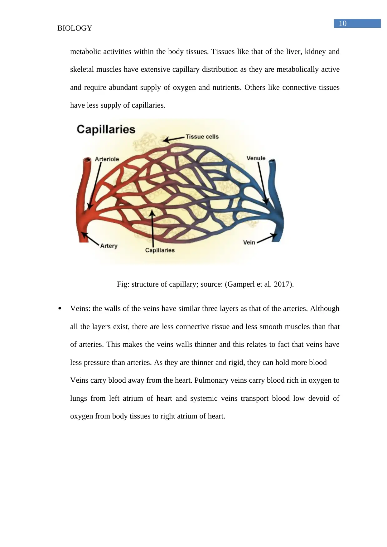
These are the smallest and most numerous of the blood vessels and form connection
between arteries and the veins. Their primary function is exchanging materials
between the blood as well as the tissue cells. Capillary distributions vary with the
Paraphrase This Document
Need a fresh take? Get an instant paraphrase of this document with our AI Paraphraser
10
BIOLOGY
metabolic activities within the body tissues. Tissues like that of the liver, kidney and
skeletal muscles have extensive capillary distribution as they are metabolically active
and require abundant supply of oxygen and nutrients. Others like connective tissues
have less supply of capillaries.
Fig: structure of capillary; source: (Gamperl et al. 2017).
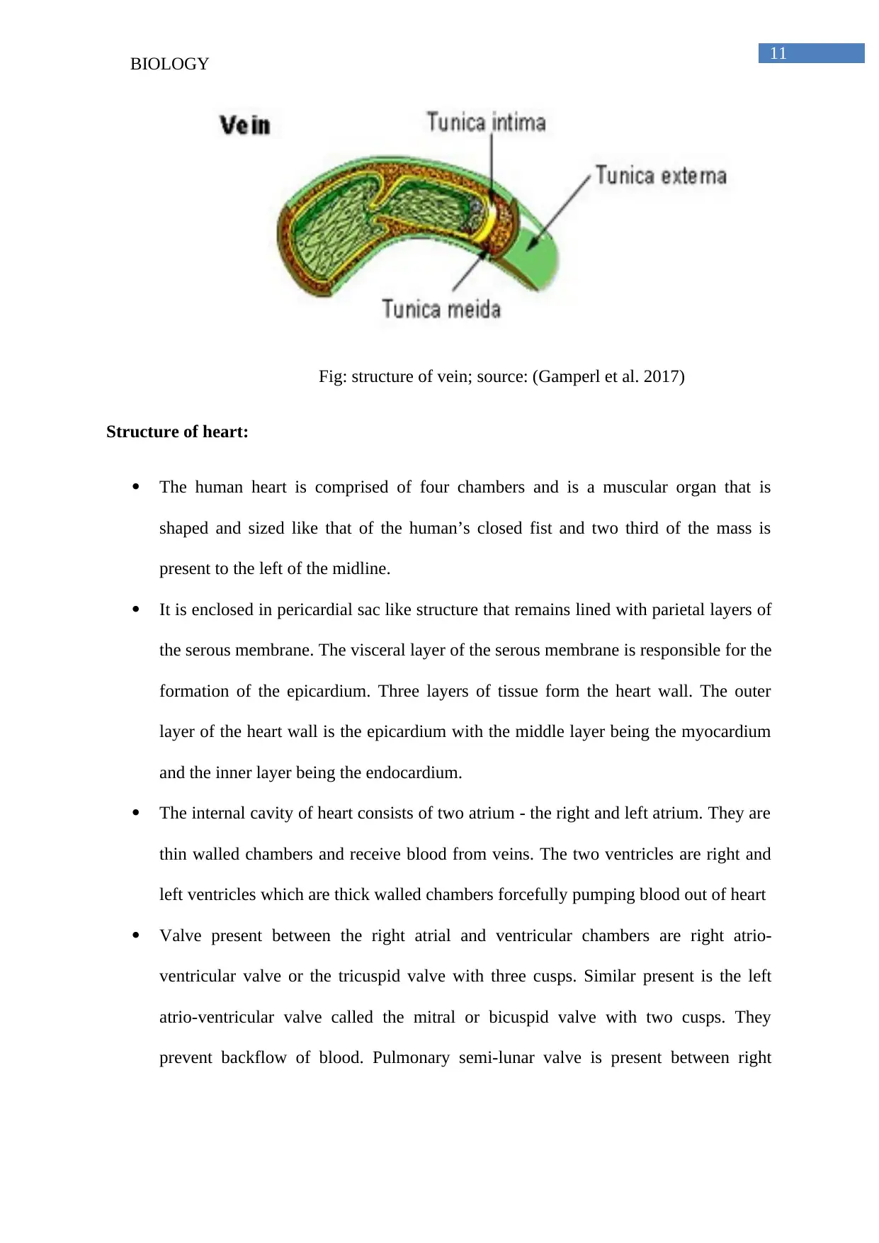
Veins: the walls of the veins have similar three layers as that of the arteries. Although
all the layers exist, there are less connective tissue and less smooth muscles than that
of arteries. This makes the veins walls thinner and this relates to fact that veins have
less pressure than arteries. As they are thinner and rigid, they can hold more blood
Veins carry blood away from the heart. Pulmonary veins carry blood rich in oxygen to
lungs from left atrium of heart and systemic veins transport blood low devoid of
oxygen from body tissues to right atrium of heart.
11
BIOLOGY
Fig: structure of vein; source: (Gamperl et al. 2017)
Structure of heart:
The human heart is comprised of four chambers and is a muscular organ that is
shaped and sized like that of the human’s closed fist and two third of the mass is
present to the left of the midline.
It is enclosed in pericardial sac like structure that remains lined with parietal layers of
the serous membrane. The visceral layer of the serous membrane is responsible for the
formation of the epicardium. Three layers of tissue form the heart wall. The outer
layer of the heart wall is the epicardium with the middle layer being the myocardium
and the inner layer being the endocardium.
The internal cavity of heart consists of two atrium - the right and left atrium. They are
thin walled chambers and receive blood from veins. The two ventricles are right and
left ventricles which are thick walled chambers forcefully pumping blood out of heart
Valve present between the right atrial and ventricular chambers are right atrio-
ventricular valve or the tricuspid valve with three cusps. Similar present is the left
atrio-ventricular valve called the mitral or bicuspid valve with two cusps. They
prevent backflow of blood. Pulmonary semi-lunar valve is present between right
⊘ This is a preview!⊘
Do you want full access?
Subscribe today to unlock all pages.

Trusted by 1+ million students worldwide
1 out of 19
Related Documents
Your All-in-One AI-Powered Toolkit for Academic Success.
+13062052269
info@desklib.com
Available 24*7 on WhatsApp / Email
![[object Object]](/_next/static/media/star-bottom.7253800d.svg)
Unlock your academic potential
Copyright © 2020–2026 A2Z Services. All Rights Reserved. Developed and managed by ZUCOL.


