logo

Biochemistry Name of the University Author

18 Pages2748 Words113 Views
   

Added on  2020-04-13

About This Document

5 BIOLOGY BIOLOGY Biology Name of the Student Name of the University Author Note Task 1 2 Answer: A 2 Answer B 2 Task 2 7 Task 3 7 Task 4 9 Answer a 9 Answer b 10 Answer c 10 Answer e 11 Task 5 12 Answer A (i) 12 Answer A (ii) 12 Answer B (i) 13 Task 6 14 Answer a 14 Answer b 15 References 18 Task 1 Answer: A A. Cell wall * Answer B Light Microscope Electron Microscope Cell components seen

Biochemistry Name of the University Author

   Added on 2020-04-13

ShareRelated Documents
Running head: BIOLOGY
Biology
Name of the Student
Name of the University
Author Note
Biochemistry Name of the University Author_1
1BIOLOGY
Table of Contents
Task 1.........................................................................................................................................2
Answer: A..............................................................................................................................2
Answer B................................................................................................................................2
Task 2.........................................................................................................................................7
Task 3.........................................................................................................................................7
Task 4.........................................................................................................................................9
Answer a.................................................................................................................................9
Answer b..............................................................................................................................10
Answer c...............................................................................................................................10
Answer e...............................................................................................................................11
Task 5.......................................................................................................................................12
Answer A (i).........................................................................................................................12
Answer A (ii)........................................................................................................................12
Answer B (i).........................................................................................................................13
Task 6.......................................................................................................................................14
Answer a...............................................................................................................................14
Answer b..............................................................................................................................15
References................................................................................................................................18
Biochemistry Name of the University Author_2
2BIOLOGY
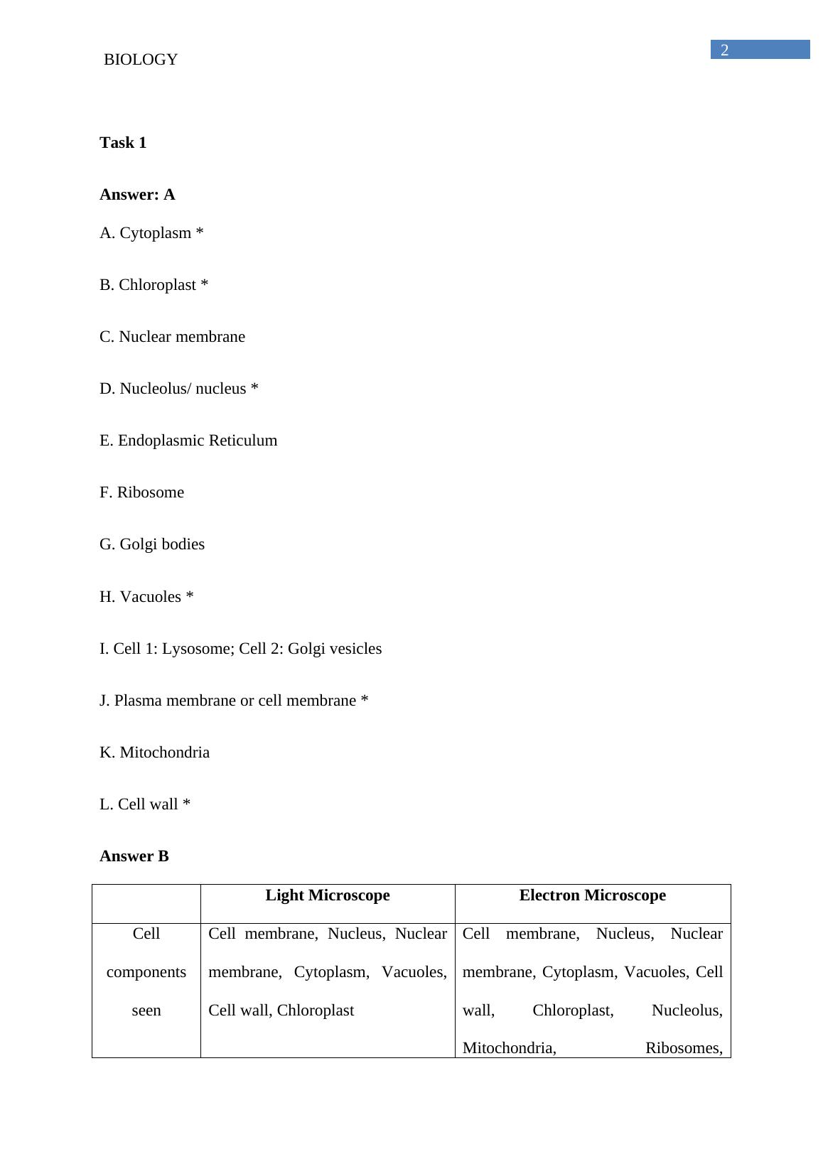
Task 1
Answer: A
A. Cytoplasm *
B. Chloroplast *
C. Nuclear membrane
D. Nucleolus/ nucleus *
E. Endoplasmic Reticulum
F. Ribosome
G. Golgi bodies
H. Vacuoles *
I. Cell 1: Lysosome; Cell 2: Golgi vesicles
J. Plasma membrane or cell membrane *
K. Mitochondria
L. Cell wall *
Answer B
Light Microscope Electron Microscope
Cell
components
seen
Cell membrane, Nucleus, Nuclear
membrane, Cytoplasm, Vacuoles,
Cell wall, Chloroplast
Cell membrane, Nucleus, Nuclear
membrane, Cytoplasm, Vacuoles, Cell
wall, Chloroplast, Nucleolus,
Mitochondria, Ribosomes,
Biochemistry Name of the University Author_3
3BIOLOGY
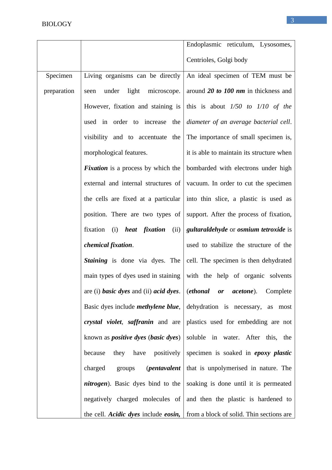
Endoplasmic reticulum, Lysosomes,
Centrioles, Golgi body
Specimen
preparation
Living organisms can be directly
seen under light microscope.
However, fixation and staining is
used in order to increase the
visibility and to accentuate the
morphological features.
Fixation is a process by which the
external and internal structures of
the cells are fixed at a particular
position. There are two types of
fixation (i) heat fixation (ii)
chemical fixation.
Staining is done via dyes. The
main types of dyes used in staining
are (i) basic dyes and (ii) acid dyes.
Basic dyes include methylene blue,
crystal violet, saffranin and are
known as positive dyes (basic dyes)
because they have positively
charged groups (pentavalent
nitrogen). Basic dyes bind to the
negatively charged molecules of
the cell. Acidic dyes include eosin,
An ideal specimen of TEM must be
around 20 to 100 nm in thickness and
this is about 1/50 to 1/10 of the
diameter of an average bacterial cell.
The importance of small specimen is,
it is able to maintain its structure when
bombarded with electrons under high
vacuum. In order to cut the specimen
into thin slice, a plastic is used as
support. After the process of fixation,
gultaraldehyde or osmium tetroxide is
used to stabilize the structure of the
cell. The specimen is then dehydrated
with the help of organic solvents
(ethonal or acetone). Complete
dehydration is necessary, as most
plastics used for embedding are not
soluble in water. After this, the
specimen is soaked in epoxy plastic
that is unpolymerised in nature. The
soaking is done until it is permeated
and then the plastic is hardened to
from a block of solid. Thin sections are
Biochemistry Name of the University Author_4

End of preview

Want to access all the pages? Upload your documents or become a member.

Related Documents
Electron Microscope - Assignment
|6
|1308
|122

Biology in Everyday Life: Cell Structure and Function
|5
|1355
|94

Cell Types in the Human Digestive System: Red Blood Cells, Kupffer Cells, Goblet Cells, Enterocytes, Chief Cells
|4
|1367
|244

Cell Biology and Biochemistry
|8
|2140
|1

Biology Mock Exam Paper with MCQs and Diagrams
|21
|3905
|65

The structure of the cell. 9 The structure of the cell
|14
|2919
|55