Biosciences Workbook Week 3: Cardiovascular System Anatomy
VerifiedAdded on 2023/06/04
|12
|3131
|169
AI Summary
This workbook aims to further reinforce knowledge and understanding of students on the systems of the body and to enable them to study at their own pace. It is also a self-assessment tool for students to identify whether learning has taken place. It encourages close reading of the lecture notes and textbook to find the relevant information. The Cardiovascular System section covers topics such as the site of the heart, the layers of the heart, the flow of blood through the heart, and more. Suitable for Biosciences for Nursing students in Year 1.
Contribute Materials
Your contribution can guide someone’s learning journey. Share your
documents today.
1
2
3
4
5 Year 1
Biosciences for nursing
Workbook week 3
Cardiovascular system
anatomy
Name Click or tap here to enter
text......................................
Student number Click or tap here to enter text.
…………...
2
3
4
5 Year 1
Biosciences for nursing
Workbook week 3
Cardiovascular system
anatomy
Name Click or tap here to enter
text......................................
Student number Click or tap here to enter text.
…………...
Secure Best Marks with AI Grader
Need help grading? Try our AI Grader for instant feedback on your assignments.
Biosciences workbook
This workbook aims to further reinforce knowledge and understanding of students on
the systems of the body and to enable them to study at their own pace. It is also a self-
assessment tool for students to identify whether learning has taken place. It encourages
close reading of the lecture notes and textbook to find the relevant information.
Learning anything new takes a lot of effort. Learning is an active process; you need to
engage with the work and practice writing the new ‘language’ down in your notebook (or
online portfolio). Pay attention to how the words are spelt for example, it is easy to
confuse atria and arterial. Eventually this language will become your own language.
Reading is a very passive activity, reading alone is not a good way to learn, you need to
engage fully by questioning everything you read. This workbook is designed to help you
with this, however, just completing the workbook is not enough, if this subject is new to
you will have to go over it many times.
Although many of the answers can be found in the lecture notes, not all can be found
here. Please use your textbook to read around the subject to extend your knowledge
and understanding. However, there is often much more in textbooks than you need to
know. In order to help you focus on what you need to know the learning outcomes for
each session are given to you.
In this workbook there is a separate section for each lecture. Please complete and
submit (workbook submission portal) the relevant section following your lectures in
preparation for the ‘live’ quiz. If you leave the work for a few weeks you will find it very
difficult to catch up.
Read you lecture notes and textbook prior to completing each section. Can you answer
the questions without looking back at your notes? Sometimes the same question is
repeated in a different way. This is deliberate as repetition helps us to remember.
Good luck!
Cardiovascular system
1. The site in the thoracic cavity that the heart occupies is called mediastinum
2. The structure that separate the right side of the heart from the left side is called
the septum
3. Which side of the heart is (a) oxygenated blood found left atrium and (b)
deoxygenated blood found left ventricle.
4. Name the tiny muscle that anchors the chordae tendinae to the inner wall of the
heart Papillary muscles.
5. Fill in the blank spaces:
The heart has 3 layers; the outermost layer is known as the Epicardium inside this layer
is a double layer called the Myocardium and Endocardium layers, which secretes an
important fluid that reduces friction during heart contraction thus allowing the heart to
contract and relax smoothly.
6. What are Chordae Tendinae and what are their functions?
Chordae tendinae are the strong fibrous connections found in between the valve leaflets
and papillary muscles of the heart. They are the thread-like bands of the fibrous tissue
that are attach on one end of the tricuspid and mitral values of the heart. It prevents
values of heart from movement by holding the flaps tightly against the strong flow of
blood. It assist the blood for the flow in one direction only. When the cusps close it
prevent the swinging back of fluid back into atrium cavity. Rupture in chordate muscles
cause potential life-threatening event which is characterized as sudden onset of
pulmonary edema or hypo-tension in patient.
7. Explain the reason why the atrial myocardium is thinner than that found in the
ventricles
Walls of the atria are thinner that the ventricle walls as they have less myocardium.
Myocardium composed of cardiac muscles fibres which enables the heart in
contractions. The myocardium found within the atria is thinnest as these chambers
primarily fill through the passive blood flow. Ventricles require thicker muscles as blood
pumped out with greater pressure from the chamber.
8. Explain why one chamber of the heart has a much thicker muscle wall
compared to the other 3 chambers, state which chamber it is and give a
reason for this difference.
The variation in thickness of the heart chamber is found due to the variation in amount
of myocardium present in them. The role of myocardium in body to generate the force
which is required by the heart to pump out the blood. Left ventricle has the thickest wall
in the heart. They require the thickest wall as they pump the oxygenated blood around
the entire body organs and tissues which requires lot of pressure. Greater pressure is
required by the left ventricle to pump out the blood.
9. Describe the flow of blood through the heart starting from the right atrium,
ending with blood leaving the left ventricle; name the chambers, valves and blood
vessels that enter and leave each chamber.
Heart is muscular organ which pumps blood from the blood vessels into the circulatory
system. Blood transport oxygen and nutrients to body. Blood enters the heart through
posterior(Inferior) Vena cava and Anterior(superior) Vena cava. They carry
deoxygenated blood from the body into right atrium of the heart. Through tricuspid valve
the blood flows from right atrium to right ventricle. When the ventricle is full then
tricuspid valve shuts to prevent the blood flow back to the atrium of the heart. Blood
from the ventricle move into pulmonary valve and then to pulmonary artery through
which blood flows to the lungs.
Pulmonary vein carries the the oxygen rich blood from the lungs to the left atrium.
Bloods flows further from the left atrium to the left ventricle with the help of the mitral
valve. Mitral valve supports the heart to prevent the blood from flowing backward into
the atrium of the heart. When the blood drain into the left ventricle, then through the
aortic valve the blood is transport to various part of the body.
10. Name the layers of the heart in the correct order starting with the outer layer
and explain each of their functions.
Heart is the organ whose main function is to pump the blood throughout the heart. Heart
contains the strong driving force which support them in pumping. It is been assist as it
compose different walls of heart. Heart walls divided into primarily 4 layers including.
They are pericardium, epicardium, myocardium and endocardium. Pericardium is the
outermost layer of the heart. When heart beats it expands to fill the blood and constricts
when blood expel out. It is the layer of the connective tissues which is filled with
lubricating fluids. These fluids support heart in reducing the friction with in pericardial
sac. It support heart in maintaining the position of the heart within the body cavity.
Epicardium is represent as the outermost layer of the heart wall. It is also know as the
visceral pericardium layer. The epicardium is responsible for delivering oxygen and
nutrients to cardiac tissue of the heart. This layer contains the coronary blood vessels
which assist heart to be oxygenated.
Myocardium is the muscular or middle layer of the heart. It primarily compose tissues of
the heart. It contains the muscular tissues which generates the contractions. These
contractions support the heart in pumping of heart. These muscular tissue are known
as cardiac muscle which contains the intercalated discs. These disc enhance rate of
electrical stimuli which generate more uniform contractions.
Secure Best Marks with AI Grader
Need help grading? Try our AI Grader for instant feedback on your assignments.
Endocardium is the innermost layer of the heart which provides protection to valves and
heart chamber. It act as the barrier between the cardiac muscle and bloodstream. It
regulates activity of cardiac muscles.
11. Describe pulmonary circulation, state where it starts and where it finishes;
name the blood vessels, heart chambers and heart valves.
Pulmonary circulation is part of blood circulation which carries oxygen-depleted blood
away from heart to lungs. It returns oxygenated blood back to the heart. Pulmonary
circulation is the movement of the blood from heart to the lungs. Pulmonary circuit
begins with the right ventricle which pumps the de-oxygenated blood through the
pulmonary veins to the lungs. Oxygen-depleted blood from body leaves systematic
circulation when it enters right atrium through superior and Inferior vena cava. Through
tricuspid valve blood is pumped into right ventricle where with pulmonary valve into the
pulmonary artery. Pulmonary artery divides further into left and right arteries through
which blood travels to lungs. In lungs gas exchange occurs through alveoli and through
pulmonary vein oxygenated blood return from the lungs to left atrium. These actions
leads to completion of the pulmonary circuit, when pulmonary circuit neds systematic
circuit begins.
12. Describe systemic circulation, state where it starts and where it finishes;
name the blood vessels, heart chambers and heart valves.
Systemic circulation is circuit of vessels which supply oxygenated blood from the left
ventricle through arteries to capillaries of the tissues in body. From these capillaries the
deoxygenated blood return form the system fo veins to the right atrium fo heart.
Oxygenated blood enters the left atrium through the pulmonary vein where the blood is
being pumped through mitral value into left ventricle. Through the aorta blood is
ascends form the left ventricles where the aorta is divided into three parts. They are
ascending aorta, aortic arch and descending aorta. When the oxygen is being delivered
then carbon dioxide richen bloods is returned to heart through system of veins. Vena
cava carry the deoxygenated blood through either superior vena cava or inferior vena
cava. The deoxygenated blood is drain back into right atrium of the heart from there
blood further travels to the pulmonary circulation.
13. Name the blood vessel(s) that carries blood away from the left ventricle and
state if it/they are arteries or veins.
The aorta is the the large artery which carries the blood from left ventricle to the arteries
of the body. They are arteries.
14. Name the blood vessel(s) that carries blood away from the right ventricle and
state if it/they are arteries or veins.
Pulmonary artery carries oxygen poor blood from right ventricles into the lungs through
which oxygen enters into the blood stream. It is a vein.
15. Name the blood vessel(s) that carries blood into the left atrium and state if
they
are arteries or veins.
Pulmonary veins bring out oxygen rich blood to the left atrium. It is a vein
16. Name the blood vessel(s) that carries blood to the right atrium and state if
it/they are arteries or veins.
Superior and inferior vena cava brings blood from body to right atrium. It is a vein.
17. Name the blood vessel(s) that carries blood to the left atrium and state if
it/they are arteries or veins.
Pulmonary veins bring out oxygen rich blood to the left atrium. It is a vein
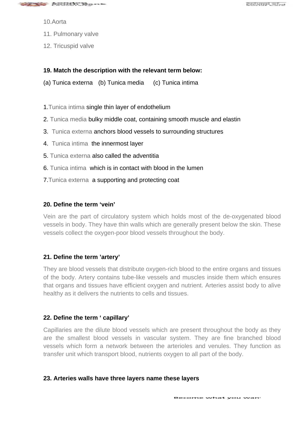
18. Draw and label a diagram of the heart showing the septum, chambers, valves,
and blood vessels entering / leaving each chamber.
Use colour to indicate whether the chambers and blood vessels carry oxygenated or
deoxygenated blood (red for oxygenated blood and blue for deoxygenated blood).
Use arrows to indicate direction of blood flow
(Do not worry about the accuracy of the outer shape of the heart, you could draw it as a
rectangle or heart shape, for example:
1.Superior Vena cava
2.Pulmonary artery
3. Pulmonary vein
4. Mitral valve
5.Aortic Valve
6. Left Ventricle
7.Right ventricle
8.Left atrium
9.Right atrium
10.Aorta
11. Pulmonary valve
12. Tricuspid valve
19. Match the description with the relevant term below:
(a) Tunica externa (b) Tunica media (c) Tunica intima
1.Tunica intima single thin layer of endothelium
2. Tunica media bulky middle coat, containing smooth muscle and elastin
3. Tunica externa anchors blood vessels to surrounding structures
4. Tunica intima the innermost layer
5. Tunica externa also called the adventitia
6. Tunica intima which is in contact with blood in the lumen
7.Tunica externa a supporting and protecting coat
20. Define the term ‘vein’
Vein are the part of circulatory system which holds most of the de-oxygenated blood
vessels in body. They have thin walls which are generally present below the skin. These
vessels collect the oxygen-poor blood vessels throughout the body.
21. Define the term ’artery’
They are blood vessels that distribute oxygen-rich blood to the entire organs and tissues
of the body. Artery contains tube-like vessels and muscles inside them which ensures
that organs and tissues have efficient oxygen and nutrient. Arteries assist body to alive
healthy as it delivers the nutrients to cells and tissues.
22. Define the term ‘ capillary’
Capillaries are the dilute blood vessels which are present throughout the body as they
are the smallest blood vessels in vascular system. They are fine branched blood
vessels which form a network between the arterioles and venules. They function as
transfer unit which transport blood, nutrients oxygen to all part of the body.
23. Arteries walls have three layers name these layers
Paraphrase This Document
Need a fresh take? Get an instant paraphrase of this document with our AI Paraphraser
Artery is the muscular tissues which is lined by the smooth tissues primarily. Three
layers of artery is the intima, media and the adventitia. Intima is inner layer lined by
smooth tissues and media is muscle layer which support the artery to handle the high
pressure of the heart. Adventitia is the outermost layer which contains the connective
tissue.
24. Vein walls have three layers name these layers
The vein consist oof the three layers namely tunica intima, tunica media and the tunica
adventitia. The innermost layer is the tunica intima which consist of flat epithelial
tissues. Middle layer is the thickest part of the wall which contains smooth blood vessels
and outermost layer is the tunica adventitia. This layer consist of connective tissues and
elastic fibres which provide vessels strength.
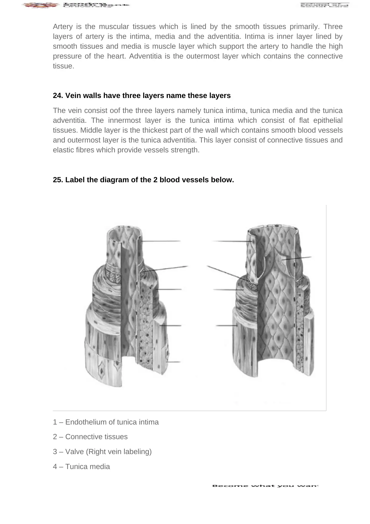
25. Label the diagram of the 2 blood vessels below.
1 – Endothelium of tunica intima
2 – Connective tissues
3 – Valve (Right vein labeling)
4 – Tunica media
5 – Tunica adventitious
(Labeling is being done in vertical manner from up to down)
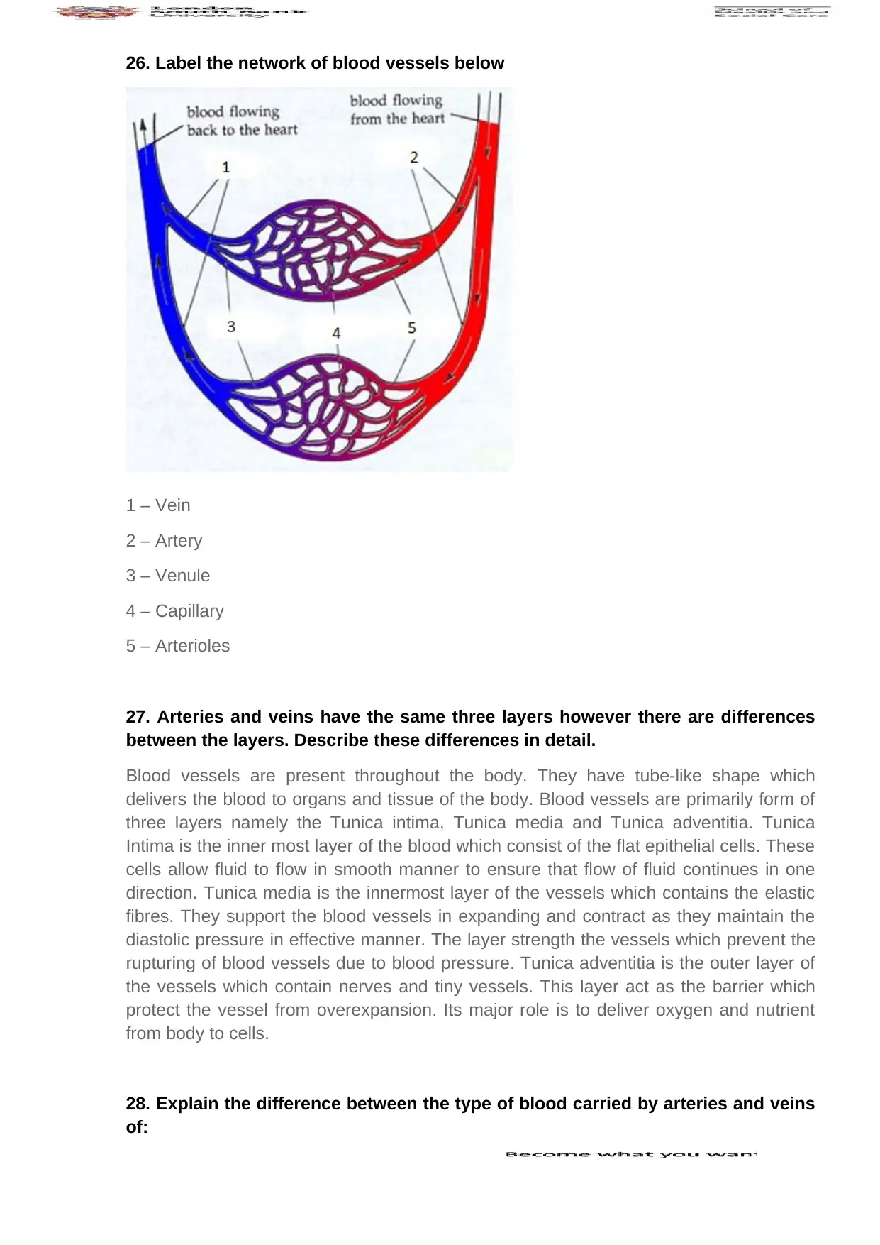
26. Label the network of blood vessels below
1 – Vein
2 – Artery
3 – Venule
4 – Capillary
5 – Arterioles
27. Arteries and veins have the same three layers however there are differences
between the layers. Describe these differences in detail.
Blood vessels are present throughout the body. They have tube-like shape which
delivers the blood to organs and tissue of the body. Blood vessels are primarily form of
three layers namely the Tunica intima, Tunica media and Tunica adventitia. Tunica
Intima is the inner most layer of the blood which consist of the flat epithelial cells. These
cells allow fluid to flow in smooth manner to ensure that flow of fluid continues in one
direction. Tunica media is the innermost layer of the vessels which contains the elastic
fibres. They support the blood vessels in expanding and contract as they maintain the
diastolic pressure in effective manner. The layer strength the vessels which prevent the
rupturing of blood vessels due to blood pressure. Tunica adventitia is the outer layer of
the vessels which contain nerves and tiny vessels. This layer act as the barrier which
protect the vessel from overexpansion. Its major role is to deliver oxygen and nutrient
from body to cells.
28. Explain the difference between the type of blood carried by arteries and veins
of:
Secure Best Marks with AI Grader
Need help grading? Try our AI Grader for instant feedback on your assignments.
a. pulmonary circulation
It begins with the pulmonary artery which carry deoxygenated blood from the heart to
the lungs. In lungs the filtration takes place and through pulmonary vein the oxygen rich
blood return back to the left atrium of heart by the lungs.
b. systemic circulation
Blood is pumped from the left ventricle of the heart through the aorta. Here further the
aorta is divided into smaller arteries, arterioles or capillaries for transport of blood all
over the body. From various part of the body, de-oxygenated blood from capillaries
merge into Venule or veins that drain the blood to vena cava. The vena cava further
drain de-oxygenated blood into the right atrium of the heart.
Multiple choice questions
1. The space occupied by the heart in the thoracic cavity is called the:
☐ Mediastinum
☐ Pericardial cavity
☐ Pleural cavity
☐ None of the above
2. Which side of the heart contains oxygenated blood?
☐ Both right and left sides
☐ Only the right side
☐ Only the left side
☐ Both the atria and the ventricles
3. Which chamber of the heart first receives oxygenated blood?
☐ The right ventricle
☐ The left atrium
☐ The left ventricle
☐ The right atrium
4. The valves that prevent back flow of blood during ventricular contraction are
the:
☐ Right and left atrioventricular valves
☐ Right atria ventricular and pulmonary valves
☐ Left atrioventricular and aortic valves
☐ Aortic and pulmonary valves
5. The valves that prevent backflow of blood during ventricular relaxation are the:
☐ Aortic and pulmonary valves
☐ Bicuspid and tricuspid valves
☐ Mitral and bicuspid valves
☐ None of the above
1 out of 12
Related Documents
Your All-in-One AI-Powered Toolkit for Academic Success.
+13062052269
info@desklib.com
Available 24*7 on WhatsApp / Email
Unlock your academic potential
© 2024 | Zucol Services PVT LTD | All rights reserved.