ProductsLogo
LogoStudy Documents
LogoAI Grader
LogoAI Answer
LogoAI Code Checker
LogoPlagiarism Checker
LogoAI Paraphraser
LogoAI Quiz
LogoAI Detector
PricingBlogAbout Us
logo

Chronic Diseases Explaining Symptoms of Mr. Jackson: Ulcerative Colitis and Crohn's Disease

Verified

Added on  2023/06/07

|8
|2308
|295
AI Summary
This article discusses the chronic diseases that explain the symptoms of Mr. Jackson, including Ulcerative Colitis and Crohn's Disease. It explains the differences between the two diseases and their diagnosis methods. The article also covers the symptoms, causes, and complications of Ulcerative Colitis and Crohn's Disease.

Contribute Materials

Your contribution can guide someone’s learning journey. Share your documents today.
Document Page
Name:
Student Number:
HPB Assignment

Secure Best Marks with AI Grader

Need help grading? Try our AI Grader for instant feedback on your assignments.
Document Page
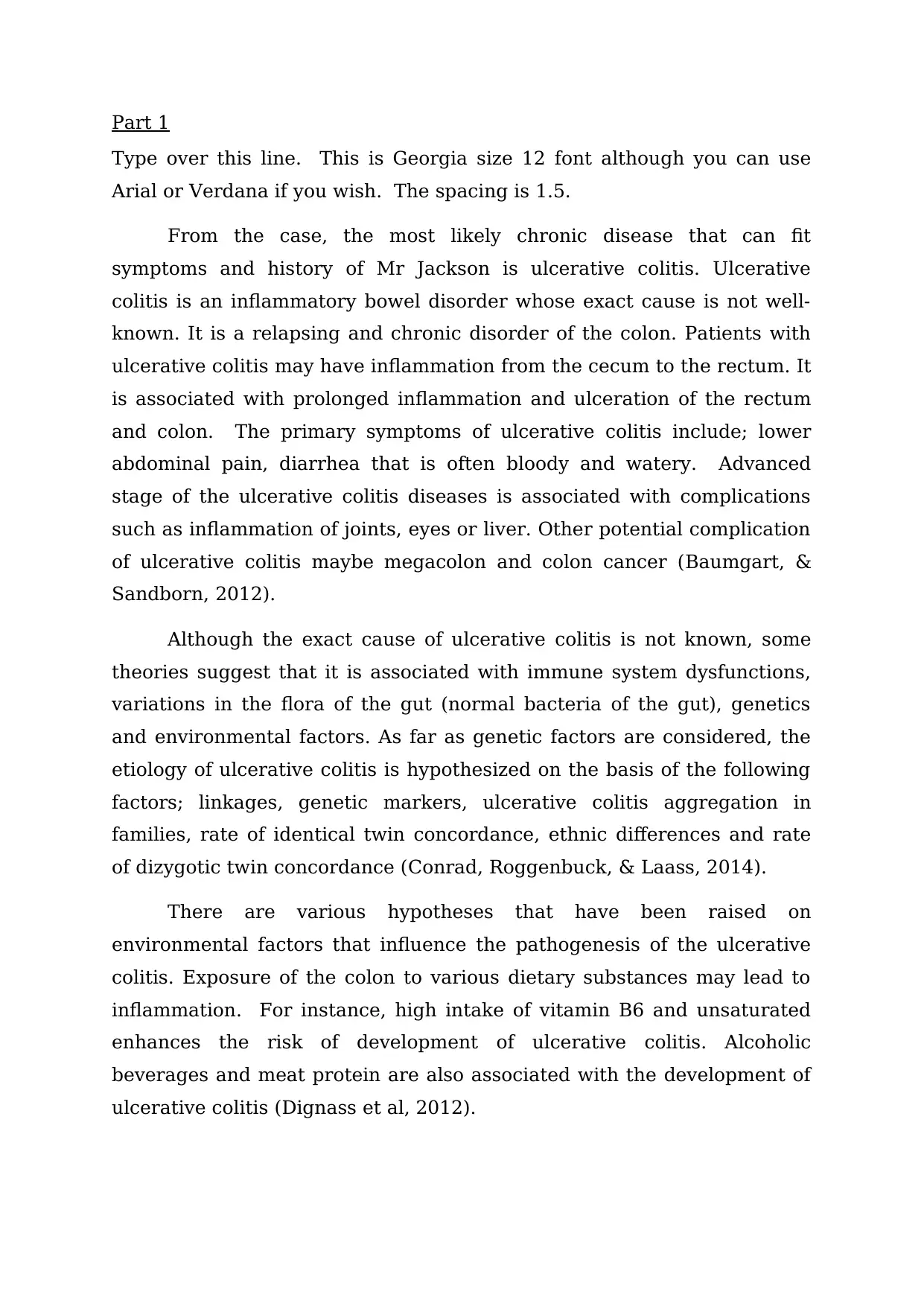
Part 1
Type over this line. This is Georgia size 12 font although you can use
Arial or Verdana if you wish. The spacing is 1.5.
From the case, the most likely chronic disease that can fit
symptoms and history of Mr Jackson is ulcerative colitis. Ulcerative
colitis is an inflammatory bowel disorder whose exact cause is not well-
known. It is a relapsing and chronic disorder of the colon. Patients with
ulcerative colitis may have inflammation from the cecum to the rectum. It
is associated with prolonged inflammation and ulceration of the rectum
and colon. The primary symptoms of ulcerative colitis include; lower
abdominal pain, diarrhea that is often bloody and watery. Advanced
stage of the ulcerative colitis diseases is associated with complications
such as inflammation of joints, eyes or liver. Other potential complication
of ulcerative colitis maybe megacolon and colon cancer (Baumgart, &
Sandborn, 2012).
Although the exact cause of ulcerative colitis is not known, some
theories suggest that it is associated with immune system dysfunctions,
variations in the flora of the gut (normal bacteria of the gut), genetics
and environmental factors. As far as genetic factors are considered, the
etiology of ulcerative colitis is hypothesized on the basis of the following
factors; linkages, genetic markers, ulcerative colitis aggregation in
families, rate of identical twin concordance, ethnic differences and rate
of dizygotic twin concordance (Conrad, Roggenbuck, & Laass, 2014).
There are various hypotheses that have been raised on
environmental factors that influence the pathogenesis of the ulcerative
colitis. Exposure of the colon to various dietary substances may lead to
inflammation. For instance, high intake of vitamin B6 and unsaturated
enhances the risk of development of ulcerative colitis. Alcoholic
beverages and meat protein are also associated with the development of
ulcerative colitis (Dignass et al, 2012).
Document Page
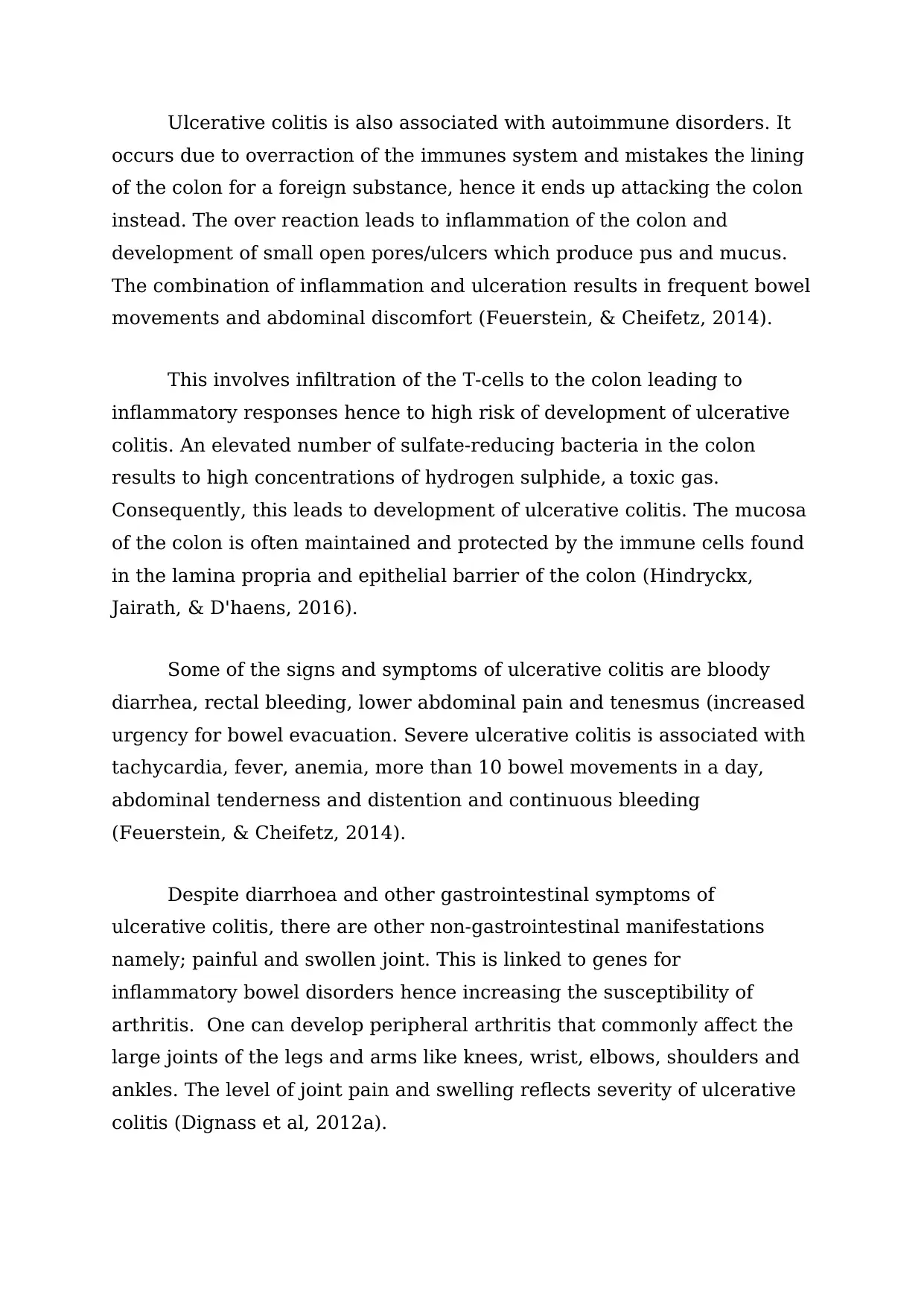
Ulcerative colitis is also associated with autoimmune disorders. It
occurs due to overraction of the immunes system and mistakes the lining
of the colon for a foreign substance, hence it ends up attacking the colon
instead. The over reaction leads to inflammation of the colon and
development of small open pores/ulcers which produce pus and mucus.
The combination of inflammation and ulceration results in frequent bowel
movements and abdominal discomfort (Feuerstein, & Cheifetz, 2014).
This involves infiltration of the T-cells to the colon leading to
inflammatory responses hence to high risk of development of ulcerative
colitis. An elevated number of sulfate-reducing bacteria in the colon
results to high concentrations of hydrogen sulphide, a toxic gas.
Consequently, this leads to development of ulcerative colitis. The mucosa
of the colon is often maintained and protected by the immune cells found
in the lamina propria and epithelial barrier of the colon (Hindryckx,
Jairath, & D'haens, 2016).
Some of the signs and symptoms of ulcerative colitis are bloody
diarrhea, rectal bleeding, lower abdominal pain and tenesmus (increased
urgency for bowel evacuation. Severe ulcerative colitis is associated with
tachycardia, fever, anemia, more than 10 bowel movements in a day,
abdominal tenderness and distention and continuous bleeding
(Feuerstein, & Cheifetz, 2014).
Despite diarrhoea and other gastrointestinal symptoms of
ulcerative colitis, there are other non-gastrointestinal manifestations
namely; painful and swollen joint. This is linked to genes for
inflammatory bowel disorders hence increasing the susceptibility of
arthritis. One can develop peripheral arthritis that commonly affect the
large joints of the legs and arms like knees, wrist, elbows, shoulders and
ankles. The level of joint pain and swelling reflects severity of ulcerative
colitis (Dignass et al, 2012a).
Document Page
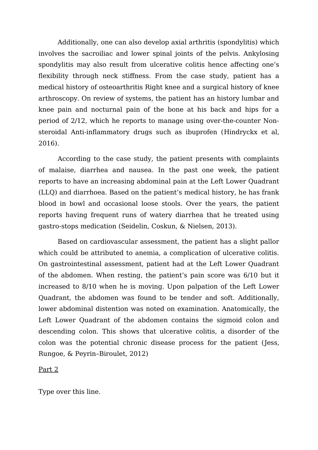
Additionally, one can also develop axial arthritis (spondylitis) which
involves the sacroiliac and lower spinal joints of the pelvis. Ankylosing
spondylitis may also result from ulcerative colitis hence affecting one’s
flexibility through neck stiffness. From the case study, patient has a
medical history of osteoarthritis Right knee and a surgical history of knee
arthroscopy. On review of systems, the patient has an history lumbar and
knee pain and nocturnal pain of the bone at his back and hips for a
period of 2/12, which he reports to manage using over-the-counter Non-
steroidal Anti-inflammatory drugs such as ibuprofen (Hindryckx et al,
2016).
According to the case study, the patient presents with complaints
of malaise, diarrhea and nausea. In the past one week, the patient
reports to have an increasing abdominal pain at the Left Lower Quadrant
(LLQ) and diarrhoea. Based on the patient’s medical history, he has frank
blood in bowl and occasional loose stools. Over the years, the patient
reports having frequent runs of watery diarrhea that he treated using
gastro-stops medication (Seidelin, Coskun, & Nielsen, 2013).
Based on cardiovascular assessment, the patient has a slight pallor
which could be attributed to anemia, a complication of ulcerative colitis.
On gastrointestinal assessment, patient had at the Left Lower Quadrant
of the abdomen. When resting, the patient’s pain score was 6/10 but it
increased to 8/10 when he is moving. Upon palpation of the Left Lower
Quadrant, the abdomen was found to be tender and soft. Additionally,
lower abdominal distention was noted on examination. Anatomically, the
Left Lower Quadrant of the abdomen contains the sigmoid colon and
descending colon. This shows that ulcerative colitis, a disorder of the
colon was the potential chronic disease process for the patient (Jess,
Rungoe, & Peyrin–Biroulet, 2012)
Part 2
Type over this line.

Secure Best Marks with AI Grader

Need help grading? Try our AI Grader for instant feedback on your assignments.
Document Page
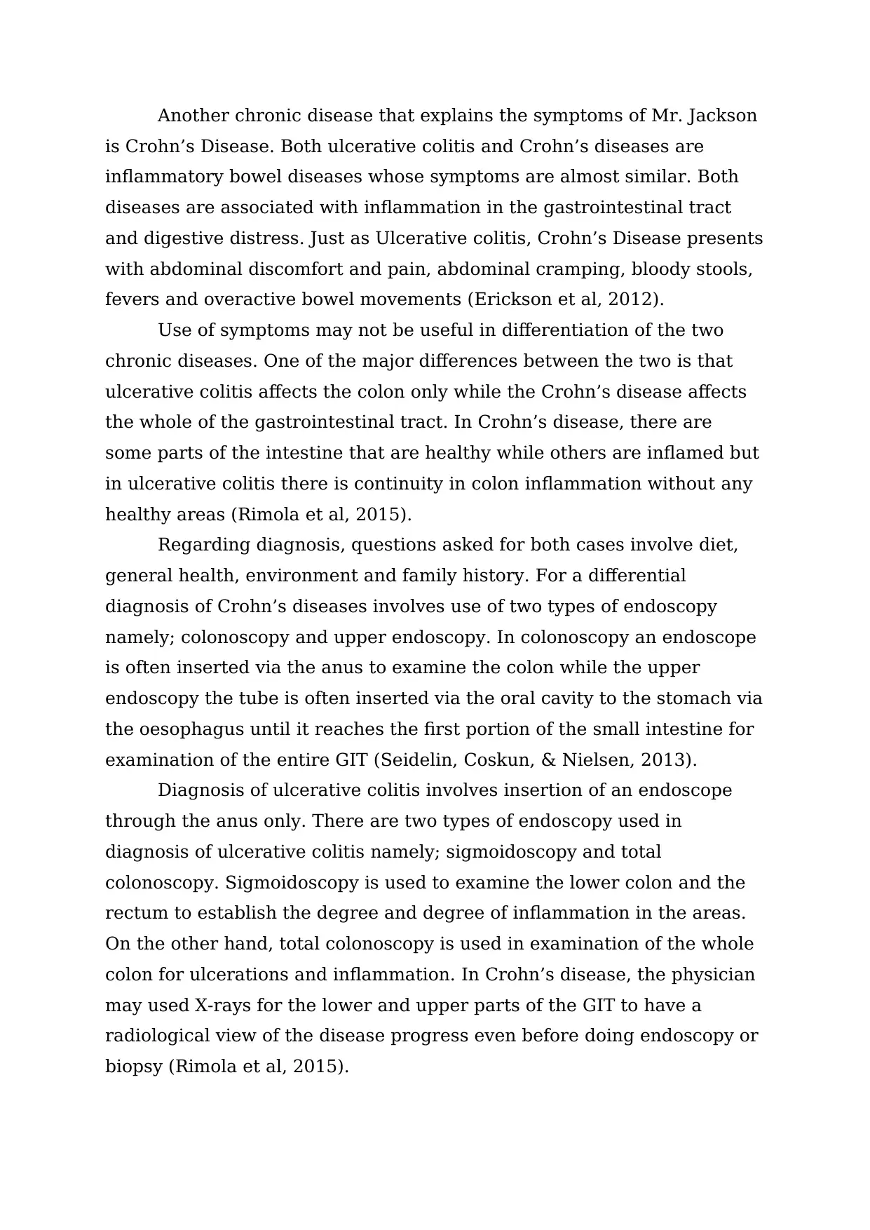
Another chronic disease that explains the symptoms of Mr. Jackson
is Crohn’s Disease. Both ulcerative colitis and Crohn’s diseases are
inflammatory bowel diseases whose symptoms are almost similar. Both
diseases are associated with inflammation in the gastrointestinal tract
and digestive distress. Just as Ulcerative colitis, Crohn’s Disease presents
with abdominal discomfort and pain, abdominal cramping, bloody stools,
fevers and overactive bowel movements (Erickson et al, 2012).
Use of symptoms may not be useful in differentiation of the two
chronic diseases. One of the major differences between the two is that
ulcerative colitis affects the colon only while the Crohn’s disease affects
the whole of the gastrointestinal tract. In Crohn’s disease, there are
some parts of the intestine that are healthy while others are inflamed but
in ulcerative colitis there is continuity in colon inflammation without any
healthy areas (Rimola et al, 2015).
Regarding diagnosis, questions asked for both cases involve diet,
general health, environment and family history. For a differential
diagnosis of Crohn’s diseases involves use of two types of endoscopy
namely; colonoscopy and upper endoscopy. In colonoscopy an endoscope
is often inserted via the anus to examine the colon while the upper
endoscopy the tube is often inserted via the oral cavity to the stomach via
the oesophagus until it reaches the first portion of the small intestine for
examination of the entire GIT (Seidelin, Coskun, & Nielsen, 2013).
Diagnosis of ulcerative colitis involves insertion of an endoscope
through the anus only. There are two types of endoscopy used in
diagnosis of ulcerative colitis namely; sigmoidoscopy and total
colonoscopy. Sigmoidoscopy is used to examine the lower colon and the
rectum to establish the degree and degree of inflammation in the areas.
On the other hand, total colonoscopy is used in examination of the whole
colon for ulcerations and inflammation. In Crohn’s disease, the physician
may used X-rays for the lower and upper parts of the GIT to have a
radiological view of the disease progress even before doing endoscopy or
biopsy (Rimola et al, 2015).
Document Page
Chromoendoscopy can be applied in Ulcerative colitis to view for
any changes in the intestinal lining and explore presence of precancerous
changes referred to as dysplasia. Additionally, for examination of the
small intestine and other parts of the GIT in Crohn’s disease, imaging
technique such as Magnetic Imaging Resonance (MRI) can be used. This
technique involves use of dyes to have a proper and clear view of the
images of the affected areas. Ulcerative colitis cannot be diagnosed using
images because it does not extend up to the small intestines but instead
it affects only the colon and rectum (Scharl, & Rogler, 2014).
Since the Crohn’s disease affects the entire GIT, its manifestations
are seen in other body parts. For example, sores in the mouth especially
between the lower lip and gums and also at the bottom and along the
sides of the tongue. Other symptoms that can manifest in Crohn’s disease
are anal tears, infections, ulcers, fissures (tears) and narrowing. Based
on manifestations, abdominal pain in ulcerative colitis is always confined
on the lower left abdominal portion while in Crohn’s disease, the
abdominal pain may occur in any part of the abdomen (Ripolles et al,
2013).
References
Baumgart, D. C., & Sandborn, W. J. (2012). Crohn's disease. The
Lancet, 380(9853), 1590-1605. Bouguen, G., Levesque, B. G.,
Feagan, B. G., Kavanaugh, A., Peyrin–Biroulet, L., Colombel, J.
F., ... & Sandborn, W. J. (2015). Treat to target: a proposed new
Document Page
paradigm for the management of Crohn's disease. Clinical
Gastroenterology and Hepatology, 13(6), 1042-1050.
Conrad, K., Roggenbuck, D., & Laass, M. W. (2014). Diagnosis and
classification of ulcerative colitis. Autoimmunity reviews, 13(4-5),
463-466.
Dignass, A., Eliakim, R., Magro, F., Maaser, C., Chowers, Y., Geboes,
K., ... & Travis, S. (2012). Second European evidence-based
consensus on the diagnosis and management of ulcerative colitis
part 1: definitions and diagnosis. Journal of Crohn's and
Colitis, 6(10), 965-990.
Dignass, A., Lindsay, J. O., Sturm, A., Windsor, A., Colombel, J. F., Allez,
M., ... & Öresland, T. (2012). Second European evidence-based
consensus on the diagnosis and management of ulcerative colitis
part 2: current management. Journal of Crohn's and Colitis, 6(10),
991-1030.
Erickson, A. R., Cantarel, B. L., Lamendella, R., Darzi, Y., Mongodin, E.
F., Pan, C., ... & Raes, J. (2012). Integrated
metagenomics/metaproteomics reveals human host-microbiota
signatures of Crohn's disease. PloS one, 7(11), e49138.
Feuerstein, J. D., & Cheifetz, A. S. (2014, November). Ulcerative colitis:
epidemiology, diagnosis, and management. In Mayo Clinic
Proceedings (Vol. 89, No. 11, pp. 1553-1563). Elsevier.
Hindryckx, P., Jairath, V., & D'haens, G. (2016). Acute severe ulcerative
colitis: from pathophysiology to clinical management. Nature
Reviews Gastroenterology & Hepatology, 13(11), 654.

Paraphrase This Document

Need a fresh take? Get an instant paraphrase of this document with our AI Paraphraser
Document Page
Jess, T., Rungoe, C., & Peyrin–Biroulet, L. (2012). Risk of colorectal
cancer in patients with ulcerative colitis: a meta-analysis of
population-based cohort studies. Clinical Gastroenterology and
Hepatology, 10(6), 639-645.
Rimola, J., Planell, N., Rodríguez, S., Delgado, S., Ordás, I., Ramírez-
Morros, A., ... & Panés, J. (2015). Characterization of inflammation
and fibrosis in Crohn’s disease lesions by magnetic resonance
imaging. The American journal of gastroenterology, 110(3), 432.
Ripolles, T., Rausell, N., Paredes, J. M., Grau, E., Martínez, M. J., &
Vizuete, J. (2013). Effectiveness of contrast-enhanced ultrasound
for characterisation of intestinal inflammation in Crohn's disease: a
comparison with surgical histopathology analysis. Journal of
Crohn's and Colitis, 7(2), 120-128.
Scharl, M., & Rogler, G. (2014). Pathophysiology of fistula formation in
Crohn's disease. World journal of gastrointestinal
pathophysiology, 5(3), 205.
Seidelin, J. B., Coskun, M., & Nielsen, O. H. (2013). Mucosal healing in
ulcerative colitis: pathophysiology and pharmacology. In Advances
in clinical chemistry (Vol. 59, pp. 101-123). Elsevier.
1 out of 8
circle_padding
hide_on_mobile
zoom_out_icon
[object Object]

Your All-in-One AI-Powered Toolkit for Academic Success.

Available 24*7 on WhatsApp / Email

[object Object]