EMG and MMG Signals for Diaphragm, Neck, and Chest Wall Muscles in COPD Patients
VerifiedAdded on 2022/10/14
|13
|3018
|126
AI Summary
This article discusses the impact of COPD on diaphragm, neck, and chest wall muscles and the use of EMG and MMG signals for non-invasive evaluation of muscle functions in COPD patients. It also highlights the importance of pulmonary rehabilitation and future prospects for research in this area.
Contribute Materials
Your contribution can guide someone’s learning journey. Share your
documents today.
Running head: CONFERENCE PAPER
CONFERENCE PAPER
Name of the Student
Name of the University
Author’s Note:
CONFERENCE PAPER
Name of the Student
Name of the University
Author’s Note:
Secure Best Marks with AI Grader
Need help grading? Try our AI Grader for instant feedback on your assignments.
1CONFERENCE PAPER
Abstract
The long-term breathing problems and weak respiratory function is known as COPD or chronic
obstructive pulmonary disorder. It's a pulmonary blockade. The patient has respiratory issues,
cough, and respiratory deficiencies. This disease is progressive and over time, the symptoms
deteriorate. Therefore everyday activities such as dressing, walking, and other simple duties
become difficult. Various causes of COPD include genetics, smoking, and pollution. However,
the most common reason for this disease is tobacco smoking, while other factors play a lesser
role. An important element in assessing patients with COPD is the analysis of EMG and MMG
signals for diaphragm, neck, and chest wall muscles. Non-invasive indicator of the experimental
and clinical significance of diaphragm, neck, and chest wall muscle functions for COPD patients.
This is performed by exploring the frequency and amplitude of muscle signals for ideal
implementation, in particular in cases of muscle contraction. For patients with respiratory
circumstances, the signals acquired with EMG and MMG are viable supplementary instruments
like EMG.
2CONFERENCE PAPER
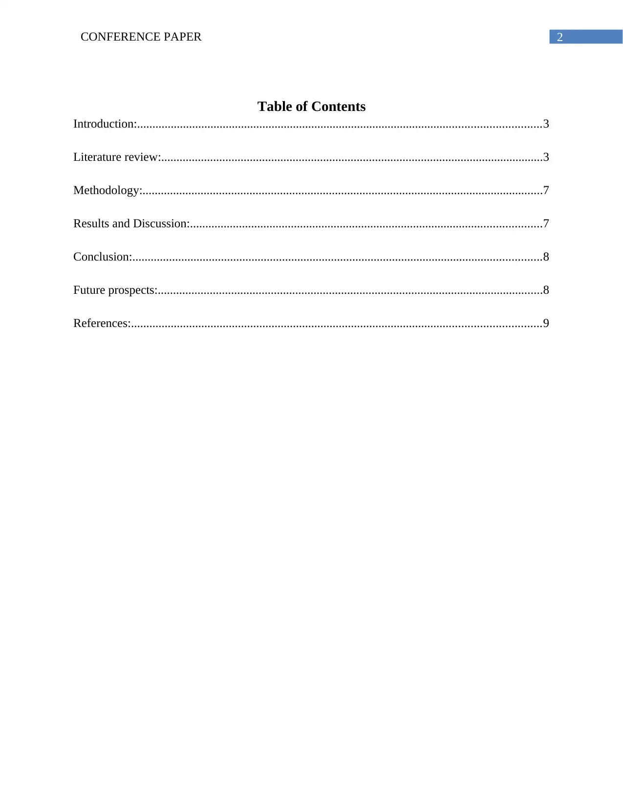
Table of Contents
Introduction:....................................................................................................................................3
Literature review:.............................................................................................................................3
Methodology:...................................................................................................................................7
Results and Discussion:...................................................................................................................7
Conclusion:......................................................................................................................................8
Future prospects:..............................................................................................................................8
References:......................................................................................................................................9
3CONFERENCE PAPER
Introduction:
COPD or Chronic obstructive pulmonary disorder is a long-term breathing difficulties and weak
respiratory function. It is a lungs-obstructive disease. The patient has breathing problems,
productive cough and breath shortages (Brij, Chatterji & Marquette, 2016, p245). The condition
is gradual and the symptoms deteriorate in the course of time. Daily activities like dressing,
walking, and other easy tasks therefore become hard. There are different COPD causes,
including genetics, cigarettes, and pollution. Tobacco smoking however, while the other
variables play a lower part, is the most prevalent reason of this disease. Low-ventilated heating
and cooking fires that have not been completely burned are responsible for air polluting that
causes this situation (Brij, Chatterji & Marquette, 2016 p250). These products have long-term
exposures to the respiratory system, particularly the lungs, which lead to inflammatory reactions.
The airways are reduced and diminished as the lung tissue is demolished. The diagnosis of
COPD is determined through trials on lung functions for inefficient air flow. Prevention is
accomplished through improved air quality both outside and indoors. The therapy involves
stopping smoking, pulmonary transplantation, and respiratory rehabilitation. The treatment of
COPD involves bronchodilators, steroids and oxygen treatment and vaccinations to avoid this
disease are also provided (Amal et al. 2017, p250).
Literature review:
The diaphragm is an anatomically below the lungs in the form of a dome. The diaphragm
contracts and plays an inspirational role, which increases the lung volume and causes the chest
cavity to grow and therefore a vacuum in which air flows into the lungs via its nose. When it
expires, the diaphragm relaxes and regains its shape, forcing air out of the lungs (McKenzie,
Secure Best Marks with AI Grader
Need help grading? Try our AI Grader for instant feedback on your assignments.
4CONFERENCE PAPER
Butler & Gandevia, 2009 p621). Due to the rise in lung volume, the diaphragm gets shorter in
COPD and therefore operates at a sub-optimal muscle fibre length. In COPD the diaphragm is
therefore weakened and its respiratory function is limited as it becomes more elastic and
powerless. This is due to modifications in diaphragm structures which lead to a loss of strength
and capacity to contract and relaxation of the muscle fibres. These patients generate minimal
force and decrease significantly the heavy sarcomere myosin chain. Severe COPD causes the
strength generation to be less calcium sensitive and has very slow cyclic kinetic cross-bridging.
The enhanced degradation of diaphragm protein triggers the severe COPD through the path of
ubiquitin - proteasis, causing contractile protein loss and diaphragm inability to produce strength
(Ottenheijm et al. 2005 p200).
As previously noted, the diaphragm muscles have been weakened in COPD, which reduces the
pressure caused by loss of fibre and elasticity. Oxidative stress and barometric damage in COPD
activate proteolytic machinery that results in individuals suffering from COPD to waste the
contractile tissue and therefore loss of power producing a diaphragm's ability (Sinderby et al.,
2001 p1641). This leads to the activation of a diaphragm which initiates the fiber type to the
oxidative type 1 which makes the overloaded diaphragm fatigue-resistant. The second technique
is by focusing on the pathway of ubiquitin protease to reduce diaphragm waste in COPD patients
(McKenzie, Butler & Gandevia, 2009 p629). Muscles and attachments of the neck are referred to
as accessory breathing muscles, when COPD has influenced the principal breathing muscle
(diaphragm). The recruitment of these muscles, however, does not completely offset the function
of the diaphragm, which results in breathing problems in individuals (Duiverman et al. 2009
p195). These muscles include scalenous bones and the muscles of the sternocleidomastoid that
help raise the shaft. Muscles of the neck are hired in the breath when a severely weakened
5CONFERENCE PAPER
COPD diaphragm is present. As indicated above, these muscles help to lift the rib cage and
depend on the breathing effort. Consequently, COPD (Orozco-Levi, 2003 p43) increases their
breathing activity. Severe COPD requires greater involvement of the throat and the attachments.
Patients with mild COPD use sternocleidomastoid muscle only in the course of practice or
physical activity while sleeping in rest. However, a lot of breathability and effort are needed in
serious COPD, which means that sternocleidomastoid muscles are hired even for a restful breath
(Cardosoet al. 2016 p0). The scalene muscles are active during quiet breathing, but the
sternocleidomastoids remain calm. The sternocleidomastoid muscles are also triggered in
enhanced respiratory volume. Therefore when individual breaths reach the maximum flow rate,
both muscles are triggered. The use of these auxiliary muscles therefore shows a sign of
breathing trouble in CBD (Heneghan, 2014, p134). The chest bones are consists of the intercostal
muscles between the ribs that form and shift the chest wall. These muscles include the thoracic
transversal, scalene and subcostal muscles, which help during the breathing phase. The muscles
of the chest wall elevate the ribs and therefore expand the thoroughfare.
Advanced airflow restrictions in COPD patients lead to lowered chest wall muscles ' ability and
functionality resulting in a lower essential ability, decrease in chest wall muscle strength and
decreased physical tolerance, resulting in decreased mobility. The pressure-generating capability
of COPD's chest wall muscle is reduced by dynamic hyperinflation (Duiverman and al. 2009
p200). Due to the destruction of the muscle fiber there is decreased elasticity and muscle strength
in the serious COPD. This leads to thorax modifications that lead to hyperinflation (O'donnell,
2006 p180). Thus, the thoracic cavity that is one of the limiting factors in serious COPD is not
expandable by the muscles of the chest wall. Emphysema present in serious COPD reduces
contact with the chest wall and reduces breathing adherence in general (Bianchi et al. 2004
6CONFERENCE PAPER
p460). The muscles of the chest wall are adjusted for endurance to COPD, although the function
is changed. Due to pulmonary hyper-inflation, the chest wall geometry and mechanics of COPD
are modified. Inspiring intercostal muscles and scalene muscles are therefore reduced. The
activation of the chest wall muscle rises as the severity of the COPD is increased. Mechanical
activation of the reduced chest wall muscles for the correction of pulmonary function is
performed in COPD patients (Orozco-Levi, 2003 p45). The neural inspirational drive increases,
which enhances the reorganization of the muscles of the chest wall.
The Electromyographic (EMG) and MMG signals of diaphragm, chest wall muscles and neck
muscles can be registered through the use of an oesophageal electrode and the trans-
diaphragmatic stress obtained through use of oesophageal and gastric samples which offer more
direct respiratory readings. The amount and pattern of air muscle activation of EMG and MMG
surface signals related to mechanical and electrical muscle activity are not invasively measured.
The EMG surface recorded above the intercostal space (best wall) evaluates the air movement of
the neuron. The findings from EMGs reflect the lung function and state of the respiratory
muscles, indicating the seriousness of the COPD, which results in higher loads, more ventilation
demands and a lower mechanical benefit (Heneghan, 2014). COPD treatment methods include
blowing, biofeedback, distraction therapeutics, guided imaging, gradual muscle relaxation,
acupuncture and other relaxation methods for the treatment of patients impacted by serious
airflow obstruction (Volpato et al., 2015). Pulmonary rehabilitation also involves important
methods for breathing muscles, breathing, and cessation of smoking, support, psychosocial
assistance, nutrition counseling, behavior training, patient training, training and optimization of
pharmacology. These methods strengthen the survivability of COPD patients, the functioning of
their muscles, and their health-related quality of life (Leupoldt et al., 2012). These operations
Paraphrase This Document
Need a fresh take? Get an instant paraphrase of this document with our AI Paraphraser
7CONFERENCE PAPER
also help to control significantly the perception of depression, anxiety and respiratory disease of
the target population. The method of gradual muscle relaxation needs tension in the target
portion of the body to be increased and then relaxed after five seconds.
Methodology:
For the purpose of this study, a small rapid evidence assessment have been conducted. In order to
do that, research article database like MedLine, Embase and Cinahl were used. Among the many
different journals, only articles from peer reviewed journals were selected. The articles were
primarily searched by the use of keywords. In some cases two or most of the keywords were
used together with the help of a Boolean operator. Inclusion and exclusion criteria were
employed during the search strategy to the narrow down the large of the literature found.
Inclusion criteria like journal published in ‘English’ language only were selected. Journals
published in any other language is excluded from the search. In addition to that, duplicates of the
articles were removed from the search. After the narrowing down the articles, all the articles
were manually evaluated by the investigators of this article.
Results and Discussion:
All total, 15 articles were selected from the literature search. COPD prevents the diaphragm
working correctly, the body compensates by expanding and tightening the chest using the neck,
back and shoulder muscles. However, the function of the diaphragm cannot be fully
compensated for by the muscles, which results in breathing disorders (Sinderby, Spahija, Beck,
Kaminski, Yan, Comtois & Sliwinski, 2001 p1637). Research has revealed that the muscles of
COPD patients attempt to adapt to chronic increase in respiratory stress and are fatigue-resistant.
The principal mechanism for hypercapnia is the diaphragm's failure. Muscle that is adequate to
8CONFERENCE PAPER
boost the core drive to support alveolar ventilation is rapidly manifest during sleep. Due to the
loss of muscle fibre and the consequent complete energy loss, diaphragm is significantly
weakened with serious COPD, causing death in COPD. Study has been conducted to determine
the impact of COPD on cardiac muscle mobility using both EMG and MMG. For example,
Heneghan case study (2014) on cervico-thoracic dysfunctional impact of COPD. EMG and
MMG are used for measurement of the movement of chest wall, muscle diaphragm and muscle
neck in patients with PDOC. From the research studies, it has been evident that the strength and
endurance of inspiratory muscle diminishes greatly in case of the patient living COPD (Lötters et
al. 2002). In the study, it has also been reported that fibres in the muscle gets damaged in the
patients with COPD. In addition to this, COPD affects the fibre in a different manner which has
been documented for the type 1 fibres (Stubbings et al. 2008).
Conclusion:
From the studies described above, it is clear that EMG and MMG is an essential, non-invasive
indicator of the experimental and clinical relevance of the diaphragm, throat, and chest wall
muscle functions used in COPD patients. This is achieved by the frequency and amplitude
exploration of the signals of the muscles, in particular, for ideal implementation during muscle
contractions. Signal EMG and MMG are a viable complement to conservative muscle evaluation
instruments like EMG for patients with respiratory circumstances. The method also produces
mechanical data on the physiological aspects of diaphragm and the muscle of the chest wall.

9CONFERENCE PAPER
Future prospects:
One of the aspects this investigation failed to address is that whether training of domiciliary
inspiratory muscle helps reduce the exacerbation of COPD among the patients admitted in the
hospital. Therefore, further studies are required to shed light in in this area.
Future prospects:
One of the aspects this investigation failed to address is that whether training of domiciliary
inspiratory muscle helps reduce the exacerbation of COPD among the patients admitted in the
hospital. Therefore, further studies are required to shed light in in this area.
Secure Best Marks with AI Grader
Need help grading? Try our AI Grader for instant feedback on your assignments.
10CONFERENCE PAPER
References:
Amal A. Abd El Aziz, Rabab A. Elwahsh, Gehan A. Abdelaal, Mohammed S. Abdullah, Rehab
A. Saad, 2017. Diaphragmatic assessment in COPD patients by different modalities, Egyptian
Journal of Chest Diseases and Tuberculosis, Volume 66, Issue 2, , Pages 247-250.
Bianchi, R., Gigliotti, F., Romagnoli, I., Lanini, B., Castellani, C., Grazzini, M. and Scano, G.,
2004. Chest wall kinematics and breathlessness during pursed-lip breathing in patients with
COPD. Chest, 125(2), pp.459-465.
Brij, S.O., Chatterji, S. and Marquette, M., 2016. Chronic Obstructive Pulmonary Disease
(COPD). In Clinical Pathways in Emergency Medicine (pp. 245-257). Springer, New Delhi.
Cardoso, D.M., Fregonezi, G.A., Jost, R.T., Gass, R., Alberton, C.L., Albuquerque, I.M., Paiva,
D.N. and Barreto, S.S., 2016. Acute effects of Expiratory Positive Airway Pressure (EPAP) on
different levels in ventilation and electrical activity of sternocleidomastoid and parasternal
muscles in Chronic Obstructive Pulmonary Disease (COPD) patients: a randomized controlled
trial. Brazilian journal of physical therapy, (AHEAD), pp.0-0.
Duiverman, M.L., de Boer, E.W.J., van Eykern, L.A., de Greef, M.H.G., Jansen, D.F., Wempe,
J.B., Kerstjens, H.A.M. and Wijkstra, P.J., 2009. Respiratory muscle activity and dyspnea during
exercise in chronic obstructive pulmonary disease. Respiratory physiology &
neurobiology, 167(2), pp.195-200.
Heneghan, N.R., 2014. Chronic obstructive pulmonary disease and cervico-thoracic
musculoskeletal dysfunction(Doctoral dissertation, University of Birmingham). Pp1-246
11CONFERENCE PAPER
Leupoldt, A. V., Fritzsche, A., Trueba, A.F., Meuret, A.E. and Ritz, T., 2012.
Behavioral medicine approaches to chronic obstructive pulmonary
disease. Annals of Behavioral Medicine, 44(1), pp.52-65.
Lötters, F., Van Tol, B., Kwakkel, G. and Gosselink, R.A.A.M., 2002. Effects of controlled
inspiratory muscle training in patients with COPD: a meta-analysis. European Respiratory
Journal, 20(3), pp.570-577.
McKenzie, D.K., Butler, J.E. and Gandevia, S.C., 2009. Respiratory muscle function and
activation in chronic obstructive pulmonary disease. Journal of applied physiology, 107(2),
pp.621-629.
Orozco-Levi, M., 2003. Structure and function of the respiratory muscles in patients with COPD:
impairment or adaptation?. European Respiratory Journal, 22(46 suppl), pp.41s-51s.
Ottenheijm, C.A., Heunks, L.M., Sieck, G.C., Zhan, W.Z., Jansen, S.M., Degens, H., De Boo, T.
and Dekhuijzen, P.R., 2005. Diaphragm dysfunction in chronic obstructive pulmonary
disease. American journal of respiratory and critical care medicine, 172(2), pp.200-205.
Sinderby, C., Spahija, J., Beck, J., Kaminski, D., Yan, S., Comtois, N. and Sliwinski, P., 2001.
Diaphragm activation during exercise in chronic obstructive pulmonary disease. American
Journal of Respiratory and Critical Care Medicine, 163(7), pp.1637-1641.
Stubbings, A.K., Moore, A.J., Dusmet, M., Goldstraw, P., West, T.G., Polkey, M.I. and Ferenczi,
M.A., 2008. Physiological properties of human diaphragm muscle fibres and the effect of
chronic obstructive pulmonary disease. The Journal of physiology, 586(10), pp.2637-2650.

12CONFERENCE PAPER
Volpato, E., Banfi, P., Rogers, S.M. and Pagnini, F., 2015. Relaxation techniques for people with
chronic obstructive pulmonary disease: a systematic review and a meta-analysis. Evidence-Based
Complementary and Alternative Medicine, 2015.
Volpato, E., Banfi, P., Rogers, S.M. and Pagnini, F., 2015. Relaxation techniques for people with
chronic obstructive pulmonary disease: a systematic review and a meta-analysis. Evidence-Based
Complementary and Alternative Medicine, 2015.
1 out of 13
Related Documents
Your All-in-One AI-Powered Toolkit for Academic Success.
+13062052269
info@desklib.com
Available 24*7 on WhatsApp / Email
![[object Object]](/_next/static/media/star-bottom.7253800d.svg)
Unlock your academic potential
© 2024 | Zucol Services PVT LTD | All rights reserved.


