MSc Project: Diabetic Retinopathy Detection Using MATLAB
VerifiedAdded on 2023/04/08
|14
|2862
|316
Project
AI Summary
This project proposal outlines an automated system for detecting Diabetic Retinopathy using MATLAB. The project aims to develop an image processing and machine learning-based solution for analyzing retinal images and identifying signs of the disease. The project's objectives include segmenting retinal images, implementing an automated classification system, and providing a hybrid solution through a MATLAB GUI interfaced with hardware. The methodology involves processing fundus images using MATLAB, segmenting them for feature extraction, and classifying them for disease detection. The intended users are doctors and hospital workers, and the project aims to reduce vision loss by enabling timely diagnosis. The project will utilize both primary data from interviews and secondary data from books and related research. The evaluation will assess the system's performance and user-friendliness, comparing it to existing products. The project deliverables include accurate optic disc localization, retinal vessel segmentation, and a user-friendly system. The project uses a block diagram to illustrate the system's processes, including image input, processing, segmentation, feature extraction, classification, and display. The project emphasizes the importance of timely diagnosis and the potential for reducing the workload associated with manual grading.

Electronics and communication engineering 1
Student name
Student ID
Course of Study
Module
Email
Project Supervisor
Student name
Student ID
Course of Study
Module
Project Supervisor
Paraphrase This Document
Need a fresh take? Get an instant paraphrase of this document with our AI Paraphraser
Electronics and communication engineering 2
TITLE:
PROJECT OUTLINE:
Problem statement
Diabetic retinopathy is a serious eye infection which can easily result in blindness hence
vision loss. Some of the health threats in Oman and most parts of the world are Diabetes mellitus
and metabolic disorder (ucchiatti, 2017). The complication of diabetes linked to the retina of the
eye is diabetic retinopathy. And if a patient has the complication he or she must undergo serious
screening of the eye done periodically. And this may be a very complication to the doctor and
also the patient.
For the biomedical engineering, automatic recognition of diseases, as well as medical imaging,
need a very high computational power to process complicated problem. Diabetic Retinopathy
is a concern which can result in blindness in a patient and it is very common in people of
Middle East countries for example Oman. Hence this research aims to propose and later
implement automatic detection system of Diabetic Retinopathy based on taking the images of
the patient eye (Wang, 2017). And for a realization of this project, a software called MATLAB
software is used to do the processing and it will be employed to boost the optometrist
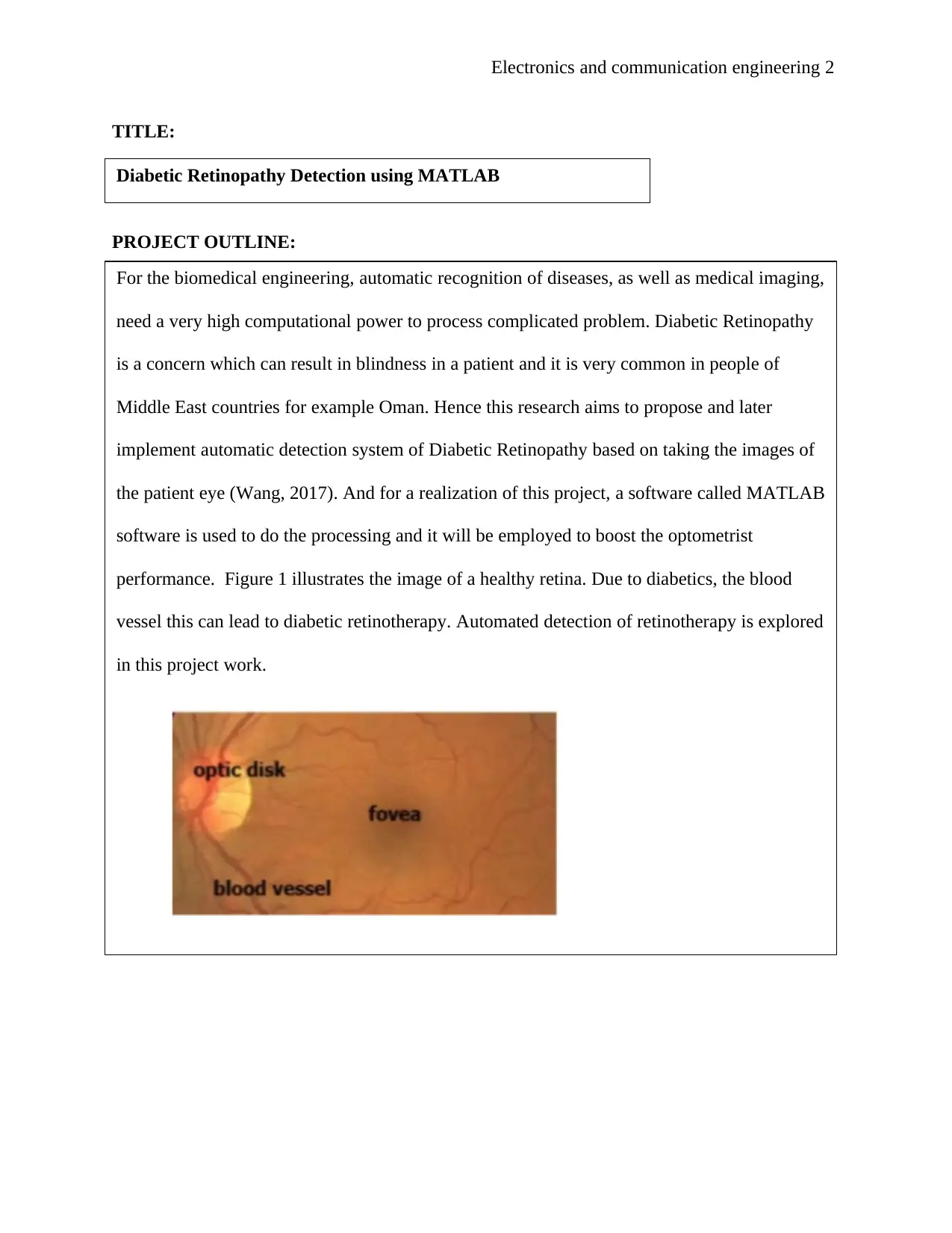
performance. Figure 1 illustrates the image of a healthy retina. Due to diabetics, the blood
vessel this can lead to diabetic retinotherapy. Automated detection of retinotherapy is explored
in this project work.
Figure 1: Showing the retina (Wang, 2017).
Diabetic Retinopathy Detection using MATLAB
Electronics and communication engineering 3
AIMS AND OBJECTIVE
The project research proposal aims to diagnose retinal captured image to help in
the identification of signs of diabetic retinopathy illustrated via dark sports in images of
the retina. The image will be processed via MATLAB which will read the image and
then process it before it quantifies the dark spots to obtain symptoms of diabetic
retinopathy.
Objective
Below are some of the objective for the realization of this project proposal;
i. To identify and segment the diabetic retinopathy.
ii. To implement an automated system that uses machine learning and image
processing to classify diabetic retinothepic healthy retinal
iii. To give a hybrid solution for diabetic retinopathy through developing
MATLAB GUI interfaced with Hardware
⊘ This is a preview!⊘
Do you want full access?
Subscribe today to unlock all pages.

Trusted by 1+ million students worldwide
Electronics and communication engineering 4
Block diagram
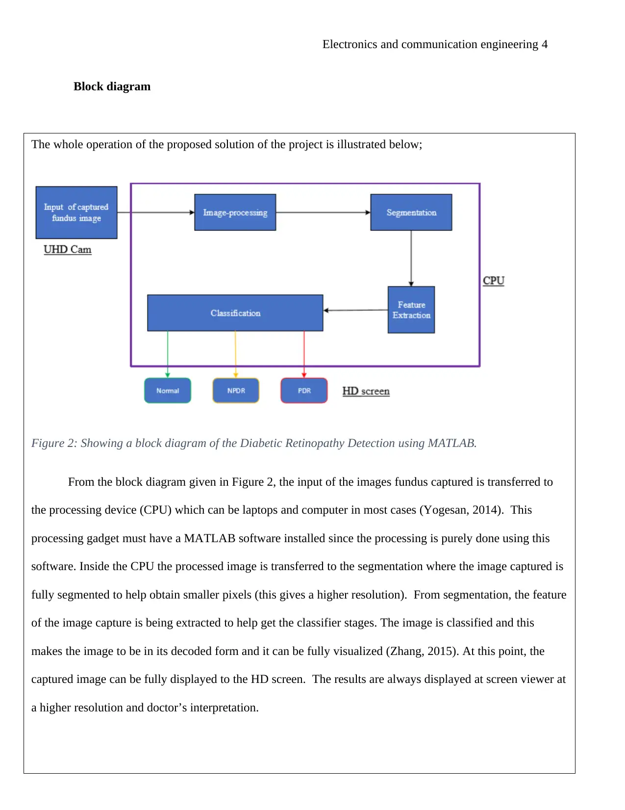
The whole operation of the proposed solution of the project is illustrated below;
Figure 2: Showing a block diagram of the Diabetic Retinopathy Detection using MATLAB.
From the block diagram given in Figure 2, the input of the images fundus captured is transferred to
the processing device (CPU) which can be laptops and computer in most cases (Yogesan, 2014). This
processing gadget must have a MATLAB software installed since the processing is purely done using this
software. Inside the CPU the processed image is transferred to the segmentation where the image captured is
fully segmented to help obtain smaller pixels (this gives a higher resolution). From segmentation, the feature
of the image capture is being extracted to help get the classifier stages. The image is classified and this
makes the image to be in its decoded form and it can be fully visualized (Zhang, 2015). At this point, the
captured image can be fully displayed to the HD screen. The results are always displayed at screen viewer at
a higher resolution and doctor’s interpretation.
Paraphrase This Document
Need a fresh take? Get an instant paraphrase of this document with our AI Paraphraser
Electronics and communication engineering 5
INTENDED USER/GROUP OF USERS AND THEIR REQUIREMENTS
This project actually helps to reduce the chances of loss of vision in most people due to diabetes.
Therefore the intended group of this project would be greatly the doctors and the hospital workers in general
(Olk, 2013). As illustrated above, this project is very important to most people in Oman and the whole world
since it will help to analyze the chances of one contacting the blindness and help to reduce the chances before
the situation is dire. When the situation is analyzed by the help of MATLAB it can be easily corrected before
it becomes so acute. Use of MATLAB will actually help in obtaining pictures of very high resolution hence
giving very accurate results of the analysis.
There are higher chances that the risk of severe loss of vision can be reduced when timely diagnosis
and treatment are done (Mason, 2011). Therefore the systematic MATLAB screening for the Diabetic
Retinopathy has been identified as a relatively low cost of health service for this condition. Live project on
Diabetic Retinopathy Detection using MATLAB will be a very crucial imaging tool for early Diabetic
Retinopathy tool for detection and this will highly reduce the workload that is associated with manual grading
and also saves time and cost of the diagnosis and treatment of this condition (Sun, 2013). Therefore this
project will satisfy the need of the timely and relatively low cost of treatment of this condition and it will help
to reduce the chances of loss of vision among the working age of citizens in Oman and other many countries
worldwide. Basically this project will be very beneficial to the people of Oman as well most individual
worldwide because of it will help in diagnosis and this can help the treatment of diabetes before it results to
loss of eye sight.
Electronics and communication engineering 6
SYSTEMS REQUIREMENTS AND PROJECT DELIVERABLES
Characteristics/properties
After the completion of this project, it is anticipated that the complete project will have the following
characteristics and properties;
i. The correct localization of the optic disc image will be 999 out of 1000 retinal images which means it
will be about 99.9 %
ii. The segmentation of retinal vessels accuracy of 20 retinal images will be around 94.2 per cent.
Process stages and deliverables
The below are the stages which will be employed in obtaining the images;
Stage one: Create a Binary Image:
The R/G intensities from the fundus images are calculated. Then about fifty-pixel medial filter scaling are
applied to R/G intensities (Acton, 2012). A threshold of about 7 per cent R/G intensities is employed to give the
binary image.
⊘ This is a preview!⊘
Do you want full access?
Subscribe today to unlock all pages.

Trusted by 1+ million students worldwide
Electronics and communication engineering 7
Stage two: Selection of the potential DHs by shape.
All the connected regions in binary images are numbered. And in every region connected the shape factor
Fs=A/Lmax are calculated. Where Lmax is the maximum distance between any two pixels in the region and A is
the area in pixels. All parts with 30 or more pixel were all labelled and factors greater than 0.54 as a potential
DH´S were perfectly shaped (Chi, 2015).
Stage three: Checking whether DHs are contained in blood vessels.
For a forty neighborhood of every remaining DH the R/G will be computed in contour steps of 0.05 ± 0.5 in the
average of R/G for every DH. In every contour selected obtain the mean absolute gradient of the neighboring
points and select the contour C which corresponding to the biggest value. The distance DC between the nearest
DH and the current contour. But DH will be rejected if DC is greater than 4.
Sources of data in the research
In this research proposal there two main sources of data which will be employed to ensure that perfect
research is perfectly done. These sources of data include the primary as well as the secondary source of data
(Wróbel, 2011). The primary sources of data are the original sources of information like an artefact, raw data,
and interviews among others. For this research proposal, we will specifically use the interview as a primary
source of data. An interview will be conducted in different hospitals where several experienced doctors in
Oman will ask to give their opinion about the discovery and the project at large. The interviewees will as well
be given chances to have positive criticism on the project and then given their views on how some
improvements can be conducted on the research project proposal (Kumar, 2018). This will for sure help a lot
to get more information and make the necessary adjustments on the project and make it better to meet the
hospitals and doctors´ requirements to help solve the above-stated problem.
While for the secondary sources of data for this project will be books. Books will help to get more
information on other related work, for example, Diabetic Retinopathy using HSV and Fuzzy among other
Paraphrase This Document
Need a fresh take? Get an instant paraphrase of this document with our AI Paraphraser
Electronics and communication engineering 8
RESEARCH
similar projects (Johnson, 2013). Through the use of books, the knowledge of MATLAB codes will be
Exudate is common defects which always occurs in the retina and these are basically bright lipid leaked from
the blood vessels, these fluid tend to be next to the puncture yielding a well-defined edge which is appropriate for the
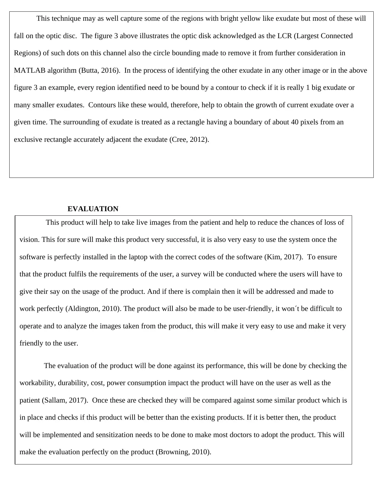
analysis using MATLAB software (Descoteaux, 2017). The diagram below illustrates a prototype of exudates on a
fundus image that is shown as a very small and light yellow regions as seen below;
Figure 3: Showing a prototype of exudates on a fundus image (Wang, 2017).
Nevertheless, there are some yellow light regions in the optic disc as illustrated in figure 3 above. Hence before a search
for exudates is done, a computer MATLAB codes and algorithms are developed for the live detection of the optic disc.
In the above diagram the yellow color links to the higher intensity of the green channel but basically the optic disc has
the highest green intensities on a particular image (Soille, 2013). Therefore an initial estimation to the optic disc is got
through sorting out the green intensity in an ascending way (that is from lowest to highest) and selecting all the pixels in
the 0.5% top intensities.
Electronics and communication engineering 9
EVALUATION
This technique may as well capture some of the regions with bright yellow like exudate but most of these will
fall on the optic disc. The figure 3 above illustrates the optic disk acknowledged as the LCR (Largest Connected
Regions) of such dots on this channel also the circle bounding made to remove it from further consideration in
MATLAB algorithm (Butta, 2016). In the process of identifying the other exudate in any other image or in the above
figure 3 an example, every region identified need to be bound by a contour to check if it is really 1 big exudate or
many smaller exudates. Contours like these would, therefore, help to obtain the growth of current exudate over a
given time. The surrounding of exudate is treated as a rectangle having a boundary of about 40 pixels from an
exclusive rectangle accurately adjacent the exudate (Cree, 2012).
This product will help to take live images from the patient and help to reduce the chances of loss of
vision. This for sure will make this product very successful, it is also very easy to use the system once the
software is perfectly installed in the laptop with the correct codes of the software (Kim, 2017). To ensure
that the product fulfils the requirements of the user, a survey will be conducted where the users will have to
give their say on the usage of the product. And if there is complain then it will be addressed and made to
work perfectly (Aldington, 2010). The product will also be made to be user-friendly, it won´t be difficult to
operate and to analyze the images taken from the product, this will make it very easy to use and make it very
friendly to the user.
The evaluation of the product will be done against its performance, this will be done by checking the
workability, durability, cost, power consumption impact the product will have on the user as well as the
patient (Sallam, 2017). Once these are checked they will be compared against some similar product which is
in place and checks if this product will be better than the existing products. If it is better then, the product
will be implemented and sensitization needs to be done to make most doctors to adopt the product. This will
make the evaluation perfectly on the product (Browning, 2010).
⊘ This is a preview!⊘
Do you want full access?
Subscribe today to unlock all pages.

Trusted by 1+ million students worldwide
Electronics and communication engineering 10
DEVELOPMENT SKILLS
This project for sure will require some useful information like the correct MATLAB codes which
will ensure that this project work as anticipated. This is very important since without the MATLAB codes
the project won´t work. Since MATLAB is a very heavy program, the project will require the use of a very
powerful laptop that is laptop from core i 5 with a good speed of above 2.4GHz. Therefore it goes without
saying that this project will require a relatively higher amount of money. More information will also be
required to understand the image captured is transferred from the patient to the display (Laptop). And it is
also very important to know how to deal with the pixels to ensure that the image obtained is high quality of a
resolution of 1216x912 and noises are highly removed from the signal transmitted to the laptop.
Basically the information on the coding of the MATLAB software I can easily learn myself and test
the codes until I obtain the correct codes which will work perfectly. Knowledge of the transmission of the
signal with very less or no noise is possible to learn hence this I can as well acquire myself.
The non-invasive method is common method employed in health sector where there is no penetration into the
human body through the use of biomedical equipment. There is no blood taken here for diagnosis. An
example of this method can be a deep palpation and percussion are non-invasive but a rectal examination is
invasive
Paraphrase This Document
Need a fresh take? Get an instant paraphrase of this document with our AI Paraphraser
Electronics and communication engineering 11
SKILL ACQUISITION
PROJECT PLANNING
The information which I may acquire myself like the knowledge on writing a perfectly MATLAB code
will be learned. I will get information from books as well as the internet to know how best to code
MATLAB for this particular project. Being that I already have knowledge in MATLAB it will be very
easy for me to get the correct codes. In some cases, I may do some interview with doctors to get their
views to know how best they would like the project to work. For the part of finance, I will seek the
assistance of sponsors to help fund my project and this will be possible if I completely defend my project
and convince them that it will work perfectly when funded.
With the increasing importance of deep learning in artificial intelligence as well as business environments,
this will highly inspire changes which always affect other systems as well as some business requirement
processes in development of software. Even though the machine learning has been considered as integral
to the toolbox analytics, specifically as a component of predictive analysis. This is set by flexibility,
increasing power as well as range of deep learning.
Electronics and communication engineering 12
GANTT CHART
No Activity Conducted Number of hours
1 Product design 10
2 Product development 100
3 Product evaluation 30
4 Development or Investigation 87
5 Final report preparation 70
6 Total number of hours 600
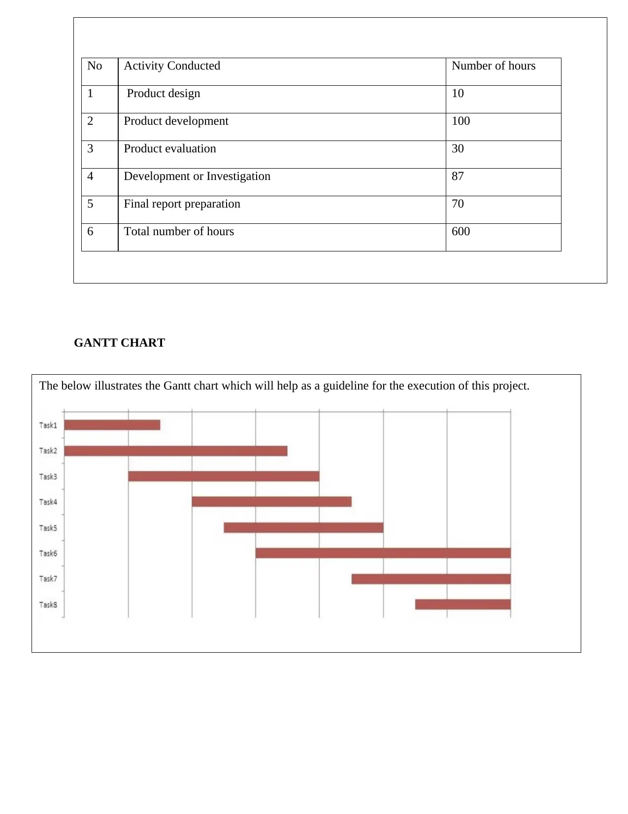
The below illustrates the Gantt chart which will help as a guideline for the execution of this project.
⊘ This is a preview!⊘
Do you want full access?
Subscribe today to unlock all pages.

Trusted by 1+ million students worldwide
1 out of 14
Your All-in-One AI-Powered Toolkit for Academic Success.
+13062052269
info@desklib.com
Available 24*7 on WhatsApp / Email
![[object Object]](/_next/static/media/star-bottom.7253800d.svg)
Unlock your academic potential
Copyright © 2020–2026 A2Z Services. All Rights Reserved. Developed and managed by ZUCOL.


