This document discusses the pathophysiology of diabetic foot ulcers in patients with type 2 diabetes, including the causes and contributing factors such as peripheral neuropathy, vasculopathy, and immunopathy. It also covers the management and nursing priorities for diabetic foot ulcers.
Contribute Materials
Your contribution can guide someone’s learning journey. Share your
documents today.
Running header: RIGHT FOOT ULCER1 Right foot ulcer Student name Student ID number Specialty area
Secure Best Marks with AI Grader
Need help grading? Try our AI Grader for instant feedback on your assignments.
Schneider2 Pathophysiology of diabetic foot ulcer Mrs. Gina has a history of type two diabetes, peripheral vascular disease and obesity which led to a right foot ulcer. Her blood glucose level was 12.6mmol/L and surrounding skin was wet from exudate, dark pink and painful to touch. Factors that caused the growth of diabetic foot ulcer in diabetic patients are divided into two. This includes causative factors and contributing factors. Causative factors include peripheral neuropathy, vasculopathy, and immunopathy. Contributing factors include atherosclerosis, and diabetes (Obaid & Eljedi, 2014). a.Neuropathy Peripheral neuropathy also called loss sensation is the impairment of nerves throughout the body. An increased hyperglycemic condition in the blood causes the secretion of sorbitol dehydrogenase and aldolase reductase. This enzyme causes the conversion of glucose to fructose and sorbitol. The accumulation of these sugar products leads to a decreased production of nerve cell myoinositol hence affecting nerve conduction. This causes a decreased peripheral sensation, vasomotor control of the pedal circulation and lastly, it damages the nerves of the little muscles in the foot (Merza & Tesfaye, 2018). When the normal functioning of the nerves is impaired it may expose an individual to minor injuries without recognizing it until it makes an ulcer. High sugar levels in the blood cause dryness and fissuring of the skin predisposing it to infection. The autonomic nervous system controls the microcirculation of the skin. Damage of the autonomic nervous system leadto the enlargement of ulcers, gangrene and lastly limb loss. b.Vasculopathy
Schneider3 Uncontrolled glucose level causes endothelial cell damage and smooth cell abnormalities. Endothelial cells damage leads to rapid growth of endothelial cells, reduced production of nitric oxide, increased blood viscosity, thickening of the basement membrane and reduced blood flow. Endothelial cells synthesize nitric oxide which causes vasodilation of blood vessels. Reduced nitric oxide production leads to a propensity for atherosclerosis, vasoconstriction of blood vessels and finally leading to ischemia. Hyperglycemia cause overproduction of thromboxane A2 causing plasma hypercoagulability. Clients with this condition manifest with rest pain, absent peripheral pulse and lastly thinning of the skin (Merza & Tesfaye, 2018). c.Immunopathy The immune system of a diabetic patient is much weaker than healthy people. The hyperglycemic condition causes overproduction of pro-inflammatory cytokines and damage of polymorphonuclear function like chemotaxis and phagocytosis. The immune system is weakened due to decreased leukocyte activity, wrong inflammatory response and lastly disturbance of cellular immunity (Merza & Tesfaye, 2018). Diminished chemotaxis of cytokines and growth factors impended wound healing by extending the inflammatory state. The presence of an open wound in a fasting state creates a catabolic state. Negative nitrogen balance results caused by protein break down. This impairment of metabolic function affects the synthesis of proteins, collagen, and fibroblast. Lack of these factors in the blood will result in prolonged healing of the wound. High blood glucose creates a favorable condition for the growth of bacteria mainly the S. Aureus and B- hemolytic streptococci (Robelledo, Soto, & Pena, 2015). Causes of the patient post-operative wound status
Schneider4 Mrs. Gina blood glucose level was 12.6mmol/L. She was unable to manage her high glucose level since she was noncompliant to medication prescribed on discharge. Her wound was wet from serous exudate output. The surrounding skin was warm, dark pink and painful to touch which was an indication of an infection. The major cause of this infection was a peripheral vascular disease caused by high blood glucose level in the blood. This condition resulted in arterial insufficiency when blood flow to the legs is reduced (Rohfling, Weidmeyer, & Little, 2017). A decrease in blood flow to the tissue may lead to tissue decay, creating a favorable environment for bacterial growth. Reduced blood flow also leads to reduced oxygen levels, nutrition, and healing rate. Lack of oxygen in the tissues prevents the neutrophils from phagocytizing bacteria and prevents the macrophages from reaching the site of infection. When the blood sugar level is out of control, there is the production of harmful molecules known as dicarbonyls. This molecule changes the structure of human beta-defensin-2 which is an infection-fighting peptide. Destruction of this peptides reduces the ability of the body to fight off bacteria and infection (Sacks, Bruns, & Macdonald, 2016). Nursing priorities and rationale Type two diabetes occurs when the glucose levels in the blood are too high. Hyperglycemia may occur for several reasons such as not taking enough insulin or eating too much. Mrs. Bail blood glucose level is 12.6mmol/L which was higher than normal. If the hypoglycemia was is untreated it would lead to diabetic ketoacidosis which may later cause unconsciousness or even death. The two main priorities of care include the following; a.Administer glucose-lowering medication.
Paraphrase This Document
Need a fresh take? Get an instant paraphrase of this document with our AI Paraphraser
Schneider5 Since the client was not compliant to medication prescribed which resulted in a high blood glucose level of 12.6mmol/L the best treatment to rapidly lower the blood glucose level is insulin pump therapy. The pump is attached to the client with a thin tubing with a needle at the end which is introduced under the skin. This pump allows insulin to flow in a rate that one can control. This means that the client can no longer give herself medication. Insulin acts by lowering blood glucose. It stimulates glucose uptake by the skeletal muscles, prevents hepatic glucose production, and lastly prevents proteolysis and lipolysis (Schetz, Wouterz, & Weekers, 2017). Metformin is administered as the first medicine. It acts by decreasing the level of glucose that the liver produces into the blood. This helps the body cells to be responsive to insulin. It’s also prescribed for clients who are obese. Sulphonylureas example glibenclamide is administered to the client to increase the amount of insulin produced by the pancreas (Buchanan, Xiang, & Ochoa, 2016). b.Monitor the foot and educate the client on the importance of foot monitoring Having diabetes means that you are at risk of developing feet problems due to damage to the nerves of the foot. This means that small cuts are not easily noticed leading to the development of foot ulcer. Close monitoring and proper foot care are important because reduced blood flow to the feet may result in decreased sensation also called neuropathy. Reduced blood flow may result in slow wound healing. This can be avoided by encouraging daily foot care this includes sterile cleaning of the wound, looking for any small cuts at the feet and lastly trimming toenails (Pendsey, 2014). Management of diabetic patients
Schneider6 Diabetes is a chronic illness that calls –for continuing medical care and client’s self- management education so as to prevent acute complication and decrease the chance of getting a long term complication. Safe nursing management of the patient include the following; 1.Management of diabetic foot ulcer The main objective in the management of diabetic ulcer is the closing of the wound to prevent further infection. The basis of diabetic wound treatment include debridement, decreasing pressure on the injured area, administering adequate antibiotics, wound treatment using a clean and moist dressing and lastly managing the bacteria by diagnosing the type of infection. a.Debridement Debridement is the process of removing nonliving material, unhealthy tissue and foreign bodies from the injured area. Debridement plays an important role in wound healing through the production of granulation tissue. The major purpose of this process is to convert a chronic wound healing environment into acute wound healing (Jupiter, Buckley, & Shibuya, 2016). Mechanical debridement is carried out by dry-wet dressing pressure irrigation and hydrotherapy. Debridement can also be done biologically by the use of sterile larvae of sericata fly which produces a proteolytic enzyme that dissolves the necrotic tissue. b.Offloading Offloading is the act of decreasing pressure on the ulcer. The major purpose of offloading is to reduce tissue trauma and enhance wound healing. Ulceration takes place on an area of the foot that gets high pressure. In addition bed, rest is a method of choice so as to decrease pressure.
Schneider7 Total contact casting is also used as an offloading method. This is done by letting the client take a walk during therapy. It is important in controlling edema that can hinder wound healing (Pataky & Conne, 2016). c.Management of infection Bacterial wounds allow bacterial entry leading to infection. Signs and symptoms of infection include pain, edema, softness, and pus. Infections are treated by oral administration of antibiotics example clindamycin. In severe infection, intravenous antibiotics are given examples include ampicillin and imipenem-cilastatin. d.Dressing A dressing is a material used to cover a wound protecting it from infections. Several types of dressing include the hydrocolloid and hydrogel. The advantages of a closed clean wound are that it creates a humid healing environment to enhance cell migration and keep away dry sores. Negative pressure wound treatment is also used to eliminate any possibility of edema formation. It acts by removing lymph in interstitial hence elevating the movement of interstitial oxygen into the cell. 2.Glycemic control Glycemic control is very important in the management of the diabetic client. High glucose levels in the blood can be controlled through the following ways; a.Stress management
Secure Best Marks with AI Grader
Need help grading? Try our AI Grader for instant feedback on your assignments.
Schneider8 Physiologic stress that may be caused by surgery or infection may contribute to high glucose level and may trigger diabetic ketoacidosis. During stress, epinephrine is produced which causes an elevation in glucose levels. In addition, during this time of emotional stress, a client with diabetes may change the time of medication, exercise, and meals. This contributes to hyperglycemia. Diabetic clients are encouraged to comply with the medication despite the emotional stress. They are also taught on methods of minimizing stress such as exercise or expressing their feelings to someone they trust (Tumilehto, Erikson, & Laakso, 2013). b.Exercise program Exercise is crucial in the management of diabetes. This is because it decreases blood glucose levels and cardiovascular risk factors. Exerting your muscles results in increased uptake glucose by skeletal muscles and also utilizes insulin (Schneider & Ruderman, 2017). Weight lifting can increase muscle tone enhancing resting metabolic rate. Since the client is obese, exercise helps in losing weight. Moreover, it reduces stress and maintains a feeling of well-being. Exercise also increases the high-density lipoprotein and reduces the low-density lipoproteins. This is important in diabetic patient because of increased risk of cardiovascular diseases (Haffner, 2014). c.Nutritional management Diet, nutrition and weight control are the basis for diabetes management. The important process in nutritional management is the control of total caloric intake so as to maintain a normal blood glucose level and maintain sensible body weight. The client is first educated on the relationship between food and insulin. Food high in fiber is recommended for diabetic patients since fiber lowers cholesterol levels in the blood. Fiber also reduces the demand for exogenous insulin by slowing down the absorption of glucose in the blood (Pan, Wang, Lin, & Jiang, 2015).
Schneider9 The client is also advised on eating a whole fruit rather than drinking juice. The fiber in the fruits slows down the absorption process. Starchy food is also advised to be taken with protein so as to slow down the absorption process lowering the glycemic index. Generally taking foods which are whole and raw will lead to lower glycemic response than chopped or cooked foods (Pan, Wang, Lin, & Jiang, 2015). References Buchanan,T., Xiang,H., & Ochoa,C. (2016). Preservation of pancreatic β-cell function and prevention of type 2 diabetes by pharmacological treatment of insulin resistance in high-risk Hispanic women.Diabetes,51, 2796-2803. Haffner,S. (2014). Management of dyslipidemia in adults with diabetes.Diabetes Care,21, 160-178. Jupiter,D., Buckley,C., & Shibuya,N. (2016). The impact of foot ulceration and amputation on mortality in diabetic patients From ulceration to death, a systematic review.Int. Wound J,13(5), 892-903. Merza,Z., & Tesfaye,S. (2018). The risk factors for diabetic foot ulceration.J. Foot Ankle Surg,13(3), 125-129. Obaid,H., & Eljedi,A. (2014). Risk factors for the development of diabetic foot ulcers in Gaza strip: a case-control study.Int. J. Diabet. Res,4(1), 1-6. Pan,X., Wang,X., Lin,J., & Jiang,Y. (2015). Effects of diet and exercise in preventing NIDDM in people with impaired glucose tolerance: the Daqing IGT and Diabetes Study.Diabetes Care,20, 537-544.
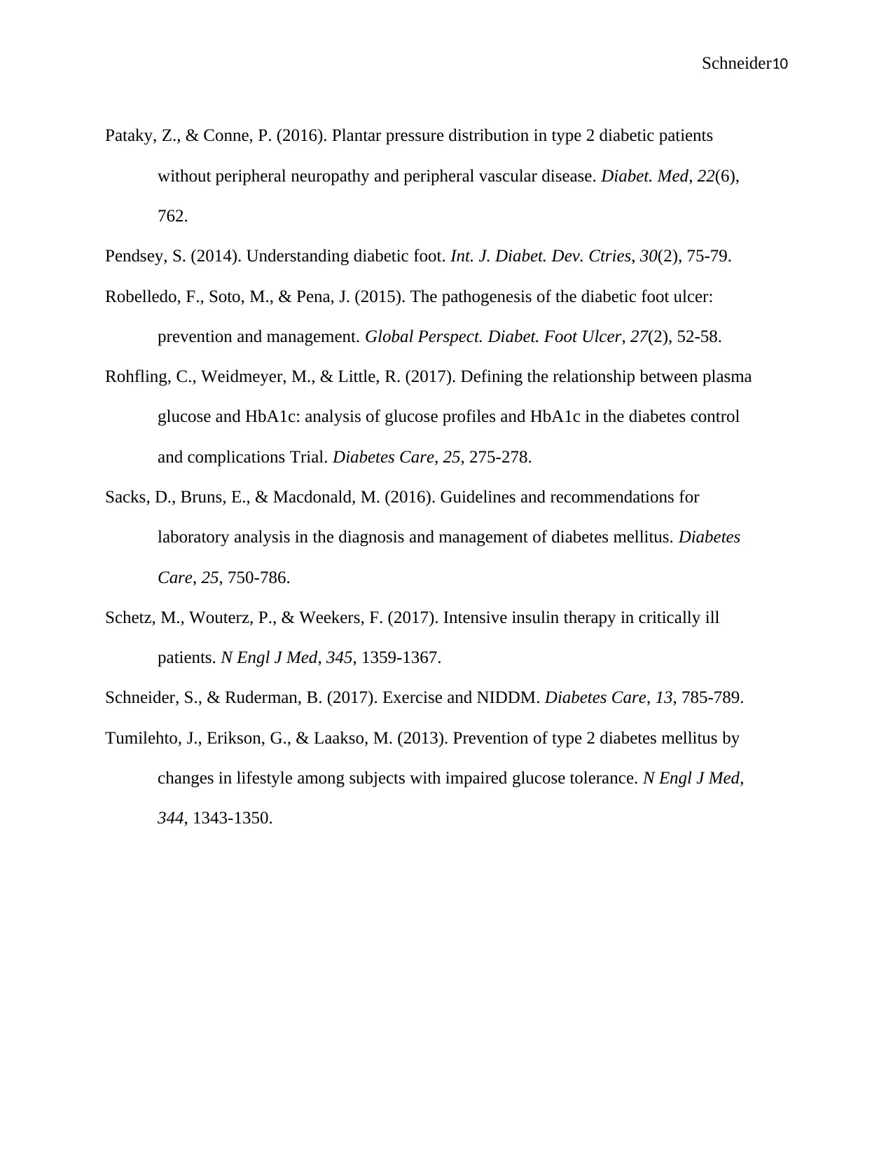
Schneider10 Pataky,Z., & Conne,P. (2016). Plantar pressure distribution in type 2 diabetic patients without peripheral neuropathy and peripheral vascular disease.Diabet. Med,22(6), 762. Pendsey,S. (2014). Understanding diabetic foot.Int. J. Diabet. Dev. Ctries,30(2), 75-79. Robelledo,F., Soto,M., & Pena,J. (2015). The pathogenesis of the diabetic foot ulcer: prevention and management.Global Perspect. Diabet. Foot Ulcer,27(2), 52-58. Rohfling,C., Weidmeyer,M., & Little,R. (2017). Defining the relationship between plasma glucose and HbA1c: analysis of glucose profiles and HbA1c in the diabetes control and complications Trial.Diabetes Care,25, 275-278. Sacks,D., Bruns,E., & Macdonald,M. (2016). Guidelines and recommendations for laboratory analysis in the diagnosis and management of diabetes mellitus.Diabetes Care,25, 750-786. Schetz,M., Wouterz,P., & Weekers,F. (2017). Intensive insulin therapy in critically ill patients.N Engl J Med,345, 1359-1367. Schneider,S., & Ruderman,B. (2017). Exercise and NIDDM.Diabetes Care,13, 785-789. Tumilehto,J., Erikson,G., & Laakso,M. (2013). Prevention of type 2 diabetes mellitus by changes in lifestyle among subjects with impaired glucose tolerance.N Engl J Med, 344, 1343-1350.