FA05 – Functional Anatomy Assignment
VerifiedAdded on 2021/06/16
|28
|10026
|30
AI Summary
Contribute Materials
Your contribution can guide someone’s learning journey. Share your
documents today.
FA05 – Functional anatomy
Assignment v1.4 (2018/01/25)
FA05 Functional anatomy
Name
Email address
Assessment
Assignment – Short answer
Assignments may include a variety of questions, this can include short or longer answer questions. These
questions are designed to test how you apply your knowledge into a real-world situation. All assignments
are completed as a Microsoft Word document and must be submitted through My eCampus for grading.
Your assessor is looking for how you apply your knowledge and how you think critically about the topic
area.
1. It is important to have a sound knowledge of posture and the involvement of musculoskeletal
anatomy and appropriate corrective actions. In the following table, you will need to identify
the postural abnormality, identify the tight and weak muscles, and provide suggestive
corrective actions. (limit 50-100words per postural abnormality)
Postural abnormality Lordosis
Tight muscles Hip flexors
Erector spinae
Weak muscles Abdominals
Hamstrings
Gluteals
Suggested corrective
exercises
(Strengthening and
Stretching)
Stretch: hip flexors and spinal extensors
Strengthen: abdominal, hamstring, and gluteals
Postural abnormality Kyphosis/Rounded Shoulders
Tight muscles Pectoralis major
Pectoralis minor
Anterior deltoids
Latissimus dorsi
Weak muscles Rhomboids
Rear deltoids
Middle/Lower trapezius
1
Secure Best Marks with AI Grader
Need help grading? Try our AI Grader for instant feedback on your assignments.
FA05 – Functional anatomy
Assignment v1.4 (2018/01/25)
Suggested corrective
exercises
(Strengthening and
Stretching)
Stretch: chest stretch, anterior deltoids, lats
Strengthen: rhomboids, rear deltoids, trapezius
Postural
abnormality
Winged Scapula
Tight muscles Pectoralis major
Pectoralis minor
Subscapularis Latissimus
dorsi
Weak muscles Serratus anterior Rhomboids
Suggested
corrective
exercises
(Strengthening
and Stretching)
Stretch: pectorals, lats, and shoulder mobility
work
Strengthen: seated row, scapula fixation work,
wall pushes (scapula pro/retraction)
2. There are five common injuries that occur as a result of poor posture. In the following table,
provide a description of each of these five conditions, outlining the impact that they have on
posture as well as the muscles involved and how they are affected. (range 50-100 words per
condition)
Injury Condition Posture and muscles involved
Sciatica spinal stenosis, the narrowing of the
spinal canal of the lower back or
degenerative disc diseases, which is a
breakdown of the discs that act as
cushions between the vertebrae. The
resulting pathology results in
compression of the sciatic nerve, which
passes under the piriformis
Tight groin muscles and weak hip
abductors
Neck pain
This is probably one of the most
common complaints in office workers
who sit at a desk and computer for
hours on end. This action leads to
aching and stiffness in the shoulders
and neck. Sitting in such a position
causes the person to fall into a slumped
position, with rounded shoulders and
the neck protruding forwards. This
eventually leads to shortening of the
Sitting at a desk or infront of
computers for hours on end results in
the larger back and neck muscles such
as Trapezius and the Rhomboids
working harder and becoming tight and
achy.
2
FA05 – Functional anatomy
Assignment v1.4 (2018/01/25)
Injury Condition Posture and muscles involved
chest muscles and weakening of the
small, postural upper back and neck
muscles, which work to pull the
shoulders back. Ultimately, this leads to
the larger back and neck muscles, i.e.
trapezius and the rhomboids, working
harder and becoming tight and achy as
they try to hold the shoulders back.
Working on correcting these
imbalances by stretching the chest
muscles and building endurance
strength in the postural muscles such
as the lower trapezius and serratus
anterior can result in a long-term fix.
Patellofemoral
knee pain This is commonly referred to as
anterior knee pain. This condition
occurs due to overuse injury, which
leads to non-specific pain at the front
of the knee.
This type of pain is aggravated by a
range of different activities including:
walking down stairs or hills and getting
up after sitting for long periods and
muscles involved are thigh bone and
patella tendon
Lower back
pain
.
.
Lower back pain has many different
causes, and the postural factors to
result in lower back pain include a leg
length difference, over pronation, and
pelvic tilts, which result in an arched
lower back.
pain typically cannot be pinpointed in
an individual. In those cases where the
lower back pain is not caused by
trauma or overuse, it is often due to
poor posture. Having poor posture
places extra strain on the muscles and
ligaments that support the lower back.
This causes certain muscles to
overwork, thereby resulting in muscle
spasms and sometimes muscle strains.
The most common postural factors to
result in lower back pain include a leg
length difference, overpronation and
pelvic tilts which result in an arched
lower back.
Shoulder
impingement These types of injuries occur when the
space in the shoulder joint is
decreased, resulting in pinching of one
or more of the tendons that pass
through this space (see winged scapula
in the following table). The reason for
the decrease in space is often related
to poor posture.
If the chest muscles are tight and the
upper back muscles such as the lower
trapezius and the serratus anterior are
weak through extended periods of
sitting slouched at a desk, then this can
cause the shoulder joint to sit in a
forwards position.
3
FA05 – Functional anatomy
Assignment v1.4 (2018/01/25)
3. There is a definitive relationship between poor posture, increased risk of injury, and muscular
deficit. Please describe how poor posture exacerbates each of the following.
1. Diminished muscle
strength and
endurance
difficulty walking; difficulty putting on socks; difficulty reaching or
extending arms above shoulder level; difficulty writing; and
difficulty handling small objects.
2. Limited flexibility Tight muscles, lower back pain, neck pain
3. Increased muscle
tension and tone
Difficulty performing physical activity, sitting, standing and lifting,
imbalanced growth of muscles..
4. Limited function Poor alignment of body muscles and joints, risk of injury
4. In the following table, several joint complexes are listed. You will need to record the main
planes of movement that the joint can work within, the movement types and the range of
motion for each movement type.
Joint complex Planes of movement Movement types Range of motion in
degrees for each
movement type
Shoulder joint
(example)
1. Sagittal Plane
2. Frontal Plane
3. Horizontal Plane
1. Flexion and extension
2. Adduction and abduction
3. Horizontal Flexion and
horizontal extension
1. 0-90 degrees
2. 0-90 degrees
3. 0-180 degrees
Elbow joint 1.Sagittal Plane 1.Flexion and Extension 0-90 degrees
Hip joint 1.Sagittal Plane
2.Frontal Plane
3.Horizontal Plane
1.Flexion and extension
2.Adduction and abduction
3.Horizontal Flexion and
horizontal extension
1. 0-90 degrees
2. 0-90 degrees
3. 0-180 degrees
Knee joint 1.Sagittal Plane 1.Flexion and Extension 1. 0-90 degrees
Ankle joint 1.Sagittal Plane 1.Plantar Flexion and Dorsi
Flexion
1. 0-90 degrees
Wrist joint 1.Sagittal Plane
2.Frontal Plane
1. Flexion and Extension
2. Adduction and Abduction
1. 0-90 degrees
2. 0-90 degrees
3. 0-180 degrees
4
Secure Best Marks with AI Grader
Need help grading? Try our AI Grader for instant feedback on your assignments.
FA05 – Functional anatomy
Assignment v1.4 (2018/01/25)
5. Skeletal muscles are arranged throughout the body in opposing pairs. During movement,
each muscle within the pair opposes the other, which allows movement to occur. In the
following table, use the same exercise and provide an example of each muscle and describe a
movement associated with it?
Muscles Movement description Muscle involved
Agonist Agonist is a prime mover in every exercise
as it is responsible for movement due to
shortening contraction.
Example: In bicep curls concentric phase
the agonist is biceps brachii.
Biceps brachii
Antagonist It opposes agonist during any exercise by
relaxing so that movement could occur.
Example: In bicep curls’ concentric phase
the triceps muscle is antagonist, as it
needs to lengthen to move elbow flexion.
Triceps
Synergist Agonist and Synergist work in coordination
to generate movement. It is a helper
muscle which indirectly helps in
movement.
Example: In biscep curl, these muscles like
brachialis and brachioradiali helps the
biceps to create movement and stabilize
elbow joint..
brachialis and brachioradiali
Fixator It stabilizes to stop unwanted movement
of an agonist’s origin. It also acts to fixate
the moving joint in one place.
Example: in bicep curl the rotator cuff
muscle acts as a guardian for the shoulder
joint.
Teres minor muscles,
Supraspinatus, Infraspinatus
5

FA05 – Functional anatomy
Assignment v1.4 (2018/01/25)
6
Assignment v1.4 (2018/01/25)
6
FA05 – Functional anatomy
Assignment v1.4 (2018/01/25)
6. Describe Wolff’s law and how its linked to bone modelling and remodelling? (limit 100 words
per factor)
Wolff’s Law was developed by a brilliant German anatomist and surgeon, Julius Wolff (1836–1902).
It states that bone in a healthy person or animal will adapt to the loads under which it is placed. If
loading on a bone increases, the bone will remodel itself over time to become stronger to resist
that type of loading. The inverse is true as well: if the loading on a bone decreases, the bone will
become less dense and weaker due to the lack of the stimulus required for continued remodelling.
7. There is a range of different conditions and lifestyles that can change the centre of gravity and
affect posture. Please complete the following table and describe how these factors result in a
change in gravity and thus affect posture. (limit 50 words per factor)
Factor that affects the
centre of gravity
Description of how posture is affected
Pregnancy As the size of belly increases in pregnancy, the centre of gravity shift
upward that effects the balance and cause lower back pain.
1.So, the rectus femoris, spinal erectors and hip flexor muscles tend to
tighten and shorten.
2. Due to under use of muscles like glutes, abdominal, obliques and
hamstring, their size tend to elongate.
Overweight Increased weight and mass cause spinal changes due to excessive force
that is put on vertebrae, muscles and supportive structures in back. It
affects muscle and joint interaction which is important to maintain
postural balance. It reduces muscle strength and lowers muscular fatigue
resistance. Obese people have erect posture that is a result of supporting
excessive load over small base of support.
Inappropriate footwear Inappropriate footwear shifts centre of gravity forward from the
beginning, applying more pressure and stress on the front of the foot. It
can lead to development of postural disorders like lumbar hyper lordosis,
knee valgus, pelvic anteversion and forward head posture. High heels
cause postural changes and body imbalance.
Poor work practices
(office worker)
Sitting position at office is a serious concern as poor posture increases
strain and stress on the body. It causes lower back and neck pain
disorder. The centre of gravity in poor sitting posture i.e. slouched sitting
posture is posterior to ischia and lumbar lordosis is reversed. In this case
body weight is less transferred to the floor through lower extremities
causing postural backache and are at higher risk of developing
musculoskeletal disorders symptoms.
7
Paraphrase This Document
Need a fresh take? Get an instant paraphrase of this document with our AI Paraphraser
FA05 – Functional anatomy
Assignment v1.4 (2018/01/25)
8. Agility is the ability to control changes in direction and body position quickly and effectively.
There are several factors that affect coordination and agility. Complete the following table,
explaining how each factor impacts coordination and agility.
Factor that affects agility Description of how it impacts agility
Fine motor skills Fine motor skills are involved in smaller movements that occur in
hands, fingers, wrists, toes and feet. They help in smaller actions
like picking up objects between finger and thumb, blinking, writing
carefully etc.
Gross motor skills It involves movement of large muscles of body like arms, torso and
legs. It is involved in activities like jumping, running, walking etc.
Children who struggles with gross motor skills have trouble with
coordination and faces hard time in activities like pedalling a bike,
climbing etc.
Hand eye skills It is the ability to track the movements of hands with eyes by
enabling the eyes to send significant signals to the brain about the
movement of hand. If hand eye coordination is poor then people
face difficulty to exercise, or other activities like writing etc.
8
FA05 – Functional anatomy
Assignment v1.4 (2018/01/25)
9. Ideal postural alignment is important for maintaining optimum health and wellbeing.
Describe the normal spinal curves, and outline the anatomical features associated with
straight line – or plumb line – running (In your answer you need to mention lordosis, kyphosis
and scoliosis). (range 50-100 words)
n order to sustain good posture – referred to as neutral posture - the muscles of the body must be
in balance to support an aligned spine. Neutral spine, does not equate to a flat spine, as there are
many natural curvatures to the vertebral column. The neck, known as the cervical spine, has a
natural anterior curve or lordosis which is necessary to balance the cranium. The thoracic spine, or
trunk, has an opposite orientation. It curves posteriorly which is known as a kyphotic curve. The
lumbar spine follows the same angle of the cervical spine, curving anteriorly, creating a second
lordotic curve in the spine. These natural curves give the spine a slight ‘S’ shape when viewed from
the side. In a neutral spine, there is balance between the musculature right-to-left and front-to-
back. When in a neutral posture, the body is in its strongest and most balanced allowing for optimal
efficiency and minimal stress on the joints and the rest of the body. Maintaining a neutral spine is a
dynamic process that is meant to transition from position to position.
10. In order to prevent injury, it is important to have an understanding of how the body may
respond to exercise if there is an injury. Complete the following table by providing an
explanation for each of the situations given.
Anatomical
situation
Description
Increase
pronation of
foot and ankle
complex
Common Problems in the Foot and Ankle Complex. ... Pronation is a normal
function that occurs when the foot rolls inward toward the midline of the body.
This movement causes the heel to collapse inward and the medial arch of
the foot to elongate and flatten.
Increase
supination of
foot and ankle
Oversupination also causes an increased external (lateral) rotation force to be
placed on the shin, knee and thigh which places additional stress on the muscles,
tendons and ... Expert interview - Oversupination explained - Podiatrist Ian
Saddler explains over pronation in this free to view sportsinjuryclinic.net video.
9
FA05 – Functional anatomy
Assignment v1.4 (2018/01/25)
Anatomical
situation
Description
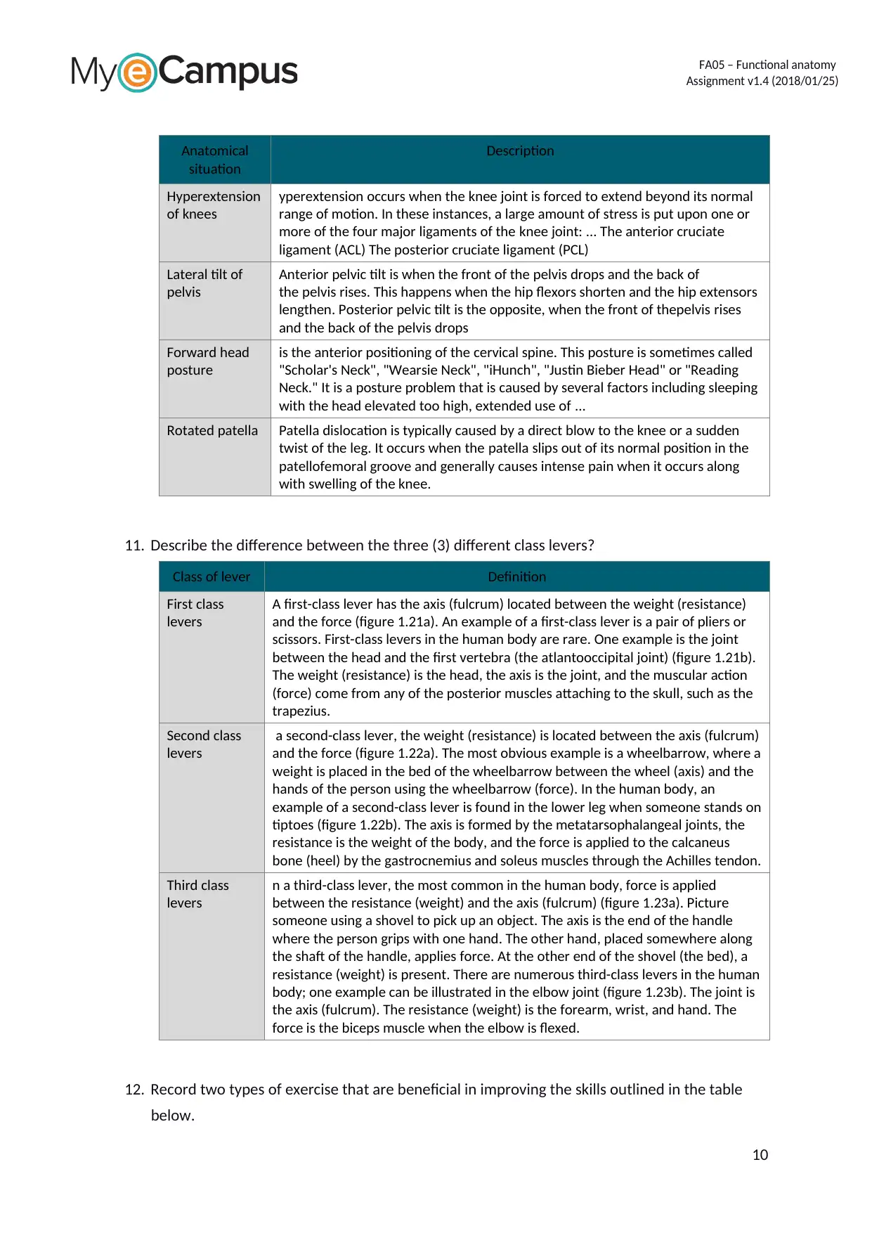
Hyperextension
of knees
yperextension occurs when the knee joint is forced to extend beyond its normal
range of motion. In these instances, a large amount of stress is put upon one or
more of the four major ligaments of the knee joint: ... The anterior cruciate
ligament (ACL) The posterior cruciate ligament (PCL)
Lateral tilt of
pelvis
Anterior pelvic tilt is when the front of the pelvis drops and the back of
the pelvis rises. This happens when the hip flexors shorten and the hip extensors
lengthen. Posterior pelvic tilt is the opposite, when the front of thepelvis rises
and the back of the pelvis drops
Forward head
posture
is the anterior positioning of the cervical spine. This posture is sometimes called
"Scholar's Neck", "Wearsie Neck", "iHunch", "Justin Bieber Head" or "Reading
Neck." It is a posture problem that is caused by several factors including sleeping
with the head elevated too high, extended use of ...
Rotated patella Patella dislocation is typically caused by a direct blow to the knee or a sudden
twist of the leg. It occurs when the patella slips out of its normal position in the
patellofemoral groove and generally causes intense pain when it occurs along
with swelling of the knee.
11. Describe the difference between the three (3) different class levers?
Class of lever Definition
First class
levers
A first-class lever has the axis (fulcrum) located between the weight (resistance)
and the force (figure 1.21a). An example of a first-class lever is a pair of pliers or
scissors. First-class levers in the human body are rare. One example is the joint
between the head and the first vertebra (the atlantooccipital joint) (figure 1.21b).
The weight (resistance) is the head, the axis is the joint, and the muscular action
(force) come from any of the posterior muscles attaching to the skull, such as the
trapezius.
Second class
levers
a second-class lever, the weight (resistance) is located between the axis (fulcrum)
and the force (figure 1.22a). The most obvious example is a wheelbarrow, where a
weight is placed in the bed of the wheelbarrow between the wheel (axis) and the
hands of the person using the wheelbarrow (force). In the human body, an
example of a second-class lever is found in the lower leg when someone stands on
tiptoes (figure 1.22b). The axis is formed by the metatarsophalangeal joints, the
resistance is the weight of the body, and the force is applied to the calcaneus
bone (heel) by the gastrocnemius and soleus muscles through the Achilles tendon.
Third class
levers
n a third-class lever, the most common in the human body, force is applied
between the resistance (weight) and the axis (fulcrum) (figure 1.23a). Picture
someone using a shovel to pick up an object. The axis is the end of the handle
where the person grips with one hand. The other hand, placed somewhere along
the shaft of the handle, applies force. At the other end of the shovel (the bed), a
resistance (weight) is present. There are numerous third-class levers in the human
body; one example can be illustrated in the elbow joint (figure 1.23b). The joint is
the axis (fulcrum). The resistance (weight) is the forearm, wrist, and hand. The
force is the biceps muscle when the elbow is flexed.
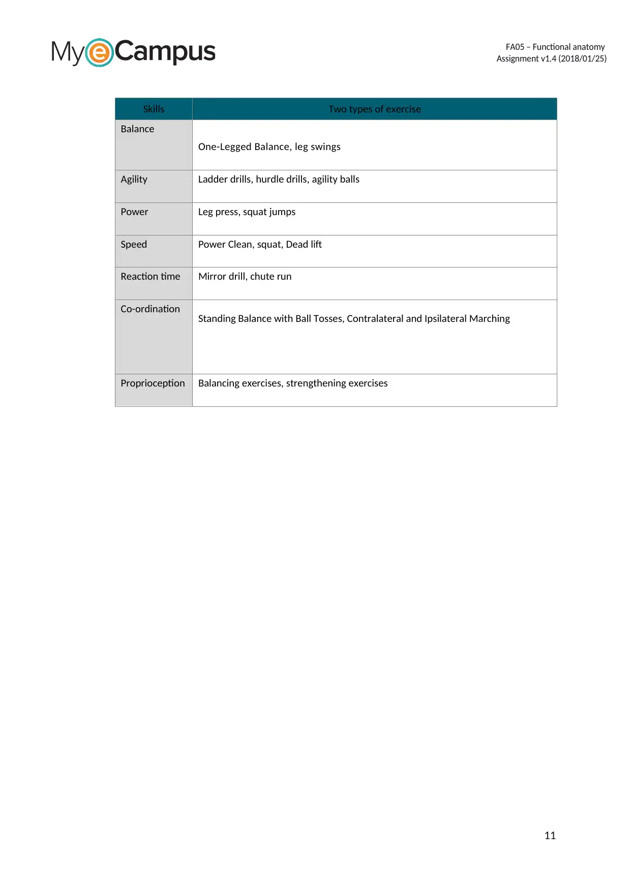
12. Record two types of exercise that are beneficial in improving the skills outlined in the table
below.
10
Secure Best Marks with AI Grader
Need help grading? Try our AI Grader for instant feedback on your assignments.
FA05 – Functional anatomy
Assignment v1.4 (2018/01/25)
Skills Two types of exercise
Balance
One-Legged Balance, leg swings
Agility Ladder drills, hurdle drills, agility balls
Power Leg press, squat jumps
Speed Power Clean, squat, Dead lift
Reaction time Mirror drill, chute run
Co-ordination Standing Balance with Ball Tosses, Contralateral and Ipsilateral Marching
Proprioception Balancing exercises, strengthening exercises
11
FA05 – Functional anatomy
Assignment v1.4 (2018/01/25)
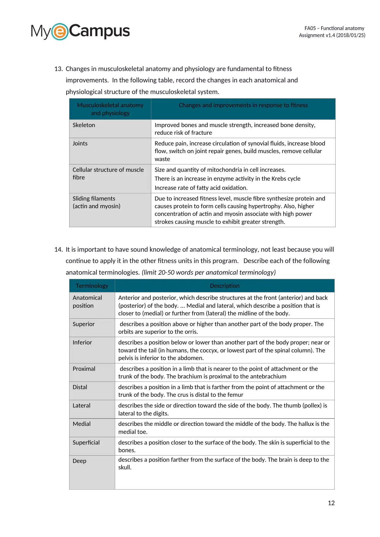
13. Changes in musculoskeletal anatomy and physiology are fundamental to fitness
improvements. In the following table, record the changes in each anatomical and
physiological structure of the musculoskeletal system.
Musculoskeletal anatomy
and physiology
Changes and improvements in response to fitness
Skeleton Improved bones and muscle strength, increased bone density,
reduce risk of fracture
Joints Reduce pain, increase circulation of synovial fluids, increase blood
flow, switch on joint repair genes, build muscles, remove cellular
waste
Cellular structure of muscle
fibre
Size and quantity of mitochondria in cell increases.
There is an increase in enzyme activity in the Krebs cycle
Increase rate of fatty acid oxidation.
Sliding filaments
(actin and myosin)
Due to increased fitness level, muscle fibre synthesize protein and
causes protein to form cells causing hypertrophy. Also, higher
concentration of actin and myosin associate with high power
strokes causing muscle to exhibit greater strength.
14. It is important to have sound knowledge of anatomical terminology, not least because you will
continue to apply it in the other fitness units in this program. Describe each of the following
anatomical terminologies. (limit 20-50 words per anatomical terminology)
Terminology Description
Anatomical
position
Anterior and posterior, which describe structures at the front (anterior) and back
(posterior) of the body. ... Medial and lateral, which describe a position that is
closer to (medial) or further from (lateral) the midline of the body.
Superior describes a position above or higher than another part of the body proper. The
orbits are superior to the orris.
Inferior describes a position below or lower than another part of the body proper; near or
toward the tail (in humans, the coccyx, or lowest part of the spinal column). The
pelvis is inferior to the abdomen.
Proximal describes a position in a limb that is nearer to the point of attachment or the
trunk of the body. The brachium is proximal to the antebrachium
Distal describes a position in a limb that is farther from the point of attachment or the
trunk of the body. The crus is distal to the femur
Lateral describes the side or direction toward the side of the body. The thumb (pollex) is
lateral to the digits.
Medial describes the middle or direction toward the middle of the body. The hallux is the
medial toe.
Superficial describes a position closer to the surface of the body. The skin is superficial to the
bones.
Deep describes a position farther from the surface of the body. The brain is deep to the
skull.
12
FA05 – Functional anatomy
Assignment v1.4 (2018/01/25)
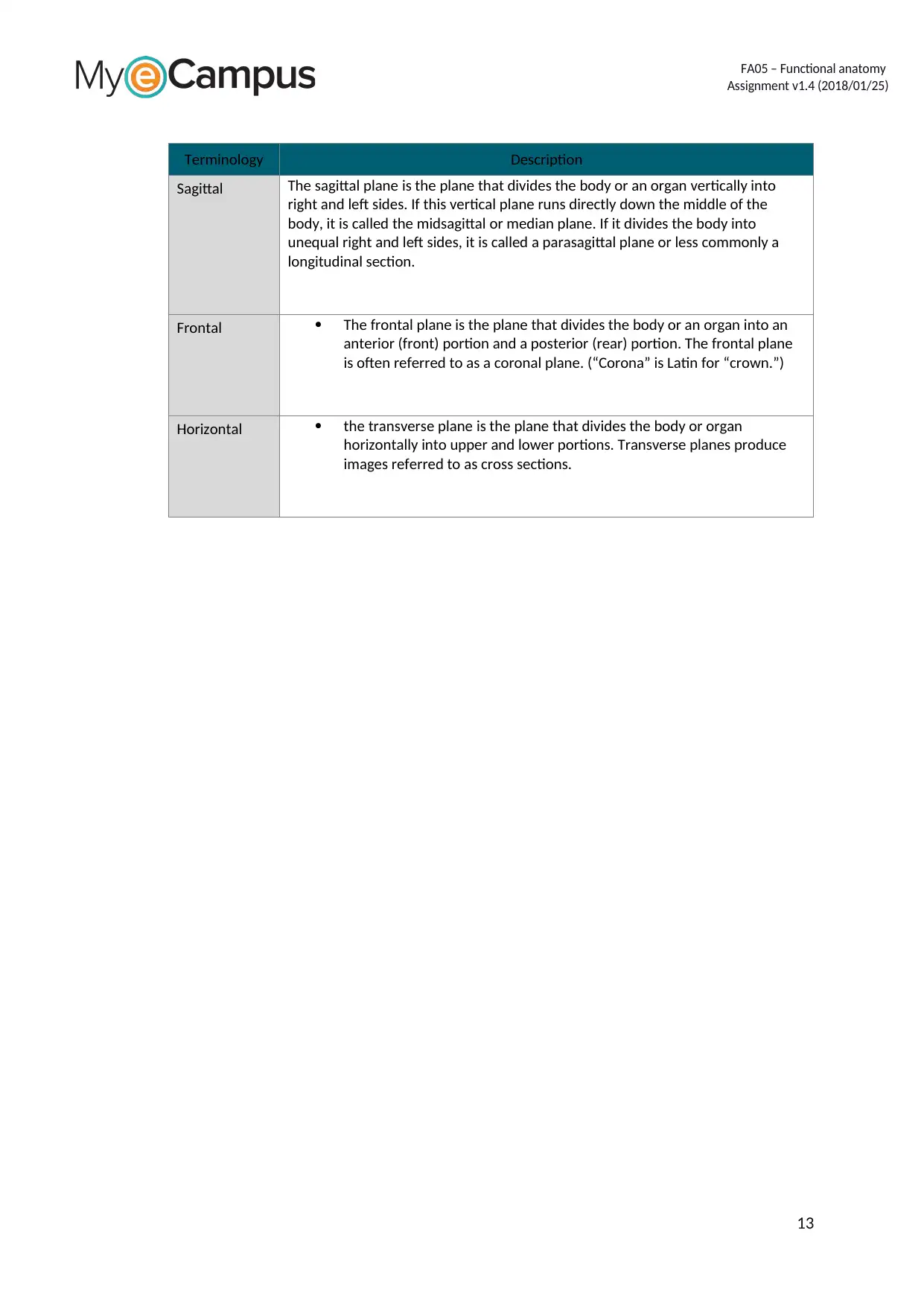
Terminology Description
Sagittal The sagittal plane is the plane that divides the body or an organ vertically into
right and left sides. If this vertical plane runs directly down the middle of the
body, it is called the midsagittal or median plane. If it divides the body into
unequal right and left sides, it is called a parasagittal plane or less commonly a
longitudinal section.
Frontal The frontal plane is the plane that divides the body or an organ into an
anterior (front) portion and a posterior (rear) portion. The frontal plane
is often referred to as a coronal plane. (“Corona” is Latin for “crown.”)
Horizontal the transverse plane is the plane that divides the body or organ
horizontally into upper and lower portions. Transverse planes produce
images referred to as cross sections.
13
Paraphrase This Document
Need a fresh take? Get an instant paraphrase of this document with our AI Paraphraser
FA05 – Functional anatomy
Assignment v1.4 (2018/01/25)
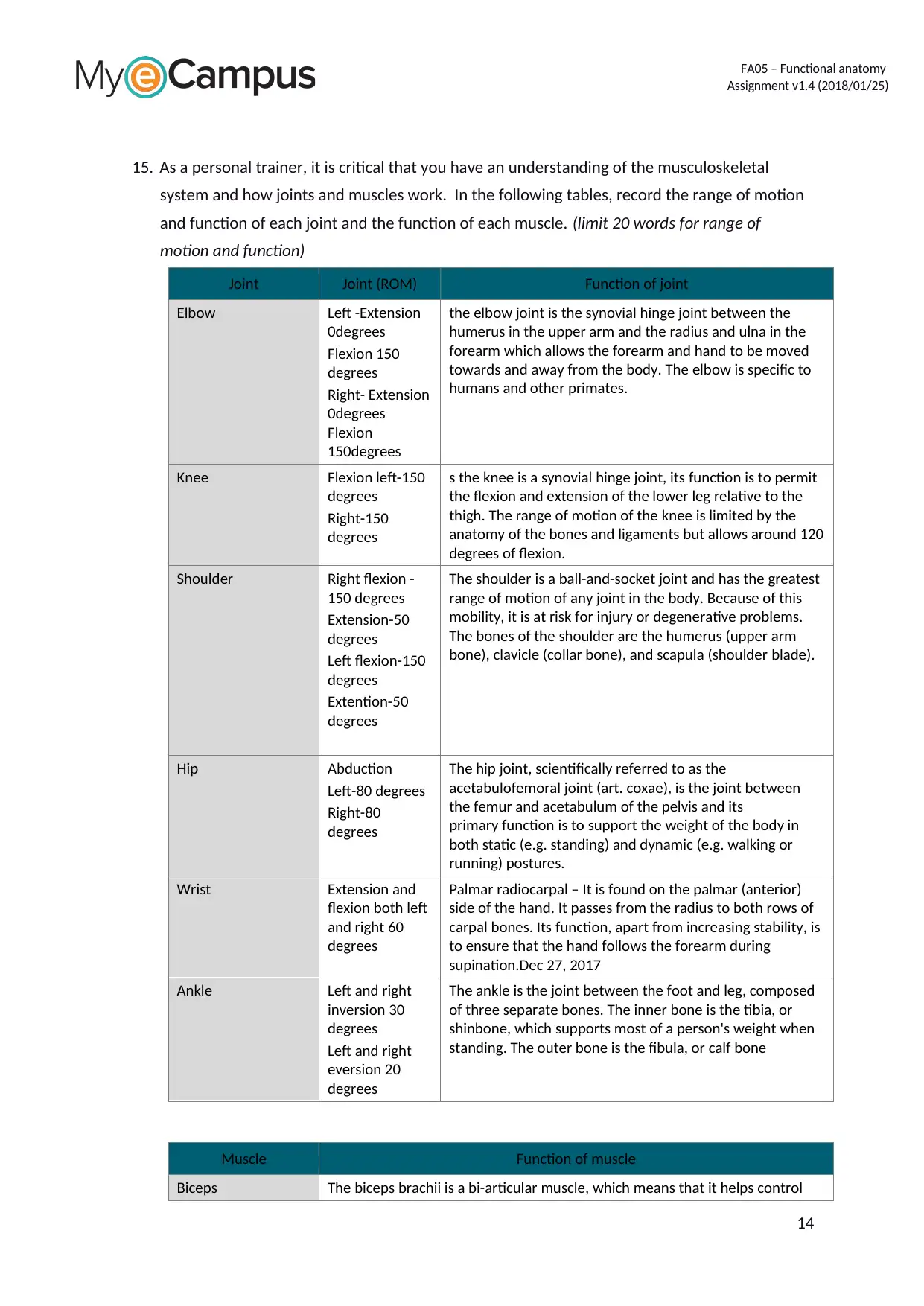
15. As a personal trainer, it is critical that you have an understanding of the musculoskeletal
system and how joints and muscles work. In the following tables, record the range of motion
and function of each joint and the function of each muscle. (limit 20 words for range of
motion and function)
Joint Joint (ROM) Function of joint
Elbow Left -Extension
0degrees
Flexion 150
degrees
Right- Extension
0degrees
Flexion
150degrees
the elbow joint is the synovial hinge joint between the
humerus in the upper arm and the radius and ulna in the
forearm which allows the forearm and hand to be moved
towards and away from the body. The elbow is specific to
humans and other primates.
Knee Flexion left-150
degrees
Right-150
degrees
s the knee is a synovial hinge joint, its function is to permit
the flexion and extension of the lower leg relative to the
thigh. The range of motion of the knee is limited by the
anatomy of the bones and ligaments but allows around 120
degrees of flexion.
Shoulder Right flexion -
150 degrees
Extension-50
degrees
Left flexion-150
degrees
Extention-50
degrees
The shoulder is a ball-and-socket joint and has the greatest
range of motion of any joint in the body. Because of this
mobility, it is at risk for injury or degenerative problems.
The bones of the shoulder are the humerus (upper arm
bone), clavicle (collar bone), and scapula (shoulder blade).
Hip Abduction
Left-80 degrees
Right-80
degrees
The hip joint, scientifically referred to as the
acetabulofemoral joint (art. coxae), is the joint between
the femur and acetabulum of the pelvis and its
primary function is to support the weight of the body in
both static (e.g. standing) and dynamic (e.g. walking or
running) postures.
Wrist Extension and
flexion both left
and right 60
degrees
Palmar radiocarpal – It is found on the palmar (anterior)
side of the hand. It passes from the radius to both rows of
carpal bones. Its function, apart from increasing stability, is
to ensure that the hand follows the forearm during
supination.Dec 27, 2017
Ankle Left and right
inversion 30
degrees
Left and right
eversion 20
degrees
The ankle is the joint between the foot and leg, composed
of three separate bones. The inner bone is the tibia, or
shinbone, which supports most of a person's weight when
standing. The outer bone is the fibula, or calf bone
Muscle Function of muscle
Biceps The biceps brachii is a bi-articular muscle, which means that it helps control
14
FA05 – Functional anatomy
Assignment v1.4 (2018/01/25)
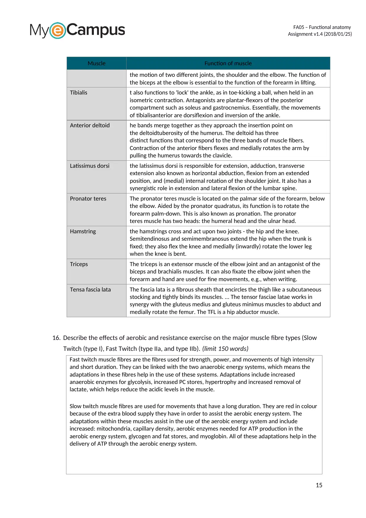
Muscle Function of muscle
the motion of two different joints, the shoulder and the elbow. The function of
the biceps at the elbow is essential to the function of the forearm in lifting.
Tibialis t also functions to 'lock' the ankle, as in toe-kicking a ball, when held in an
isometric contraction. Antagonists are plantar-flexors of the posterior
compartment such as soleus and gastrocnemius. Essentially, the movements
of tibialisanterior are dorsiflexion and inversion of the ankle.
Anterior deltoid he bands merge together as they approach the insertion point on
the deltoidtuberosity of the humerus. The deltoid has three
distinct functions that correspond to the three bands of muscle fibers.
Contraction of the anterior fibers flexes and medially rotates the arm by
pulling the humerus towards the clavicle.
Latissimus dorsi the latissimus dorsi is responsible for extension, adduction, transverse
extension also known as horizontal abduction, flexion from an extended
position, and (medial) internal rotation of the shoulder joint. It also has a
synergistic role in extension and lateral flexion of the lumbar spine.
Pronator teres The pronator teres muscle is located on the palmar side of the forearm, below
the elbow. Aided by the pronator quadratus, its function is to rotate the
forearm palm-down. This is also known as pronation. The pronator
teres muscle has two heads: the humeral head and the ulnar head.
Hamstring the hamstrings cross and act upon two joints - the hip and the knee.
Semitendinosus and semimembranosus extend the hip when the trunk is
fixed; they also flex the knee and medially (inwardly) rotate the lower leg
when the knee is bent.
Triceps The triceps is an extensor muscle of the elbow joint and an antagonist of the
biceps and brachialis muscles. It can also fixate the elbow joint when the
forearm and hand are used for fine movements, e.g., when writing.
Tensa fascia lata The fascia lata is a fibrous sheath that encircles the thigh like a subcutaneous
stocking and tightly binds its muscles. ... The tensor fasciae latae works in
synergy with the gluteus medius and gluteus minimus muscles to abduct and
medially rotate the femur. The TFL is a hip abductor muscle.
16. Describe the effects of aerobic and resistance exercise on the major muscle fibre types (Slow
Twitch (type I), Fast Twitch (type IIa, and type IIb). (limit 150 words)
Fast twitch muscle fibres are the fibres used for strength, power, and movements of high intensity
and short duration. They can be linked with the two anaerobic energy systems, which means the
adaptations in these fibres help in the use of these systems. Adaptations include increased
anaerobic enzymes for glycolysis, increased PC stores, hypertrophy and increased removal of
lactate, which helps reduce the acidic levels in the muscle.
Slow twitch muscle fibres are used for movements that have a long duration. They are red in colour
because of the extra blood supply they have in order to assist the aerobic energy system. The
adaptations within these muscles assist in the use of the aerobic energy system and include
increased: mitochondria, capillary density, aerobic enzymes needed for ATP production in the
aerobic energy system, glycogen and fat stores, and myoglobin. All of these adaptations help in the
delivery of ATP through the aerobic energy system.
15
FA05 – Functional anatomy
Assignment v1.4 (2018/01/25)
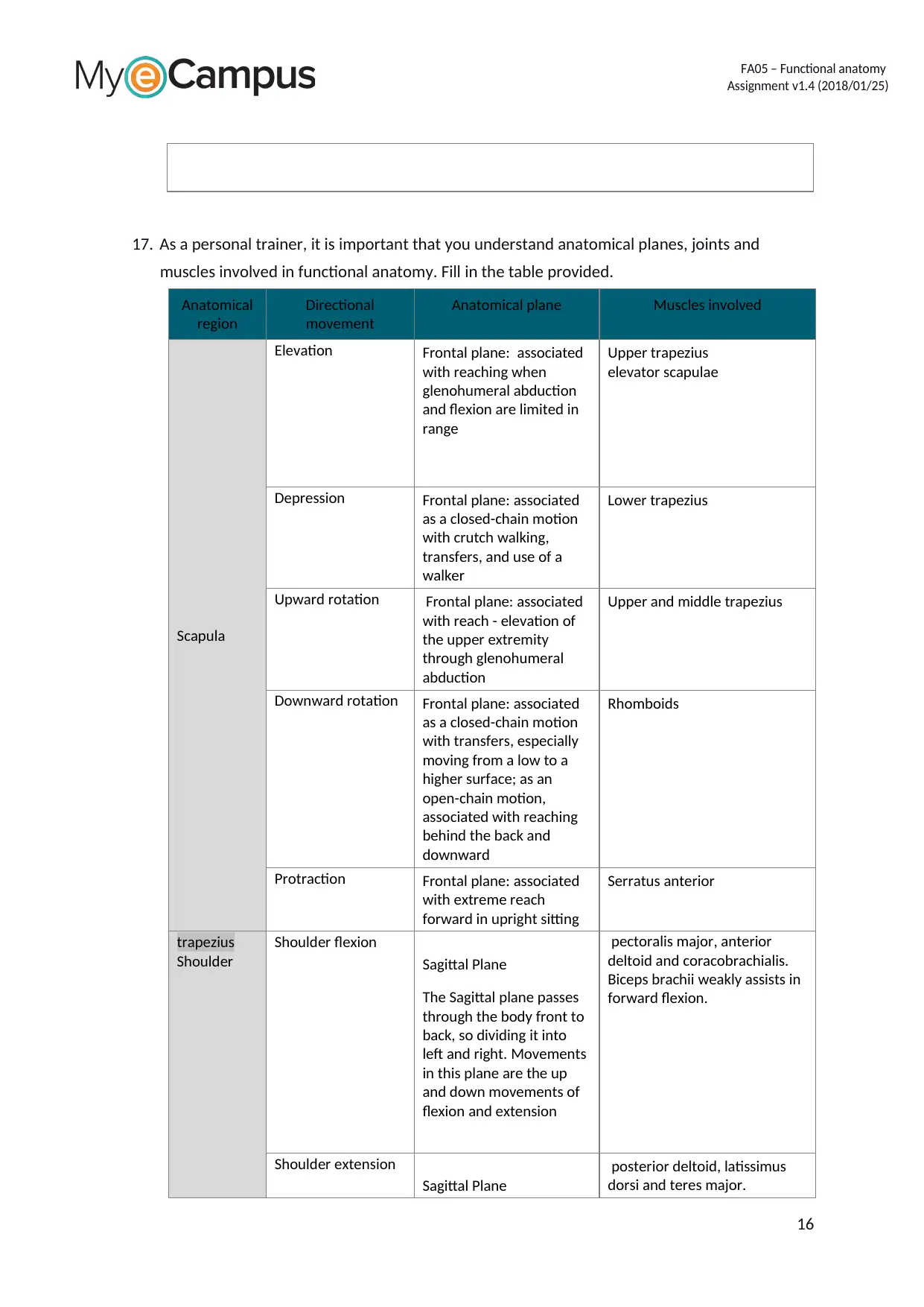
17. As a personal trainer, it is important that you understand anatomical planes, joints and
muscles involved in functional anatomy. Fill in the table provided.
Anatomical
region
Directional
movement
Anatomical plane Muscles involved
Scapula
Elevation Frontal plane: associated
with reaching when
glenohumeral abduction
and flexion are limited in
range
Upper trapezius
elevator scapulae
Depression Frontal plane: associated
as a closed-chain motion
with crutch walking,
transfers, and use of a
walker
Lower trapezius
Upward rotation Frontal plane: associated
with reach - elevation of
the upper extremity
through glenohumeral
abduction
Upper and middle trapezius
Downward rotation Frontal plane: associated
as a closed-chain motion
with transfers, especially
moving from a low to a
higher surface; as an
open-chain motion,
associated with reaching
behind the back and
downward
Rhomboids
Protraction Frontal plane: associated
with extreme reach
forward in upright sitting
Serratus anterior
trapezius
Shoulder
Shoulder flexion
Sagittal Plane
The Sagittal plane passes
through the body front to
back, so dividing it into
left and right. Movements
in this plane are the up
and down movements of
flexion and extension
pectoralis major, anterior
deltoid and coracobrachialis.
Biceps brachii weakly assists in
forward flexion.
Shoulder extension
Sagittal Plane
posterior deltoid, latissimus
dorsi and teres major.
16
Secure Best Marks with AI Grader
Need help grading? Try our AI Grader for instant feedback on your assignments.
FA05 – Functional anatomy
Assignment v1.4 (2018/01/25)
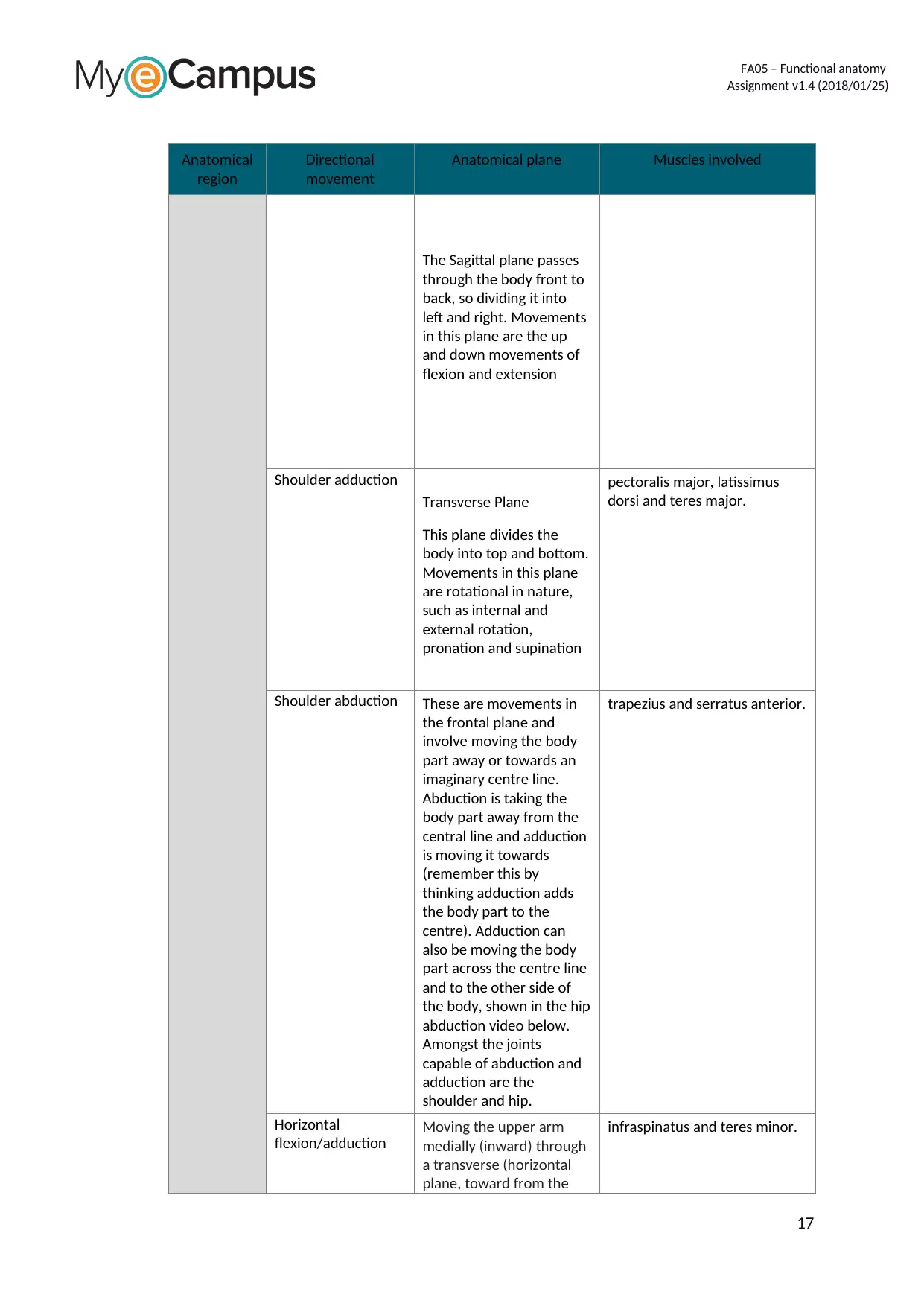
Anatomical
region
Directional
movement
Anatomical plane Muscles involved
The Sagittal plane passes
through the body front to
back, so dividing it into
left and right. Movements
in this plane are the up
and down movements of
flexion and extension
Shoulder adduction
Transverse Plane
This plane divides the
body into top and bottom.
Movements in this plane
are rotational in nature,
such as internal and
external rotation,
pronation and supination
pectoralis major, latissimus
dorsi and teres major.
Shoulder abduction These are movements in
the frontal plane and
involve moving the body
part away or towards an
imaginary centre line.
Abduction is taking the
body part away from the
central line and adduction
is moving it towards
(remember this by
thinking adduction adds
the body part to the
centre). Adduction can
also be moving the body
part across the centre line
and to the other side of
the body, shown in the hip
abduction video below.
Amongst the joints
capable of abduction and
adduction are the
shoulder and hip.
trapezius and serratus anterior.
Horizontal
flexion/adduction
Moving the upper arm
medially (inward) through
a transverse (horizontal
plane, toward from the
infraspinatus and teres minor.
17
FA05 – Functional anatomy
Assignment v1.4 (2018/01/25)
Anatomical
region
Directional
movement
Anatomical plane Muscles involved
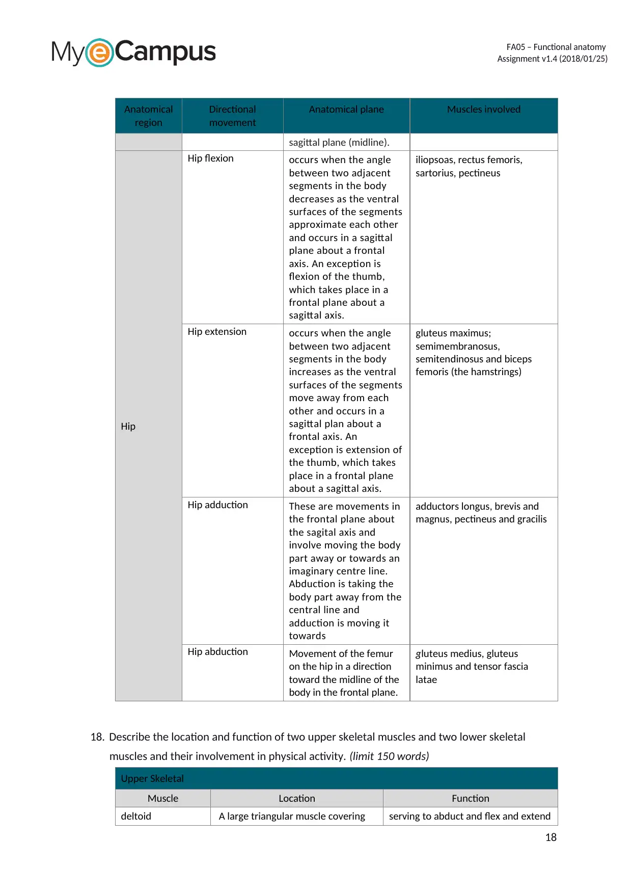
sagittal plane (midline).
Hip
Hip flexion occurs when the angle
between two adjacent
segments in the body
decreases as the ventral
surfaces of the segments
approximate each other
and occurs in a sagittal
plane about a frontal
axis. An exception is
flexion of the thumb,
which takes place in a
frontal plane about a
sagittal axis.
iliopsoas, rectus femoris,
sartorius, pectineus
Hip extension occurs when the angle
between two adjacent
segments in the body
increases as the ventral
surfaces of the segments
move away from each
other and occurs in a
sagittal plan about a
frontal axis. An
exception is extension of
the thumb, which takes
place in a frontal plane
about a sagittal axis.
gluteus maximus;
semimembranosus,
semitendinosus and biceps
femoris (the hamstrings)
Hip adduction These are movements in
the frontal plane about
the sagital axis and
involve moving the body
part away or towards an
imaginary centre line.
Abduction is taking the
body part away from the
central line and
adduction is moving it
towards
adductors longus, brevis and
magnus, pectineus and gracilis
Hip abduction Movement of the femur
on the hip in a direction
toward the midline of the
body in the frontal plane.
gluteus medius, gluteus
minimus and tensor fascia
latae
18. Describe the location and function of two upper skeletal muscles and two lower skeletal
muscles and their involvement in physical activity. (limit 150 words)
Upper Skeletal
Muscle Location Function
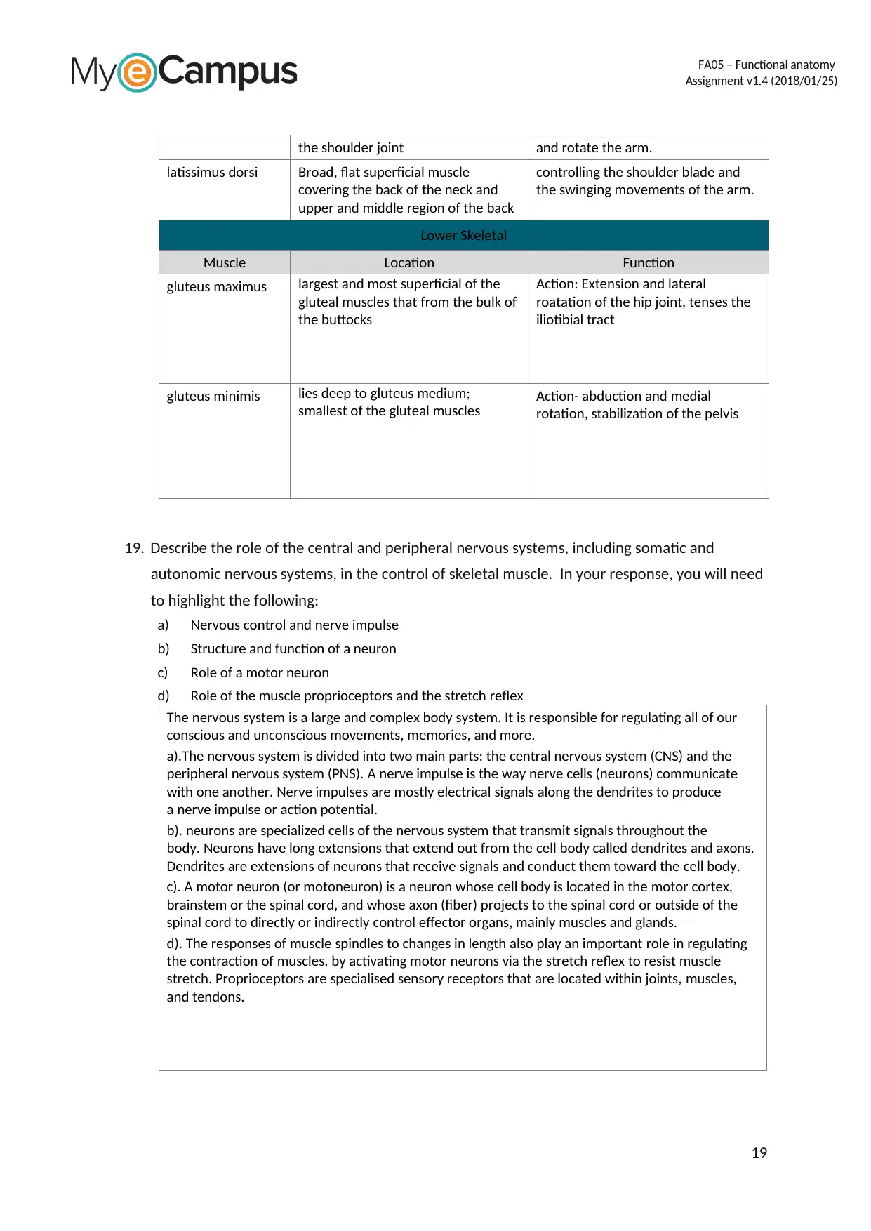
deltoid A large triangular muscle covering serving to abduct and flex and extend
18
FA05 – Functional anatomy
Assignment v1.4 (2018/01/25)
the shoulder joint and rotate the arm.
latissimus dorsi Broad, flat superficial muscle
covering the back of the neck and
upper and middle region of the back
controlling the shoulder blade and
the swinging movements of the arm.
Lower Skeletal
Muscle Location Function
gluteus maximus largest and most superficial of the
gluteal muscles that from the bulk of
the buttocks
Action: Extension and lateral
roatation of the hip joint, tenses the
iliotibial tract
gluteus minimis lies deep to gluteus medium;
smallest of the gluteal muscles
Action- abduction and medial
rotation, stabilization of the pelvis
19. Describe the role of the central and peripheral nervous systems, including somatic and
autonomic nervous systems, in the control of skeletal muscle. In your response, you will need
to highlight the following:
a) Nervous control and nerve impulse
b) Structure and function of a neuron
c) Role of a motor neuron
d) Role of the muscle proprioceptors and the stretch reflex
The nervous system is a large and complex body system. It is responsible for regulating all of our
conscious and unconscious movements, memories, and more.
a).The nervous system is divided into two main parts: the central nervous system (CNS) and the
peripheral nervous system (PNS). A nerve impulse is the way nerve cells (neurons) communicate
with one another. Nerve impulses are mostly electrical signals along the dendrites to produce
a nerve impulse or action potential.
b). neurons are specialized cells of the nervous system that transmit signals throughout the
body. Neurons have long extensions that extend out from the cell body called dendrites and axons.
Dendrites are extensions of neurons that receive signals and conduct them toward the cell body.
c). A motor neuron (or motoneuron) is a neuron whose cell body is located in the motor cortex,
brainstem or the spinal cord, and whose axon (fiber) projects to the spinal cord or outside of the
spinal cord to directly or indirectly control effector organs, mainly muscles and glands.
d). The responses of muscle spindles to changes in length also play an important role in regulating
the contraction of muscles, by activating motor neurons via the stretch reflex to resist muscle
stretch. Proprioceptors are specialised sensory receptors that are located within joints, muscles,
and tendons.
19
Paraphrase This Document
Need a fresh take? Get an instant paraphrase of this document with our AI Paraphraser
FA05 – Functional anatomy
Assignment v1.4 (2018/01/25)
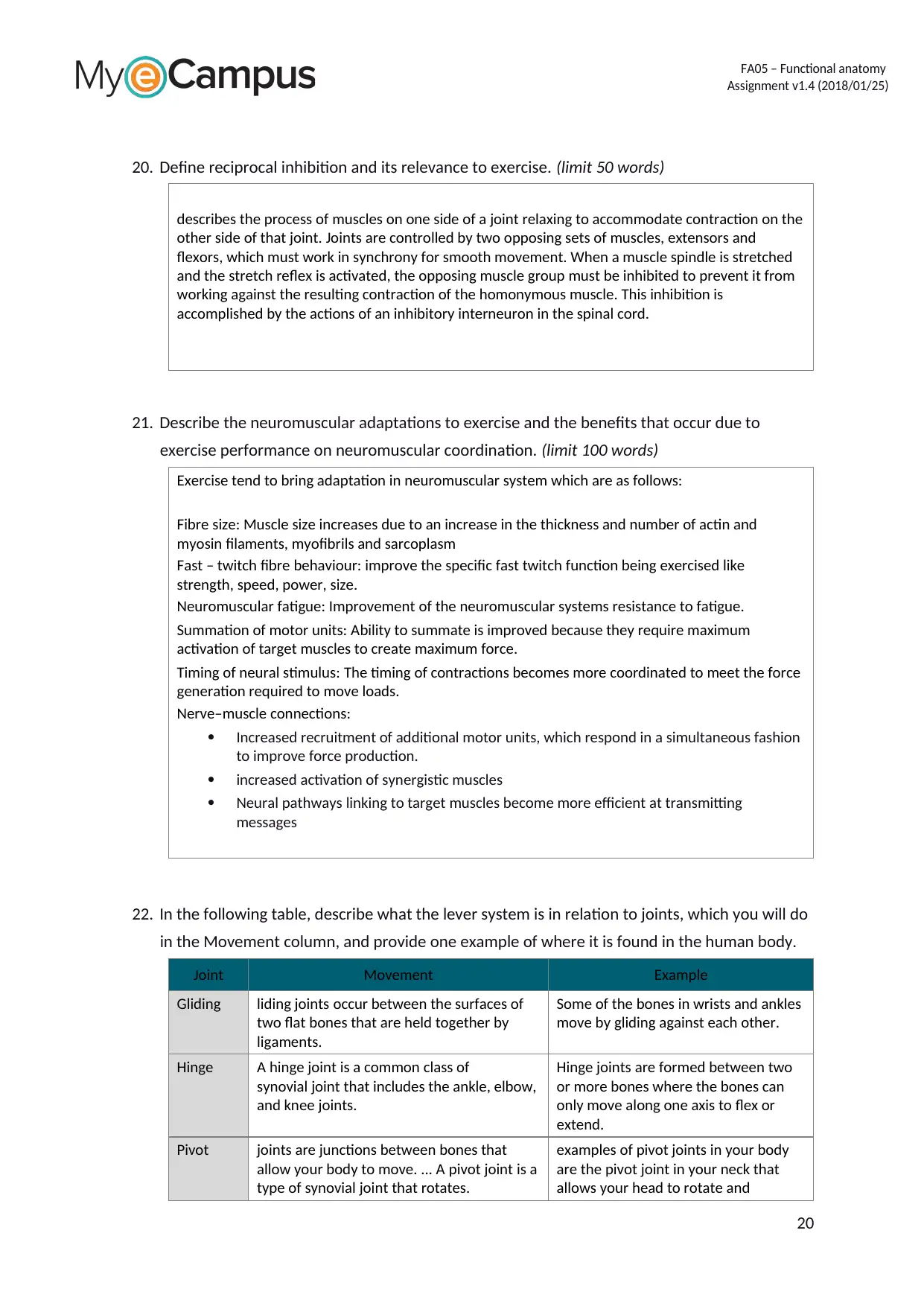
20. Define reciprocal inhibition and its relevance to exercise. (limit 50 words)
describes the process of muscles on one side of a joint relaxing to accommodate contraction on the
other side of that joint. Joints are controlled by two opposing sets of muscles, extensors and
flexors, which must work in synchrony for smooth movement. When a muscle spindle is stretched
and the stretch reflex is activated, the opposing muscle group must be inhibited to prevent it from
working against the resulting contraction of the homonymous muscle. This inhibition is
accomplished by the actions of an inhibitory interneuron in the spinal cord.
21. Describe the neuromuscular adaptations to exercise and the benefits that occur due to
exercise performance on neuromuscular coordination. (limit 100 words)
Exercise tend to bring adaptation in neuromuscular system which are as follows:
Fibre size: Muscle size increases due to an increase in the thickness and number of actin and
myosin filaments, myofibrils and sarcoplasm
Fast – twitch fibre behaviour: improve the specific fast twitch function being exercised like
strength, speed, power, size.
Neuromuscular fatigue: Improvement of the neuromuscular systems resistance to fatigue.
Summation of motor units: Ability to summate is improved because they require maximum
activation of target muscles to create maximum force.
Timing of neural stimulus: The timing of contractions becomes more coordinated to meet the force
generation required to move loads.
Nerve–muscle connections:
Increased recruitment of additional motor units, which respond in a simultaneous fashion
to improve force production.
increased activation of synergistic muscles
Neural pathways linking to target muscles become more efficient at transmitting
messages
22. In the following table, describe what the lever system is in relation to joints, which you will do
in the Movement column, and provide one example of where it is found in the human body.
Joint Movement Example
Gliding liding joints occur between the surfaces of
two flat bones that are held together by
ligaments.
Some of the bones in wrists and ankles
move by gliding against each other.
Hinge A hinge joint is a common class of
synovial joint that includes the ankle, elbow,
and knee joints.
Hinge joints are formed between two
or more bones where the bones can
only move along one axis to flex or
extend.
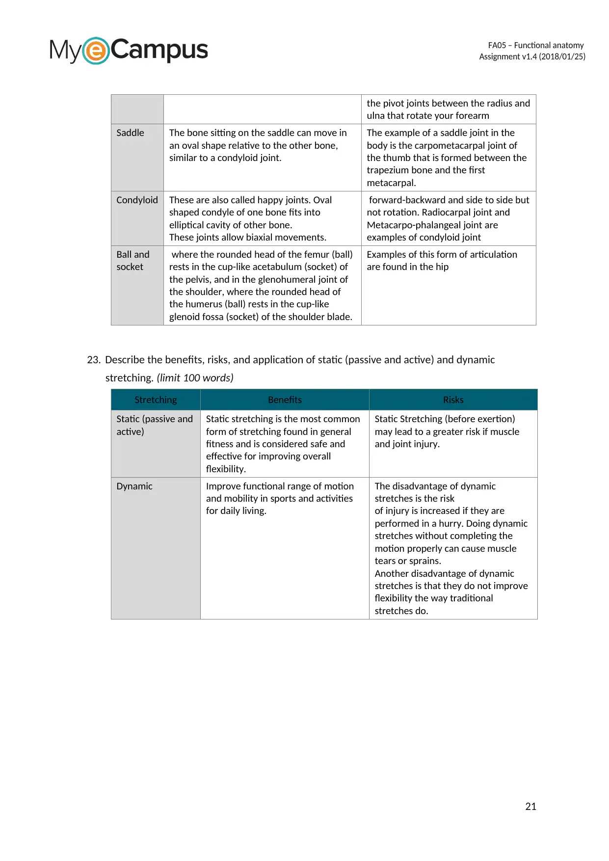
Pivot joints are junctions between bones that
allow your body to move. ... A pivot joint is a
type of synovial joint that rotates.
examples of pivot joints in your body
are the pivot joint in your neck that
allows your head to rotate and
20
FA05 – Functional anatomy
Assignment v1.4 (2018/01/25)
the pivot joints between the radius and
ulna that rotate your forearm
Saddle The bone sitting on the saddle can move in
an oval shape relative to the other bone,
similar to a condyloid joint.
The example of a saddle joint in the
body is the carpometacarpal joint of
the thumb that is formed between the
trapezium bone and the first
metacarpal.
Condyloid These are also called happy joints. Oval
shaped condyle of one bone fits into
elliptical cavity of other bone.
These joints allow biaxial movements.
forward-backward and side to side but
not rotation. Radiocarpal joint and
Metacarpo-phalangeal joint are
examples of condyloid joint
Ball and
socket
where the rounded head of the femur (ball)
rests in the cup-like acetabulum (socket) of
the pelvis, and in the glenohumeral joint of
the shoulder, where the rounded head of
the humerus (ball) rests in the cup-like
glenoid fossa (socket) of the shoulder blade.
Examples of this form of articulation
are found in the hip
23. Describe the benefits, risks, and application of static (passive and active) and dynamic
stretching. (limit 100 words)
Stretching Benefits Risks
Static (passive and
active)
Static stretching is the most common
form of stretching found in general
fitness and is considered safe and
effective for improving overall
flexibility.
Static Stretching (before exertion)
may lead to a greater risk if muscle
and joint injury.
Dynamic Improve functional range of motion
and mobility in sports and activities
for daily living.
The disadvantage of dynamic
stretches is the risk
of injury is increased if they are
performed in a hurry. Doing dynamic
stretches without completing the
motion properly can cause muscle
tears or sprains.
Another disadvantage of dynamic
stretches is that they do not improve
flexibility the way traditional
stretches do.
21
FA05 – Functional anatomy
Assignment v1.4 (2018/01/25)
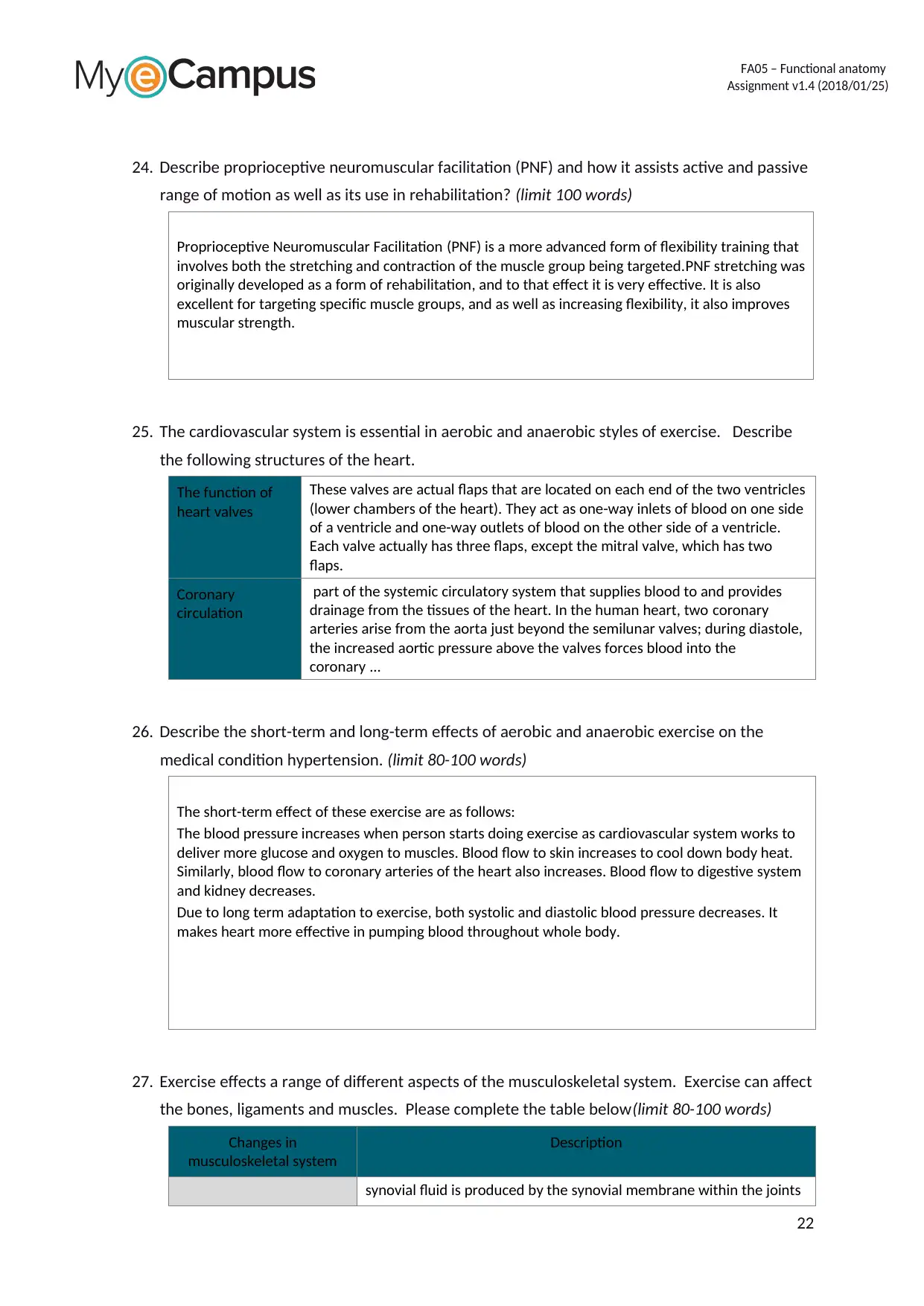
24. Describe proprioceptive neuromuscular facilitation (PNF) and how it assists active and passive
range of motion as well as its use in rehabilitation? (limit 100 words)
Proprioceptive Neuromuscular Facilitation (PNF) is a more advanced form of flexibility training that
involves both the stretching and contraction of the muscle group being targeted.PNF stretching was
originally developed as a form of rehabilitation, and to that effect it is very effective. It is also
excellent for targeting specific muscle groups, and as well as increasing flexibility, it also improves
muscular strength.
25. The cardiovascular system is essential in aerobic and anaerobic styles of exercise. Describe
the following structures of the heart.
The function of
heart valves
These valves are actual flaps that are located on each end of the two ventricles
(lower chambers of the heart). They act as one-way inlets of blood on one side
of a ventricle and one-way outlets of blood on the other side of a ventricle.
Each valve actually has three flaps, except the mitral valve, which has two
flaps.
Coronary
circulation
part of the systemic circulatory system that supplies blood to and provides
drainage from the tissues of the heart. In the human heart, two coronary
arteries arise from the aorta just beyond the semilunar valves; during diastole,
the increased aortic pressure above the valves forces blood into the
coronary ...
26. Describe the short-term and long-term effects of aerobic and anaerobic exercise on the
medical condition hypertension. (limit 80-100 words)
The short-term effect of these exercise are as follows:
The blood pressure increases when person starts doing exercise as cardiovascular system works to
deliver more glucose and oxygen to muscles. Blood flow to skin increases to cool down body heat.
Similarly, blood flow to coronary arteries of the heart also increases. Blood flow to digestive system
and kidney decreases.
Due to long term adaptation to exercise, both systolic and diastolic blood pressure decreases. It
makes heart more effective in pumping blood throughout whole body.
27. Exercise effects a range of different aspects of the musculoskeletal system. Exercise can affect
the bones, ligaments and muscles. Please complete the table below(limit 80-100 words)
Changes in
musculoskeletal system
Description
synovial fluid is produced by the synovial membrane within the joints
22
Secure Best Marks with AI Grader
Need help grading? Try our AI Grader for instant feedback on your assignments.
FA05 – Functional anatomy
Assignment v1.4 (2018/01/25)
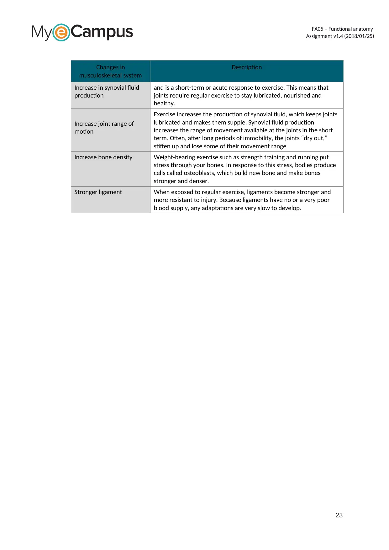
Changes in
musculoskeletal system
Description
Increase in synovial fluid
production
and is a short-term or acute response to exercise. This means that
joints require regular exercise to stay lubricated, nourished and
healthy.
Increase joint range of
motion
Exercise increases the production of synovial fluid, which keeps joints
lubricated and makes them supple. Synovial fluid production
increases the range of movement available at the joints in the short
term. Often, after long periods of immobility, the joints “dry out,”
stiffen up and lose some of their movement range
Increase bone density Weight-bearing exercise such as strength training and running put
stress through your bones. In response to this stress, bodies produce
cells called osteoblasts, which build new bone and make bones
stronger and denser.
Stronger ligament When exposed to regular exercise, ligaments become stronger and
more resistant to injury. Because ligaments have no or a very poor
blood supply, any adaptations are very slow to develop.
23
FA05 – Functional anatomy
Assignment v1.4 (2018/01/25)
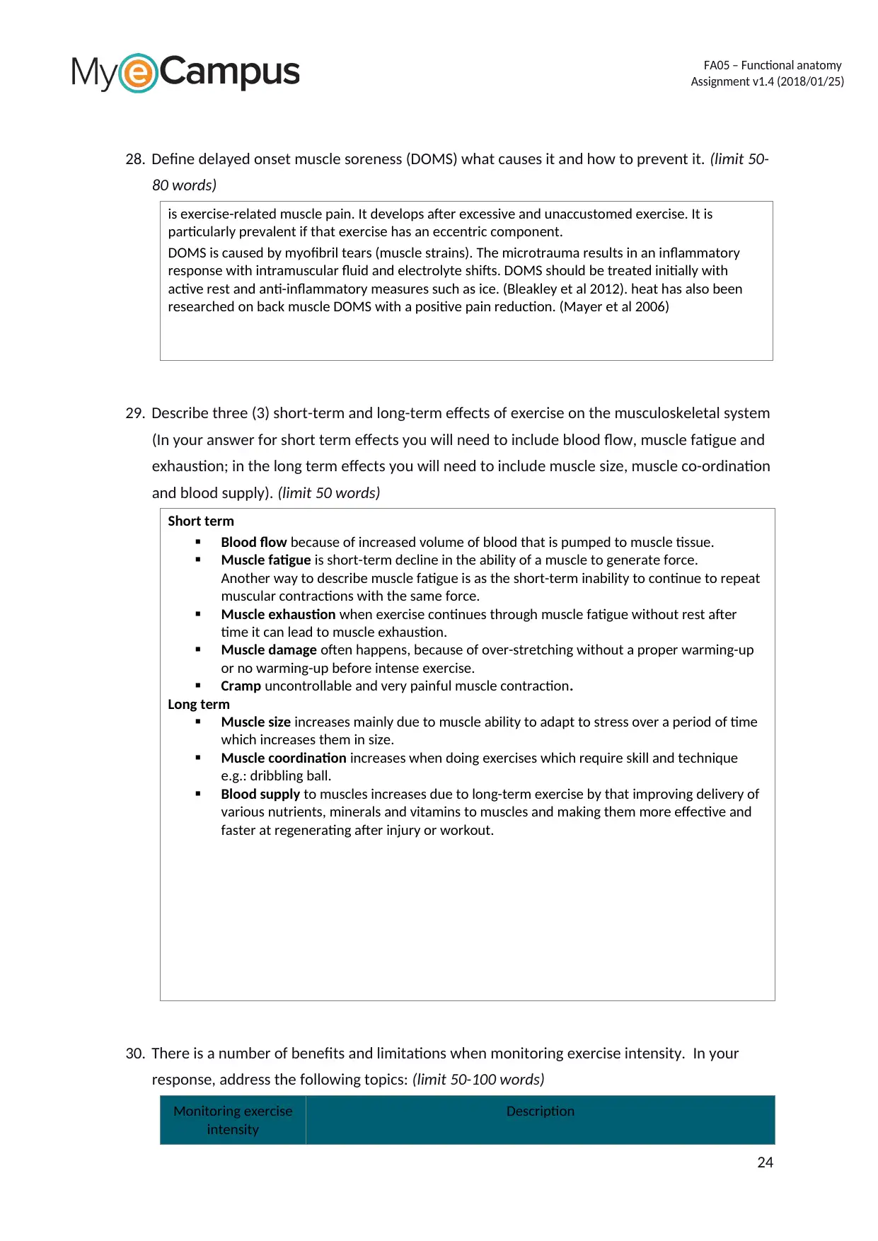
28. Define delayed onset muscle soreness (DOMS) what causes it and how to prevent it. (limit 50-
80 words)
is exercise-related muscle pain. It develops after excessive and unaccustomed exercise. It is
particularly prevalent if that exercise has an eccentric component.
DOMS is caused by myofibril tears (muscle strains). The microtrauma results in an inflammatory
response with intramuscular fluid and electrolyte shifts. DOMS should be treated initially with
active rest and anti-inflammatory measures such as ice. (Bleakley et al 2012). heat has also been
researched on back muscle DOMS with a positive pain reduction. (Mayer et al 2006)
29. Describe three (3) short-term and long-term effects of exercise on the musculoskeletal system
(In your answer for short term effects you will need to include blood flow, muscle fatigue and
exhaustion; in the long term effects you will need to include muscle size, muscle co-ordination
and blood supply). (limit 50 words)
Short term
Blood flow because of increased volume of blood that is pumped to muscle tissue.
Muscle fatigue is short-term decline in the ability of a muscle to generate force.
Another way to describe muscle fatigue is as the short-term inability to continue to repeat
muscular contractions with the same force.
Muscle exhaustion when exercise continues through muscle fatigue without rest after
time it can lead to muscle exhaustion.
Muscle damage often happens, because of over-stretching without a proper warming-up
or no warming-up before intense exercise.
Cramp uncontrollable and very painful muscle contraction.
Long term
Muscle size increases mainly due to muscle ability to adapt to stress over a period of time
which increases them in size.
Muscle coordination increases when doing exercises which require skill and technique
e.g.: dribbling ball.
Blood supply to muscles increases due to long-term exercise by that improving delivery of
various nutrients, minerals and vitamins to muscles and making them more effective and
faster at regenerating after injury or workout.
30. There is a number of benefits and limitations when monitoring exercise intensity. In your
response, address the following topics: (limit 50-100 words)
Monitoring exercise
intensity
Description
24
FA05 – Functional anatomy
Assignment v1.4 (2018/01/25)
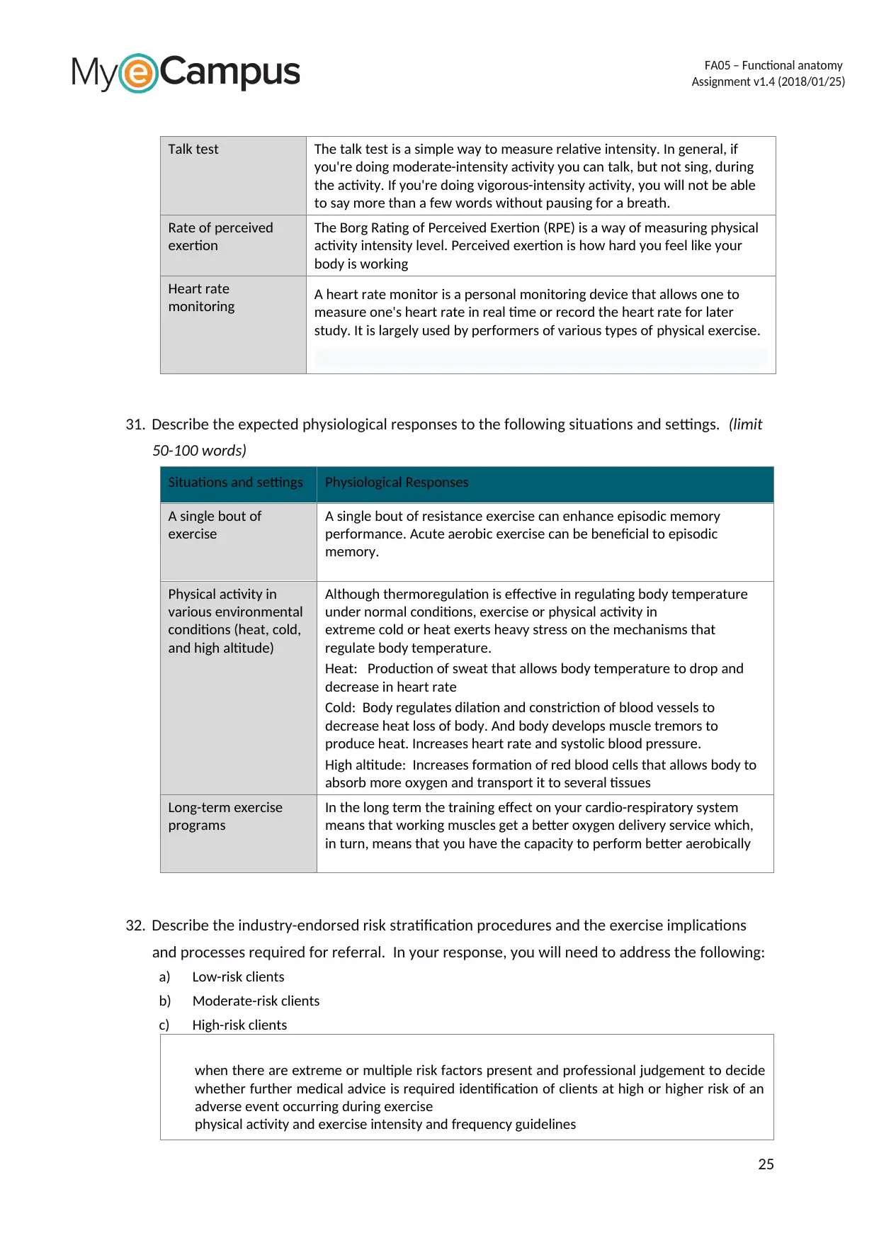
Talk test The talk test is a simple way to measure relative intensity. In general, if
you're doing moderate-intensity activity you can talk, but not sing, during
the activity. If you're doing vigorous-intensity activity, you will not be able
to say more than a few words without pausing for a breath.
Rate of perceived
exertion
The Borg Rating of Perceived Exertion (RPE) is a way of measuring physical
activity intensity level. Perceived exertion is how hard you feel like your
body is working
Heart rate
monitoring A heart rate monitor is a personal monitoring device that allows one to
measure one's heart rate in real time or record the heart rate for later
study. It is largely used by performers of various types of physical exercise.
31. Describe the expected physiological responses to the following situations and settings. (limit
50-100 words)
Situations and settings Physiological Responses
A single bout of
exercise
A single bout of resistance exercise can enhance episodic memory
performance. Acute aerobic exercise can be beneficial to episodic
memory.
Physical activity in
various environmental
conditions (heat, cold,
and high altitude)
Although thermoregulation is effective in regulating body temperature
under normal conditions, exercise or physical activity in
extreme cold or heat exerts heavy stress on the mechanisms that
regulate body temperature.
Heat: Production of sweat that allows body temperature to drop and
decrease in heart rate
Cold: Body regulates dilation and constriction of blood vessels to
decrease heat loss of body. And body develops muscle tremors to
produce heat. Increases heart rate and systolic blood pressure.
High altitude: Increases formation of red blood cells that allows body to
absorb more oxygen and transport it to several tissues
Long-term exercise
programs
In the long term the training effect on your cardio-respiratory system
means that working muscles get a better oxygen delivery service which,
in turn, means that you have the capacity to perform better aerobically
32. Describe the industry-endorsed risk stratification procedures and the exercise implications
and processes required for referral. In your response, you will need to address the following:
a) Low-risk clients
b) Moderate-risk clients
c) High-risk clients
when there are extreme or multiple risk factors present and professional judgement to decide
whether further medical advice is required identification of clients at high or higher risk of an
adverse event occurring during exercise
physical activity and exercise intensity and frequency guidelines
25
Paraphrase This Document
Need a fresh take? Get an instant paraphrase of this document with our AI Paraphraser
FA05 – Functional anatomy
Assignment v1.4 (2018/01/25)
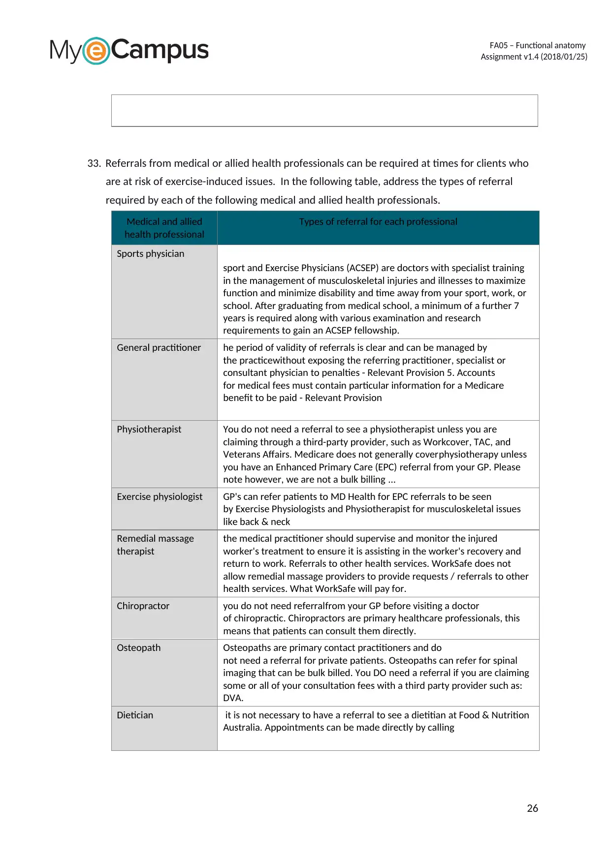
33. Referrals from medical or allied health professionals can be required at times for clients who
are at risk of exercise-induced issues. In the following table, address the types of referral
required by each of the following medical and allied health professionals.
Medical and allied
health professional
Types of referral for each professional
Sports physician
sport and Exercise Physicians (ACSEP) are doctors with specialist training
in the management of musculoskeletal injuries and illnesses to maximize
function and minimize disability and time away from your sport, work, or
school. After graduating from medical school, a minimum of a further 7
years is required along with various examination and research
requirements to gain an ACSEP fellowship.
General practitioner he period of validity of referrals is clear and can be managed by
the practicewithout exposing the referring practitioner, specialist or
consultant physician to penalties - Relevant Provision 5. Accounts
for medical fees must contain particular information for a Medicare
benefit to be paid - Relevant Provision
Physiotherapist You do not need a referral to see a physiotherapist unless you are
claiming through a third-party provider, such as Workcover, TAC, and
Veterans Affairs. Medicare does not generally coverphysiotherapy unless
you have an Enhanced Primary Care (EPC) referral from your GP. Please
note however, we are not a bulk billing ...
Exercise physiologist GP's can refer patients to MD Health for EPC referrals to be seen
by Exercise Physiologists and Physiotherapist for musculoskeletal issues
like back & neck
Remedial massage
therapist
the medical practitioner should supervise and monitor the injured
worker's treatment to ensure it is assisting in the worker's recovery and
return to work. Referrals to other health services. WorkSafe does not
allow remedial massage providers to provide requests / referrals to other
health services. What WorkSafe will pay for.
Chiropractor you do not need referralfrom your GP before visiting a doctor
of chiropractic. Chiropractors are primary healthcare professionals, this
means that patients can consult them directly.
Osteopath Osteopaths are primary contact practitioners and do
not need a referral for private patients. Osteopaths can refer for spinal
imaging that can be bulk billed. You DO need a referral if you are claiming
some or all of your consultation fees with a third party provider such as:
DVA.
Dietician it is not necessary to have a referral to see a dietitian at Food & Nutrition
Australia. Appointments can be made directly by calling
26
FA05 – Functional anatomy
Assignment v1.4 (2018/01/25)
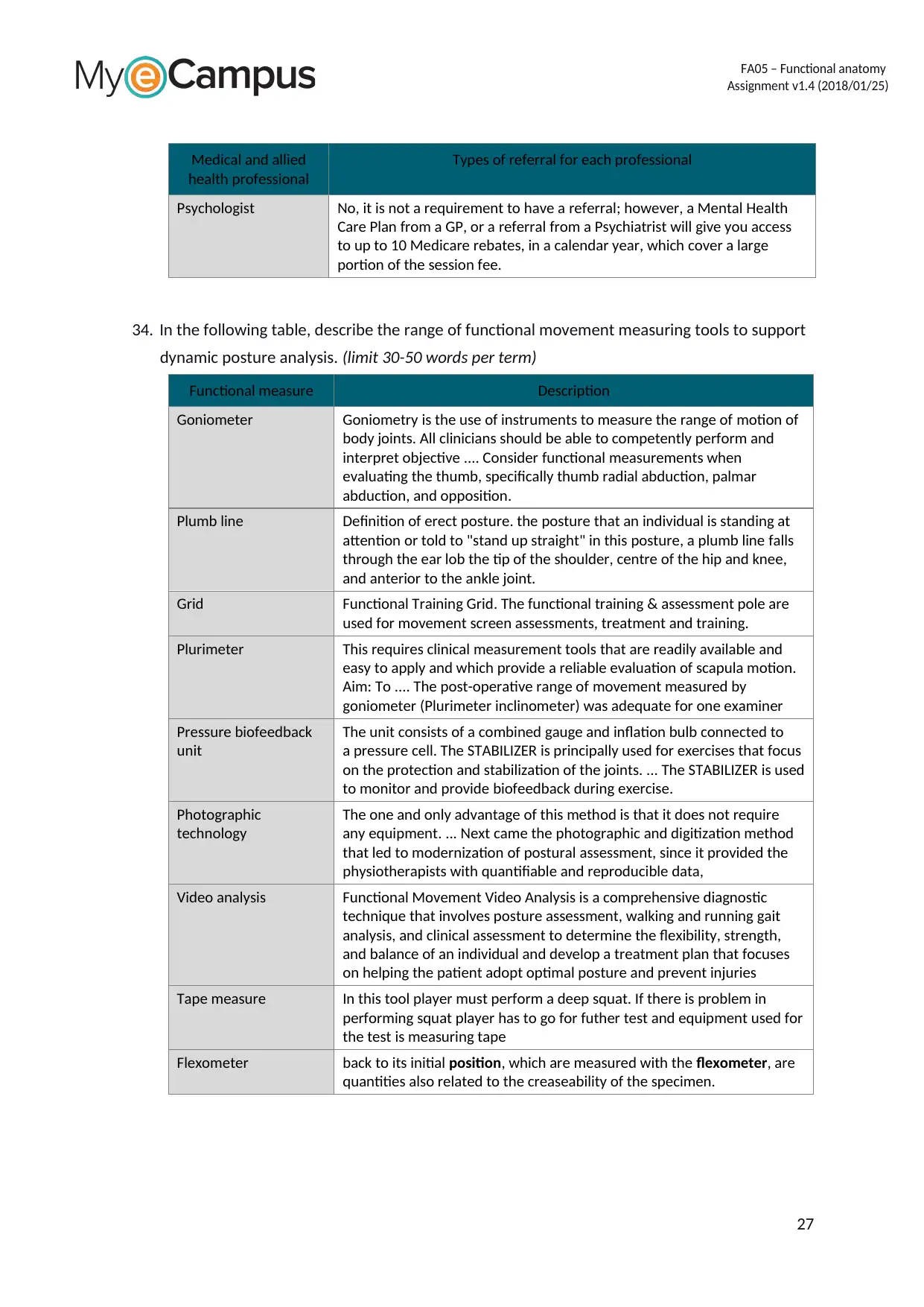
Medical and allied
health professional
Types of referral for each professional
Psychologist No, it is not a requirement to have a referral; however, a Mental Health
Care Plan from a GP, or a referral from a Psychiatrist will give you access
to up to 10 Medicare rebates, in a calendar year, which cover a large
portion of the session fee.
34. In the following table, describe the range of functional movement measuring tools to support
dynamic posture analysis. (limit 30-50 words per term)
Functional measure Description
Goniometer Goniometry is the use of instruments to measure the range of motion of
body joints. All clinicians should be able to competently perform and
interpret objective .... Consider functional measurements when
evaluating the thumb, specifically thumb radial abduction, palmar
abduction, and opposition.
Plumb line Definition of erect posture. the posture that an individual is standing at
attention or told to "stand up straight" in this posture, a plumb line falls
through the ear lob the tip of the shoulder, centre of the hip and knee,
and anterior to the ankle joint.
Grid Functional Training Grid. The functional training & assessment pole are
used for movement screen assessments, treatment and training.
Plurimeter This requires clinical measurement tools that are readily available and
easy to apply and which provide a reliable evaluation of scapula motion.
Aim: To .... The post-operative range of movement measured by
goniometer (Plurimeter inclinometer) was adequate for one examiner
Pressure biofeedback
unit
The unit consists of a combined gauge and inflation bulb connected to
a pressure cell. The STABILIZER is principally used for exercises that focus
on the protection and stabilization of the joints. ... The STABILIZER is used
to monitor and provide biofeedback during exercise.
Photographic
technology
The one and only advantage of this method is that it does not require
any equipment. ... Next came the photographic and digitization method
that led to modernization of postural assessment, since it provided the
physiotherapists with quantifiable and reproducible data,
Video analysis Functional Movement Video Analysis is a comprehensive diagnostic
technique that involves posture assessment, walking and running gait
analysis, and clinical assessment to determine the flexibility, strength,
and balance of an individual and develop a treatment plan that focuses
on helping the patient adopt optimal posture and prevent injuries
Tape measure In this tool player must perform a deep squat. If there is problem in
performing squat player has to go for futher test and equipment used for
the test is measuring tape
Flexometer back to its initial position, which are measured with the flexometer, are
quantities also related to the creaseability of the specimen.
27
FA05 – Functional anatomy
Assignment v1.4 (2018/01/25)
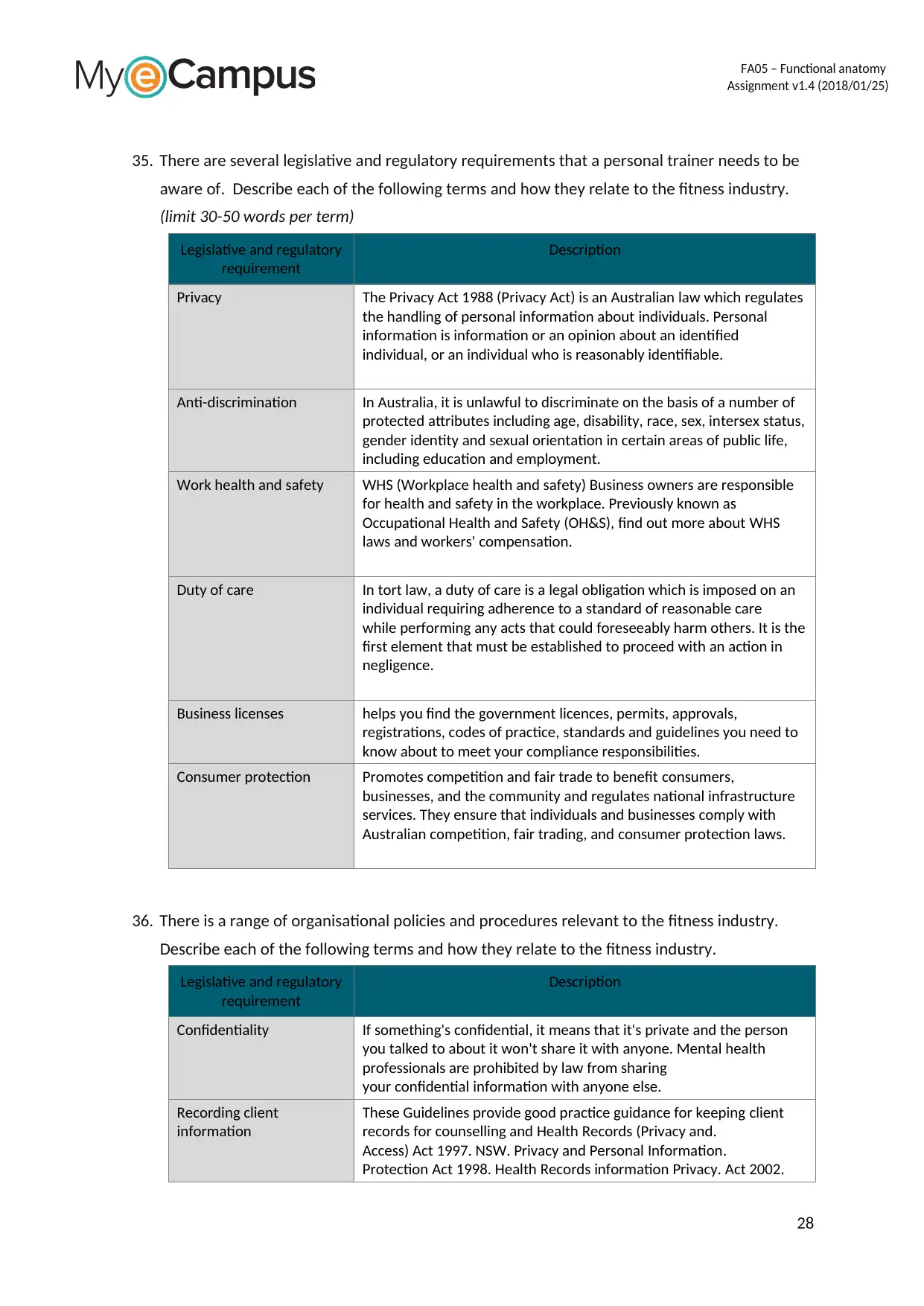
35. There are several legislative and regulatory requirements that a personal trainer needs to be
aware of. Describe each of the following terms and how they relate to the fitness industry.
(limit 30-50 words per term)
Legislative and regulatory
requirement
Description
Privacy The Privacy Act 1988 (Privacy Act) is an Australian law which regulates
the handling of personal information about individuals. Personal
information is information or an opinion about an identified
individual, or an individual who is reasonably identifiable.
Anti-discrimination In Australia, it is unlawful to discriminate on the basis of a number of
protected attributes including age, disability, race, sex, intersex status,
gender identity and sexual orientation in certain areas of public life,
including education and employment.
Work health and safety WHS (Workplace health and safety) Business owners are responsible
for health and safety in the workplace. Previously known as
Occupational Health and Safety (OH&S), find out more about WHS
laws and workers' compensation.
Duty of care In tort law, a duty of care is a legal obligation which is imposed on an
individual requiring adherence to a standard of reasonable care
while performing any acts that could foreseeably harm others. It is the
first element that must be established to proceed with an action in
negligence.
Business licenses helps you find the government licences, permits, approvals,
registrations, codes of practice, standards and guidelines you need to
know about to meet your compliance responsibilities.
Consumer protection Promotes competition and fair trade to benefit consumers,
businesses, and the community and regulates national infrastructure
services. They ensure that individuals and businesses comply with
Australian competition, fair trading, and consumer protection laws.
36. There is a range of organisational policies and procedures relevant to the fitness industry.
Describe each of the following terms and how they relate to the fitness industry.
Legislative and regulatory
requirement
Description
Confidentiality If something's confidential, it means that it's private and the person
you talked to about it won't share it with anyone. Mental health
professionals are prohibited by law from sharing
your confidential information with anyone else.
Recording client
information
These Guidelines provide good practice guidance for keeping client
records for counselling and Health Records (Privacy and.
Access) Act 1997. NSW. Privacy and Personal Information.
Protection Act 1998. Health Records information Privacy. Act 2002.
28
1 out of 28
Related Documents
Your All-in-One AI-Powered Toolkit for Academic Success.
+13062052269
info@desklib.com
Available 24*7 on WhatsApp / Email
![[object Object]](/_next/static/media/star-bottom.7253800d.svg)
Unlock your academic potential
© 2024 | Zucol Services PVT LTD | All rights reserved.


