Comparative Cytology: Cardiac and Skeletal Muscle
VerifiedAdded on 2020/05/08
|8
|1338
|117
AI Summary
This assignment delves into a comparative study of cardiac and skeletal muscle tissues. It explores their similarities and differences in structure, function, and histology. The document highlights the unique characteristics of each muscle type, emphasizing their crucial roles in physiological processes. From the self-sustaining nature of cardiac muscle to the intricate control mechanisms governing skeletal muscle movement, this assignment provides a comprehensive understanding of these vital tissues.
Contribute Materials
Your contribution can guide someone’s learning journey. Share your
documents today.
Running head: FUNDAMENTALS OF SCIENCE
Task 5- Fundamentals of science
Name of the Student
Name of the University
Author Note
Task 5- Fundamentals of science
Name of the Student
Name of the University
Author Note
Secure Best Marks with AI Grader
Need help grading? Try our AI Grader for instant feedback on your assignments.
FUNDAMENTALS OF SCIENCE 1
Comparative cytology (Cardiac and Skeletal muscles)
The ability of animals to voluntarily carry out their actions with the help of muscles
distinguishes them from other organisms. This action is generally brought about by changes in
muscle cells length, by the process of muscle contraction. The three types of muscle,
distinguished on the basis of their structure and function are: skeletal, cardiac and smooth
muscle. This assignment will compare the structures of skeletal and cardiac muscle and will
illustrate their structural, histological and functional differences.
Cardiac muscles are found only in the heart. During systolic phase, they contract and
pump blood away from the heart, to distant body parts. This supplies necessary oxygen to all
cells. On the other hand, the skeletal muscles remain attached to tendons, which in turn are
connected to bones. Contractions of these muscles make the tendons pull on the bones. This
leads to movement of the arms 1.
There are few similarities between cardiac and skeletal muscles. Both of them contain t-
tubules that help in conduction of action potential. These action potentials trigger calcium flow
that facilitates muscle contraction. There is more number of t-tubules in skeletal fibres. However,
they are wider in the cardiac muscles.
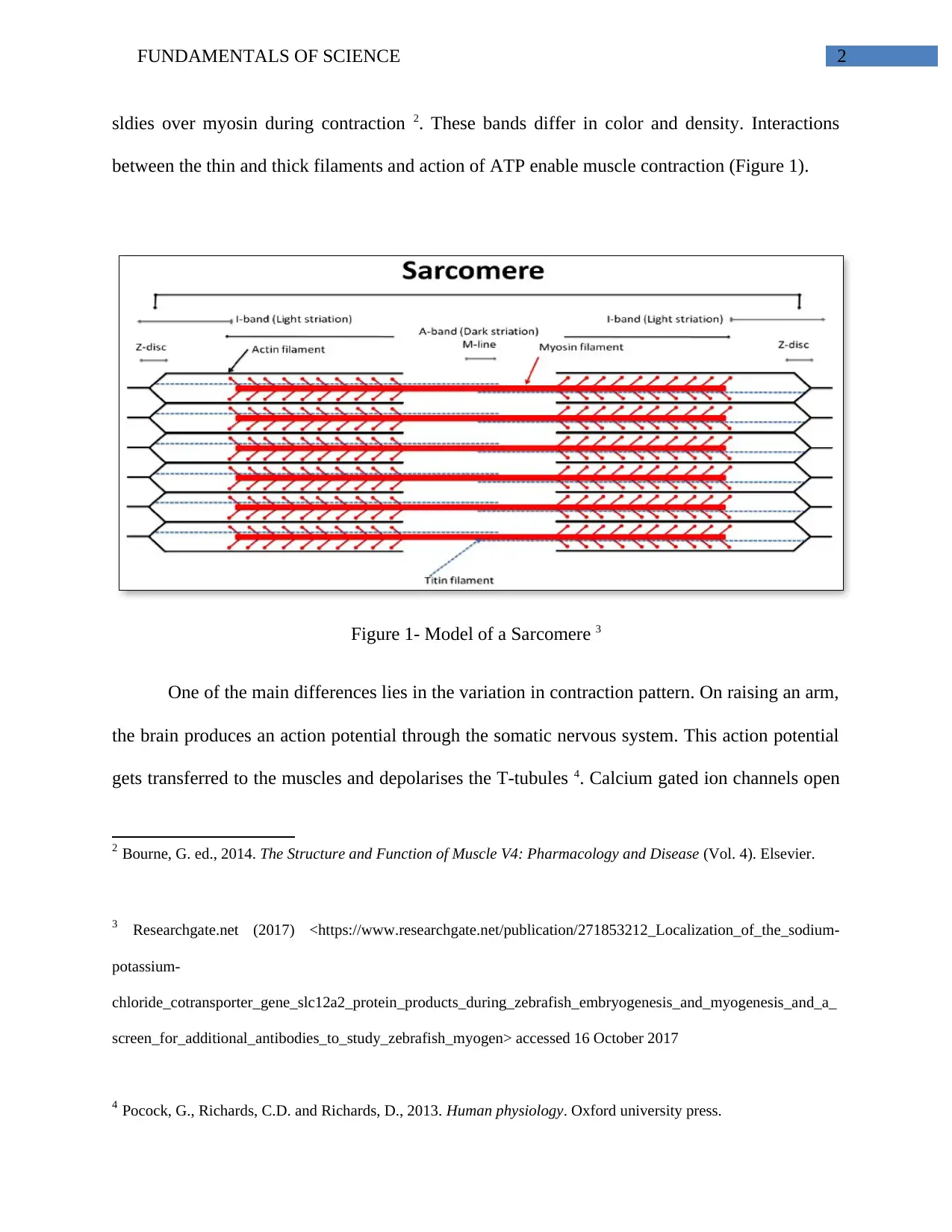
Another common trait is their striated structure. They have thin, thick and elastic
filaments that form sarcomeres. They are formed of actin and myosin myofilaments and the actin
1 Hall, J.E., 2015. Guyton and Hall Textbook of Medical Physiology E-Book. Elsevier Health Sciences.
FUNDAMENTALS OF SCIENCE 2
sldies over myosin during contraction 2. These bands differ in color and density. Interactions
between the thin and thick filaments and action of ATP enable muscle contraction (Figure 1).
Figure 1- Model of a Sarcomere 3
One of the main differences lies in the variation in contraction pattern. On raising an arm,
the brain produces an action potential through the somatic nervous system. This action potential
gets transferred to the muscles and depolarises the T-tubules 4. Calcium gated ion channels open
2 Bourne, G. ed., 2014. The Structure and Function of Muscle V4: Pharmacology and Disease (Vol. 4). Elsevier.
3 Researchgate.net (2017) <https://www.researchgate.net/publication/271853212_Localization_of_the_sodium-
potassium-
chloride_cotransporter_gene_slc12a2_protein_products_during_zebrafish_embryogenesis_and_myogenesis_and_a_
screen_for_additional_antibodies_to_study_zebrafish_myogen> accessed 16 October 2017
4 Pocock, G., Richards, C.D. and Richards, D., 2013. Human physiology. Oxford university press.
FUNDAMENTALS OF SCIENCE 3
and this leads to actin and myosin sliding past each other. Thus, the skeletal muscles contract and
the bone get lifted. Therefore, a nervous control is essential to the functioning of skeletal muscle.
Cardiac muscles are also regulated by the nervous system. However, cardiac muscle
contractions are involuntary. These muscles are to the autonomic nervous system. Unlike skeletal
muscles, muscle contraction generating action potentials are originated in the myogenic cells of
the heart. These cells are located in the SAN (Sino Atrial Node), which produces a pacemaker
potential and sets the intrinsic rhythm of heart. This ANS therefore modulates the heart rate 5.
The rate increases by the action of sympathetic nervous system during fight or flight reaction and
the gets slowed down under the effect of the parasympathetic nervous system.
Skeletal muscles are made up of long, thin cylindrical fibres. Each fibre is innervated by
a somatic alpha motor neuron. Upon entering the muscle, the axon branches and gets connected
to single muscle fibres 6.
In cardiac muscle, gap junctions and adherens junctions link the fibres together. They
strengthen the structure of cardiac muscles to avoid tear of fibres during forceful contractions.
They also allow passage of electrical signals between muscle cells, to ensure that all muscles
show synchronised contraction. Unsynchronised contractions cause damage to cardiac muscles.
5 Canale, E.D., Campbell, G.R. and Smolich, J.J., 2012. Cardiac muscle (Vol. 2). Springer Science & Business
Media.
6 Frontera, W.R. and Ochala, J., 2015. Skeletal muscle: a brief review of structure and function. Calcified tissue
international, 96(3), pp.183-195.
Secure Best Marks with AI Grader
Need help grading? Try our AI Grader for instant feedback on your assignments.
FUNDAMENTALS OF SCIENCE 4
Cardiac and skeletal muscles react to single action potential and produce a single twitch
response. Lon increasing the frequency of these signals, summation occurs in the skeletal muscle
and may lead to tetanus when the refractory period becomes shorter.
However, action potential duration is longer in cardiac muscles, due to slow activation of
calcium channels and presence of longer T-tubules. Cardiac muscles fail to show tetanus and this
enables suficient time to sufficiently fill up the heart with blood before arrival of next action
potential. Furthermore, they do not tire much, unlike skeletal muscles. This proves beneficial,
because tired cardiac muscles may lead to angina and other serious cardiovascular conditions.
Cardiac muscles need more ATP due to continuous functioning of the heart. This
accounts for the presence of more mitochondria, when compared to skeletal muscles. Moreover,
cardiac muscles undergo oxidative phosphorylation to produce ATP that is required for sliding of
actin over myosin, for muscle contraction 7. Thus, they require a supply of respiratory substrates
and oxygen for aerobic respiration. On the other hand, less contraction of skeletal muscles
account for the presence of low number of mitochondria.
The two muscles differ greatly in the length of muscle fibres as well. While a cardiac
fibre can be of 100Âμm length, length of a skeletal fibre usually ranges from few millimetres to
10cm. Moreover, one nucleus is present in cardiac muscle fibres compared to many nuclei along
skeletal muscle fibres. The huge length of skeletal muscle fibre serves as the prerequisite for the
presence of many nuclei 8.
7 Sherwood, L., 2015. Human physiology: from cells to systems. Cengage learning.
8 SIRS, E.M.O., 2013. SKELETAL MUSCLE FUNCTION. Oxygen Transport to Tissue XIX, 428, p.333.
FUNDAMENTALS OF SCIENCE 5
Other differences also exist between cardiac and skeletal muscles. The heart is myogenic
in nature. It is capable of generating cardiac contractions without the action of nervous input.
Thus, it is self-sufficient. However, skeletal muscles are controlled by the nervous system. It is
also essential that the cardiac muscles in the heart should function without any obstructions. A
minor problem in contraction can severely damage the organ and lead to death. Both these
muscle types are important for physiological functioning of humans as well as other animals.
Thus, it can be concluded that the cardiac muscles play a vital role in our existence. They
form a bulk of the entire mass of the heart and are specialised to pump blood throughout our
body. The high endurance and contractile strength of these muscles help in circulation of oxygen
and nutrients during our lifetime. If the cardiac muscles fail to operate, we would not be able to
survive. Skeletal muscles, on the other hand allow us to interact with the surrounding
environment. As stated above, while both the similarities between both the muscles are evident,
the differences in their structure, function and histology are prominent. They operate as
functional parts of our body. If the muscles do not function efficiently, we will not be able to
move or perform daily activities. Therefore, the inherent mechanism of both the tissue types is
essential to maintain our physiological system.
FUNDAMENTALS OF SCIENCE 6
References
Bourne, G. ed., 2014. The Structure and Function of Muscle V4: Pharmacology and
Disease (Vol. 4). Elsevier.
Canale, E.D., Campbell, G.R. and Smolich, J.J., 2012. Cardiac muscle (Vol. 2). Springer Science
& Business Media.
Frontera, W.R. and Ochala, J., 2015. Skeletal muscle: a brief review of structure and
function. Calcified tissue international, 96(3), pp.183-195.
Pocock, G., Richards, C.D. and Richards, D., 2013. Human physiology. Oxford university press.
Researchgate.net (2017)
<https://www.researchgate.net/publication/271853212_Localization_of_the_sodium-potassium-
chloride_cotransporter_gene_slc12a2_protein_products_during_zebrafish_embryogenesis_and_
myogenesis_and_a_screen_for_additional_antibodies_to_study_zebrafish_myogen> accessed 16
October 2017
Sherwood, L., 2015. Human physiology: from cells to systems. Cengage learning.
SIRS, E.M.O., 2013. SKELETAL MUSCLE FUNCTION. Oxygen Transport to Tissue XIX, 428,
p.333.
Hall, J.E., 2015. Guyton and Hall Textbook of Medical Physiology E-Book. Elsevier Health
Sciences.
Paraphrase This Document
Need a fresh take? Get an instant paraphrase of this document with our AI Paraphraser

FUNDAMENTALS OF SCIENCE 7
1 out of 8
Related Documents
Your All-in-One AI-Powered Toolkit for Academic Success.
+13062052269
info@desklib.com
Available 24*7 on WhatsApp / Email
![[object Object]](/_next/static/media/star-bottom.7253800d.svg)
Unlock your academic potential
© 2024 | Zucol Services PVT LTD | All rights reserved.


