This article discusses the process of fusion protein expression, the role of PCR in making his tag fusion protein, and the advantages of using a hexahistidine tag for protein purification. It covers topics such as cloning, PCR primer design, ligation, transformation, and purification methods.
Contribute Materials
Your contribution can guide someone’s learning journey. Share your
documents today.
Running head: FUSION PROTEIN Fusion protein Name of the student: Name of the university: Author note:
Secure Best Marks with AI Grader
Need help grading? Try our AI Grader for instant feedback on your assignments.
1FUSION PROTEIN Fusion protein: Many cloning vectors have been designed in the field of recombinant technology so that protein being expressed further fused another protein called tag. Tags are used for facilitating protein purification (Wang, 2016). Examples of these tags are glutathione-S transferase, maltose binding or multiple binding residues. This paper will illustrate the process of histamine tagged fusion protein and the role of PCR in fusion protein expression. Process: The tag vectors are usually designed for coding sequence for an amino acid sequence cleaved by a specific protease which is inserted between coding sequence and tag to incorporate and tag can be cleaved off. In this context, polyhistidine fusion for protein purification can be used. In order to make a fusion protein, the gene of interest which will encode the protein is first need to insert into a vector (Frank, 2018). pBAD vectors can be used for producing the fashion protein having six histidine-tags (Khan et al., 2016).The gene of interest is inserted into the vector by using a ligase enzyme. Role of PCR in making his tag fusion protein: Designing primer: For the synthesis of the fusion protein were used s PCR methods are used. The polymerase chain reaction is another process of cloning of amplification of any DNA sequence for experimental purpose.InPCR method, the primer can be used which have reparative histidine codes (CAT or CAC) right next to stop codon or start codon in addition to the several 18 to 20 bases from one end of the DNA. The best way of putting histamine tag to the protein is to include six histamine tags to either N terminal or C terminal (Apsana et al., 2016).
2FUSION PROTEIN In case of N terminal region can be, the primer sequence would be his sequence "CATCATCACCACCACCAT” after ATG. (i.e. start coding of the gene ) in forwarding primer. In case of C terminal protein, the antisense strand of above-mentioned sequences would be used and it can be added to just before sequencing in reverse primer just before the stop codon of the gene (Zhao et al., 2016). The C terminal and N terminal protein were used for obtaining better enables us to detect by Western blotting (Nasrabadi et al., 2016). Sometimes a linker of gly-gly- gly can be used between the histamine tags and protein of interest so that the activity of the protein should be affected. After designing a primer, running PCR to amplify insert DNA using traditional PCR method where Taq polymerase is used. After PCR cycle isolation of DNA can be done using gel electrophoresis where agarose gel is used to obtain the DNA purified DNA. The DNA bands of choice can be isolated from the gel through excision procedure using the scalpel (Ortiz et al., 2016). Ligate: Ligation of the insert into the Vector can be done using standard ligation procedure where recipient plasmid to insert ratio of approximately 1:3 can be used for the successful ligation. The optimal ligation temperature is 25 degree which is an optimal temperature for the ligase enzyme (Wang, 2016). Transformation: After ligation, transformation can be done into competent cells where vector with insert can be transformed. After transformation, the pore plate can be done to obtain the colony with an insert of his tag within vectors (Frank, 2018). Cells can be induced for the synthesis of the fusion protein.
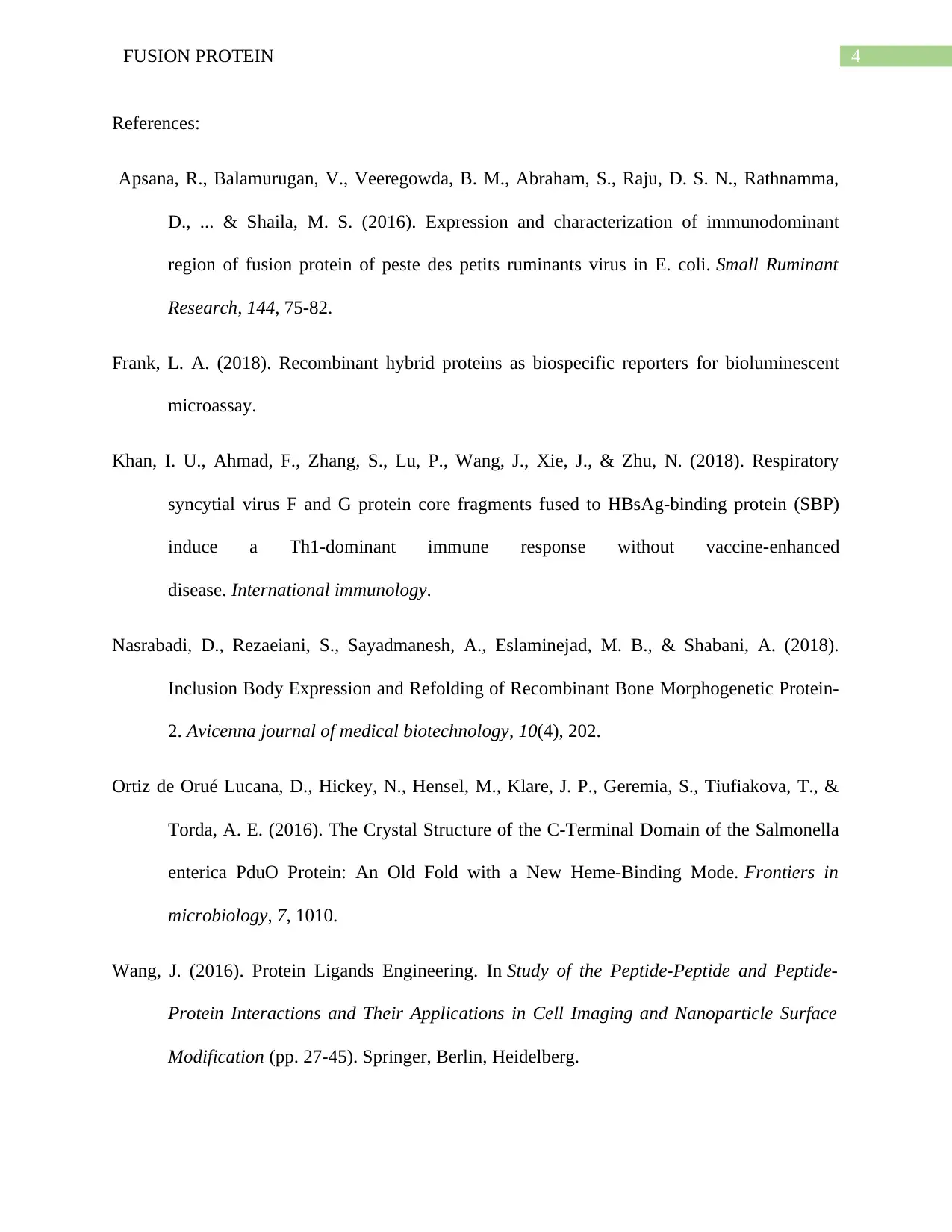
3FUSION PROTEIN In this case, western blotting needs to be done by using SDS PAGE.After inducing synthesis, the cells need to be lysed and the viscosity of the lysate needs to be reduced by nuclease treatment.To facilitate assay of the fusion proteins, short antibody recognition sequence can be incorporated into the tag which will further enhance purification (Wang, 2016). These, antibiotic can be further detected by western blotting, antibody carrying appropriate epitope. A polyhistidine tag at the C terminus can function for both assay and purification. It can be further purified through the affinity chromatography where imidazole can be given as a competitor of his tag to obtain the protein (Zhao et al., 2016). Imidazole competes with his tag for binding with the metal charged resin in the chromatography and thus it is used to elute protein from the column. Ni2+ is most commonly used for his-tag purification because it gives a high yield of proteins (Frank, 2018).. Advantage: Hexahistidine tag is much smaller and therefore provides high yields of tagged protein. Another advantage of this protein is that it doesn’t interfere with the structure and function of the recombinant protein (Zhao et al., 2016). Figure: gene sequence of fusion protein Source: (Frank, 2018).
Paraphrase This Document
Need a fresh take? Get an instant paraphrase of this document with our AI Paraphraser
4FUSION PROTEIN References: Apsana, R., Balamurugan, V., Veeregowda, B. M., Abraham, S., Raju, D. S. N., Rathnamma, D., ... & Shaila, M. S. (2016). Expression and characterization of immunodominant region of fusion protein of peste des petits ruminants virus in E. coli.Small Ruminant Research,144, 75-82. Frank, L. A. (2018). Recombinant hybrid proteins as biospecific reporters for bioluminescent microassay. Khan, I. U., Ahmad, F., Zhang, S., Lu, P., Wang, J., Xie, J., & Zhu, N. (2018). Respiratory syncytial virus F and G protein core fragments fused to HBsAg-binding protein (SBP) induceaTh1-dominantimmuneresponsewithoutvaccine-enhanced disease.International immunology. Nasrabadi, D., Rezaeiani, S., Sayadmanesh, A., Eslaminejad, M. B., & Shabani, A. (2018). Inclusion Body Expression and Refolding of Recombinant Bone Morphogenetic Protein- 2.Avicenna journal of medical biotechnology,10(4), 202. Ortiz de Orué Lucana, D., Hickey, N., Hensel, M., Klare, J. P., Geremia, S., Tiufiakova, T., & Torda, A. E. (2016). The Crystal Structure of the C-Terminal Domain of the Salmonella enterica PduO Protein: An Old Fold with a New Heme-Binding Mode.Frontiers in microbiology,7, 1010. Wang, J. (2016). Protein Ligands Engineering. InStudy of the Peptide-Peptide and Peptide- Protein Interactions and Their Applications in Cell Imaging and Nanoparticle Surface Modification(pp. 27-45). Springer, Berlin, Heidelberg.
5FUSION PROTEIN Zhao,D.,Huang,Z.,Liu,J.,Ma,L.,&He,J.(2018).Expression,purification,and characterization of N-terminal His-tagged proteins with mutations in zinc finger 3 of zinc finger protein ZNF191 (243–368).Preparative Biochemistry and Biotechnology, 1-6.