Detailed Report: Exploring Various Medical Imaging Modalities
VerifiedAdded on 2020/04/13
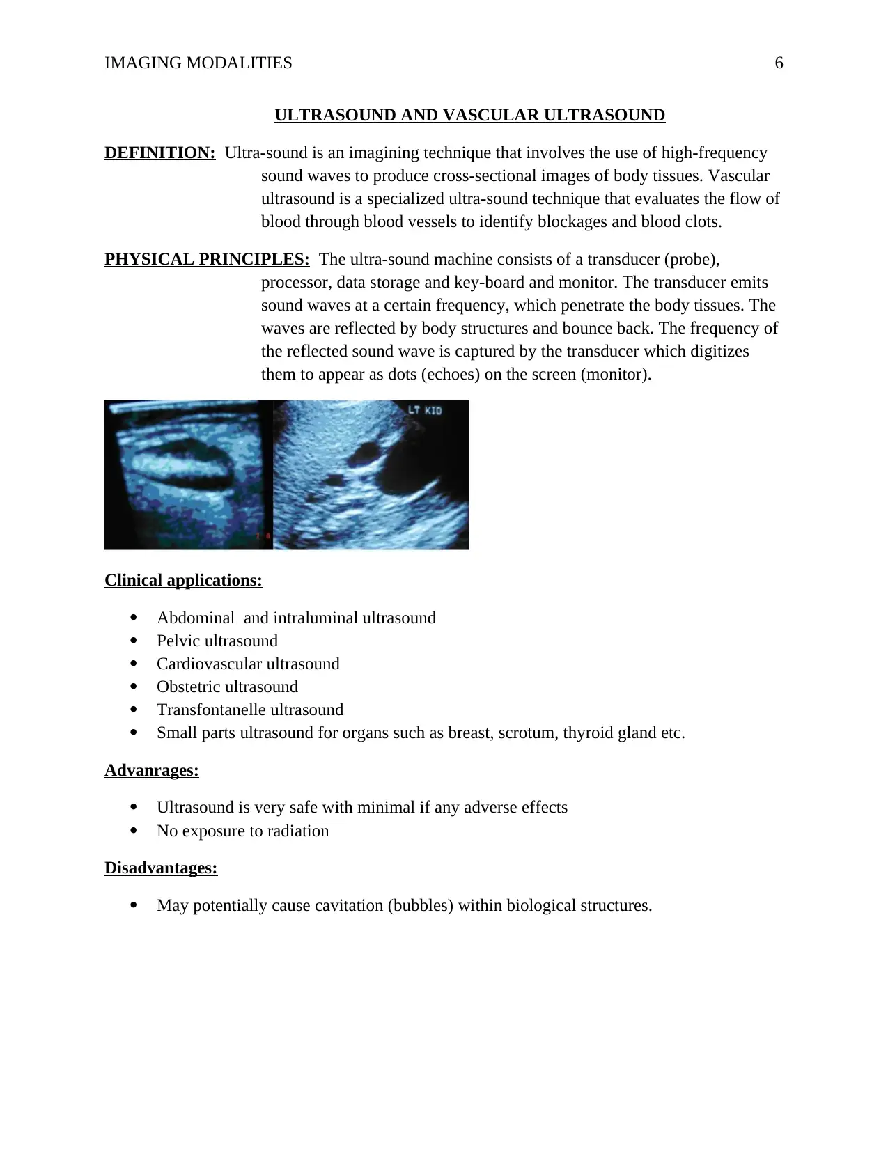
|7
|1251
|286
Report
AI Summary
This report provides a comprehensive overview of various medical imaging modalities used in healthcare. It begins with X-rays, detailing their history, physical principles, medical applications, advantages, and disadvantages. The report then moves on to Computed Tomography (CT) scans, explaining their principles, clinical applications, and benefits. Following this, it covers Magnetic Resonance Imaging (MRI) scans, highlighting their physical principles, clinical applications, and advantages. The report further delves into Nuclear Medicine, describing its principles, clinical applications, and the advantages and disadvantages associated with it. Finally, it examines Ultrasound and Vascular Ultrasound, detailing their physical principles, clinical applications, advantages, and disadvantages. The report utilizes references to support the information presented.

1 out of 7
Related Documents


Your All-in-One AI-Powered Toolkit for Academic Success.
+13062052269
info@desklib.com
Available 24*7 on WhatsApp / Email
![[object Object]](/_next/static/media/star-bottom.7253800d.svg)
Copyright © 2020–2026 A2Z Services. All Rights Reserved. Developed and managed by ZUCOL.