CT Scan: Composition, Biophysical Features, and Functions
Added on 2023-05-29
8 Pages1660 Words281 Views
CT Scan 1
CT SCAN
By (Name of Student)
The Name of the Class (course)
Professor (Tutor)
The Name of the School (University)
The City and State where it is located
Date
CT SCAN
By (Name of Student)
The Name of the Class (course)
Professor (Tutor)
The Name of the School (University)
The City and State where it is located
Date

CT Scan 2
Introduction
Computerized axial tomography and computerized tomography scans are unique X-ray machines
that generate cross-sectional body images with the use of computers and X-rays. Computerized
tomography was autonomously designed by an engineer from Britain called Dr. Allan Cormack
and Godfrey Hounsfield. This machine is relied upon for medical diagnosis of illnesses. The
cr0sss-sectional body images are utilized by computerized tomography machine using biological
tissues relying on the X-rays absorption (Van Dessel, et al., 2017). The signals are determined in
Hounsfield which is a random scale that shows the difference in the attenuation coefficient of the
features of biological tissues. With technology, the CT tests may be conducted faster as the
enhancements enable Doctors to diagnose easily due to the images with high resolutions. There
has been a substantial increase in the detection of the specificity and sensitivity of illnesses. This
article outlines the CT test machine and its composition, biophysical features as well as its
functions (Kohli et al., 2017).
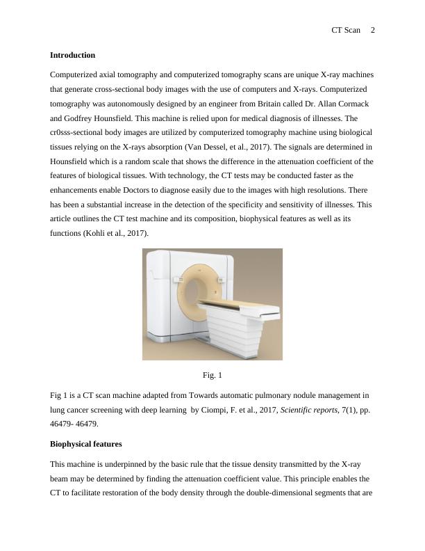
Fig. 1
Fig 1 is a CT scan machine adapted from Towards automatic pulmonary nodule management in
lung cancer screening with deep learning by Ciompi, F. et al., 2017, Scientific reports, 7(1), pp.
46479- 46479.
Biophysical features
This machine is underpinned by the basic rule that the tissue density transmitted by the X-ray
beam may be determined by finding the attenuation coefficient value. This principle enables the
CT to facilitate restoration of the body density through the double-dimensional segments that are
Introduction
Computerized axial tomography and computerized tomography scans are unique X-ray machines
that generate cross-sectional body images with the use of computers and X-rays. Computerized
tomography was autonomously designed by an engineer from Britain called Dr. Allan Cormack
and Godfrey Hounsfield. This machine is relied upon for medical diagnosis of illnesses. The
cr0sss-sectional body images are utilized by computerized tomography machine using biological
tissues relying on the X-rays absorption (Van Dessel, et al., 2017). The signals are determined in
Hounsfield which is a random scale that shows the difference in the attenuation coefficient of the
features of biological tissues. With technology, the CT tests may be conducted faster as the
enhancements enable Doctors to diagnose easily due to the images with high resolutions. There
has been a substantial increase in the detection of the specificity and sensitivity of illnesses. This
article outlines the CT test machine and its composition, biophysical features as well as its
functions (Kohli et al., 2017).
Fig. 1
Fig 1 is a CT scan machine adapted from Towards automatic pulmonary nodule management in
lung cancer screening with deep learning by Ciompi, F. et al., 2017, Scientific reports, 7(1), pp.
46479- 46479.
Biophysical features
This machine is underpinned by the basic rule that the tissue density transmitted by the X-ray
beam may be determined by finding the attenuation coefficient value. This principle enables the
CT to facilitate restoration of the body density through the double-dimensional segments that are
CT Scan 3
perpendicular to the acquisition system's axis (Rashid et al., 2017). The X-ray tube released N
photons which attenuate as they go through the biological function layers and the information
used to develop scanned object (Van Dessel et al., 2017). Generally, there are two absorption
processes which include Compton effect and the photoelectric effect. This feature is
characterized by one coefficient, mju.
When using the CT machine, the X-rays emitters are rotated all over the patient while the
detector is put in opposite sides of the diameters which collects up the body part images, in this
case, the detector and the beam moving in a synchronized manner. The detectors of the scanner
do not generate images. They determine the X-rays thin beams that are transmitted via a
complete body scan. The images of particular parts are taken at various angles which enables for
the retrieval of data inside the third dimension. The CT detector produces measurements that are
equivalent to the attenuation coefficient value (Oh, et al., 2017). The computer applies unique
mathematical algorithms to reconstruct images to achieve the patient's tomographic images
relying on raw scan information (Oh, et al., 2017).
One can find the proportionate attenuation coefficient to the amount of irradiated tissue if the X-
ray is converted in to quasi-monochromatic or monochromatic with the most suitable filters
using the basic absorption formula of X-rays in practice (Rashid, et al., 2017). The location
determines the emitted intensity I (x) of the photons beams obtained. Whereby, I(x) is less while
the body is greatly radio-opaque. The sectional image of the Irradiated X-ray object is restored
from a huge amount of attenuation coefficient values. It collects all the information received
from the material’s elementary volumes via the detectors (Tu et al., 2018).
The CT image scanner involves the element’s square matrix (pixel) every single of it
representing a voxel of the patient’s tissue. The measurement of the attenuated coefficient for
every proportionate voxel to the pixels must be estimated while processing the image. Every
point of the image is bordered by a star with halo-shape which degrades the difference and blurs
the object's boundary. The filter procedure works in a way that the obtained negative value
amounts to the projection filtered while it is eliminated when projected backward thus producing
the image (Moifo et al., 2017). Before information is displayed on the screen, the CT values are
generated from the conventional rescaling presented in Hounsfield Units. Therefore, the PC
End of preview
Want to access all the pages? Upload your documents or become a member.
Related Documents
Computerized Axial Tomography Scanninglg...
|20
|4569
|318
Understanding the Formation and Reconstruction of CT Projection Data in Medical Imaginglg...
|7
|1490
|377
Biophysical Procedures Used in Medical Studieslg...
|6
|1099
|425
Computed Tomographylg...
|9
|1982
|38
Quality assurance and quality control in Computed tomography.lg...
|13
|3446
|4
Principles of Computed Tomographylg...
|18
|4870
|20