Analysis of Inflammatory Cytokine using Flow Cytometry, q-PCR and Elisa Test on Mice Immune System
VerifiedAdded on 2023/06/07
|17
|4024
|474
AI Summary
This study focuses on the analysis of inflammatory cytokine using Flow Cytometry, q-PCR and Elisa Test on mice immune system. It discusses the importance of q-PCR and Flow Cytometry/Elisa tests in biomedical sciences and their role in detecting and diagnosing problems in the human body. The study also highlights the significance of IL2 inflammatory mediators. The materials and methods used in the study are discussed in detail along with the results and discussions.
Contribute Materials
Your contribution can guide someone’s learning journey. Share your
documents today.
MEDICAL IMAGING AND DRUG
DELIVERY
Analysis of inflammatory cytokine using Flow cytometry,
q- PCR and Elisa test on mice immune system
DELIVERY
Analysis of inflammatory cytokine using Flow cytometry,
q- PCR and Elisa test on mice immune system
Secure Best Marks with AI Grader
Need help grading? Try our AI Grader for instant feedback on your assignments.

Table of contents
1. Abstract
2. Introduction
3. Materials and Methods
4. Results
5. Discussions
6. Conclusions
7. References
1. Abstract
2. Introduction
3. Materials and Methods
4. Results
5. Discussions
6. Conclusions
7. References

Abstract
n biomedical fi eld the primary step before any treatment of medication is testing of disease esting isI , . T very
necessary to fi nd the real cause behind any symptoms or disease rom a list of different testing. F methods in
biomedical sciences q CR, -P and lowF cytometry lisa/E are two important methods.
mmunologicalI memory is the ability of immune system to identify the disease causing outer agents. hisT
memory is responsible for further activation of antibodies to counter that outer attack.
n recent years we have heard a lot about R CR test R CR is an abbreviation of reverseI , T-P . T- P transcription
polymerase chain reaction. During C V DO I -19, almost every person heard about this name. hisT test is
different from q CR however-P , it is also a kind of CRP .
low cytometry is another test that is mostly used to evaluate fl uids and their characteristics in ourF body.
t isI necessary to fi nd the details of different constituents in a fl uid. Apart from testing, analysis of the test
results is also necessary as at this step we recognize the issues and the important parameters that are
responsible for the cause in our body.
n this study low cytometry CR and SA were carried out following the stimulation of cells AllI , F , P , ELI .
circumstances had the same statistical significance with values range from toP- 0.038589 (0.003) 0.000034
for(0.0193) each of the studies. CD3/28 and CD3/28/200 na veï cells, on the other hand, show a
substantial increase in while CD is down regulated e pression in memory cells was foundTNF-, 3 - . TNF x to
be at the same level in CD and CD cells indings from this study p value3/28 3/28/200 . F ( =0.579026,
show that there is statistical significance in the presence of the0.390750, 0.01195, 0.00007) IL2
inflammatory mediators aive cells spanning CD and CD showed an enhanced. N T 3/28 3/28/200
e pressionx of IL-2.
Introduction
low cytometry test is used to study the fl uid particles of our bodyF Chow (2021) lisa test is a. E test to
detect antibodies in human body lisa is an abbreviation for enzyme linked immunosorbent. E assay lisa. E
test is used in diagnosing many diseases like V rotavirus ika virus Syphills etcHI , , Z , . Woodall, R. T.
(2020) low cytometry is used in research for a number of purposes like cell. F counting sorting detecting, ,
micro organisms diagnosing bone marrow and blood cancers etc hese- , . T tests are very useful in detecting
and diagnosing problems in human body. All the problems where fl uid fl ow study is required fl ow,
cytometry is used to determine the parameters and its effects After. complete blood count C C( B ) test, this
test is commonly used as a follow up- test Anderson Gwenin, & Gwenin,(2019).
q CR test is a quantitative test q CR is an abbreviation for quantitative polymerase chain reaction-P . -P . hisT
technology is used for measuring D A using CR CR is a common method forN P . P amplifying D A hisN . T
test is a modern methodology for studying gene e pression nx . I this test, nucleic acids are detected
rapidly that are responsible for the disease in the body.
Memory and cells constitute basis of immunological memory yet a lot is still unknown about theirT B ,
biology his study focuses on cell responses using a model of antigen. T T stimulation based on antibody
attached to artificial devices. hisT was a cross sectional study and

conducted at niversityU of ullH . thicalE approval was obtained from thicalE review board. nI this study,
memory cells differentiated in vitro from alb c mice spleen and lymph nodes compared to na veB / ( ), ï cells,
freshly harvested from alb c mouse spleens and lymph nodes After stimulation of cells fl owB / . , cytometry,
CRP and SAELI were done.
ne of CD s more common names is MRC An average human CD is kDa in size CDO -200' OX-2. -200 41-47 . -
is an intracellular signaling protein with two immunoglobulin superfamily domains he genes that200 . T
code for CD are located on chromosome q-200 3. (3 12-13) Khanal & et.al., (2020 tracellular). Ex
immunoglobulin superfamily domain single V C short cytoplasmic tail and a single transmembrane( + ), ,
region devoid of signaling motif make up the molecule s structural components At its terminal' . N-
immunoglobulin like domain names CD s terminal CC faces are the primary point of contact, 200' N- GF ′
between CD and its receptor Residues and which are typically found in200 . E44, I71, T73, E75, I133,
CD R s200 1' CCGF ′ region, play an important role in this interaction, despite the fact that E44 is positioned
outside of this area, within the so called- A strand, in the V like- domain Zarei & et.al., (2021).
C controls CD e pression at the transcriptional level and are all under their/EBP- -200 x . TNF-, IFN-IL-6, IL-1
control as are a slew of other molecules CD e pression is induced by and through, . -200 x TNF- IFN- NF-
kappa S A and R hen it comes to D A human and mouse CD cells share an estimatedB, T T1, I F-1. W N , -200
similarity while the protein content is estimated at percent81.7% , 77.5 Liu & et.al.,(2018)
raph pad prism software is a sophisticated software as it contains various tools to interpret andG analyze
the data t is easier to plot curve and data in this software or analysis it contains various. I . F , inbuilt tools
like anova test, parametric test etc. that provides an easy way to do the analysis of data set inter(W ,
n our scenario we have collected data from two teams for these tests and analyzed2013). I , these data
set using the graph pad prism software n addition to analysis data representation is also. I , quite easier in
graph pad prism software Zhou, T. & et.al., (2022).
Materials and methods
Chemical, Reagents, and buffers
Different materials are required to do these tests Apart from materials instruments are needed for. ,
carrying out these test like computer and related apparatus e will discuss in details about methods. W
used to do these tests one by one.
Memory cells were differentiated in vitro two three weeks ahead by seeding solenocytes with anti CD/ , - 3 and
anti CD activating antibodies to simulate signal and respectively for a week maintained at- 28 ( 1 2, ) , C in37° 5%
CO2 in well plates with complete R M penicillin streptomycin glutamine and CS24- P I ( , , 5% F ), then
washed and seeded in culture with memory cell cytokines and in complete R M for, IL-7 IL-2 P I additional
weeks oth na ve and memory cells were used in this as primary cell lines2-3 . B ï T . Microscopy of different
markers i e CD CD CD CD and CD CD CD were also done by using. ., 28, 03, 3 + 28, 3+ 28+ 200 eubergerN
chamber.
Animals
he animals were maintained and used under strict ethical conditions according to internationalT
recommendations for animal welfare set by Committee Members, nternational(I Society on o icologyT x , 1992).
n this study memory cells differentiated in vitro from alb c mice spleen and lymph nodesI , B / ( ), compared to
na veï cells, freshly harvested from alb c mouseB / spleens and lymph nodes.
Mice were bled at and hours and sera were separated and stored at C until0, 1/4, 1/2, 1, 2, 4, 8 24 , −20°
Secure Best Marks with AI Grader
Need help grading? Try our AI Grader for instant feedback on your assignments.

use.
Test Details
q-PCR ( uantitativeQ polymerase chain reaction)
Materials needed: luorescentF dyes, D AN binding dyes, real ti me CRP instrument, CRP analysis software, CRP
prime assays CR plastics and, P , CR reagentsP
Method CR chemicals and unique or custom primers are used to start up amplification reactions An: P .
instrument s proprietary software analyses the acquired data from real ti me CR devices he' - P . T
incorporation of a fl uorescent reporter molecule in each reaction well enables real ti me detection of- CRP
products which increases in fl uorescence as the amount of product D A increases D A binding, N . N - dyes
and fl uorescently labelled primers or probes specific to certain sequences are among the fl uorescence
chemistries used for this purpose Haribabu & et.al., (2019) hermal cyclers fi tt ed with fl uorescence. T
detector modules are used to monitor the fl uorescence signal as it amplifies. nI order to quantify the
quantity of amplicon created in each cycle the fl uorescence detected is correlated to the overall amount,
of amplicon.
Flow Cytometry
Materials needed: luorescentF dye, laser beam, detector consisting of forward scatter and side scatter, fl ow
cytometer blood bone, / marrow sample
A fl ow cell is part of the fl uidics system where the sample fl uid is introduced articles are aligned in a, . P single
stream as they move through a ti ny channel into the laser intercept light beam by using sheath( ) fl uid.
aser interrogation is made possible by hydrodynamic focusing which isolates and analyses aL , single cell.
asersL are the most common light source for the optical system since they produce a precise wavelength
and frequency of light Sharifi & et.al., (2019) his is when at least one laser beam is used to pass. T
through the particles asers come in a wide variety of wavelengths from ultraviolet to far red and in a. L , - ,
wide range of power levels. Any fl uorescent probes that have been attached to antibodies and are
compatible with the laser beam are e cited and produce light or fl uoresce at the designatedx ( )
wavelengths as a result of the laser beam interrogation.
Results
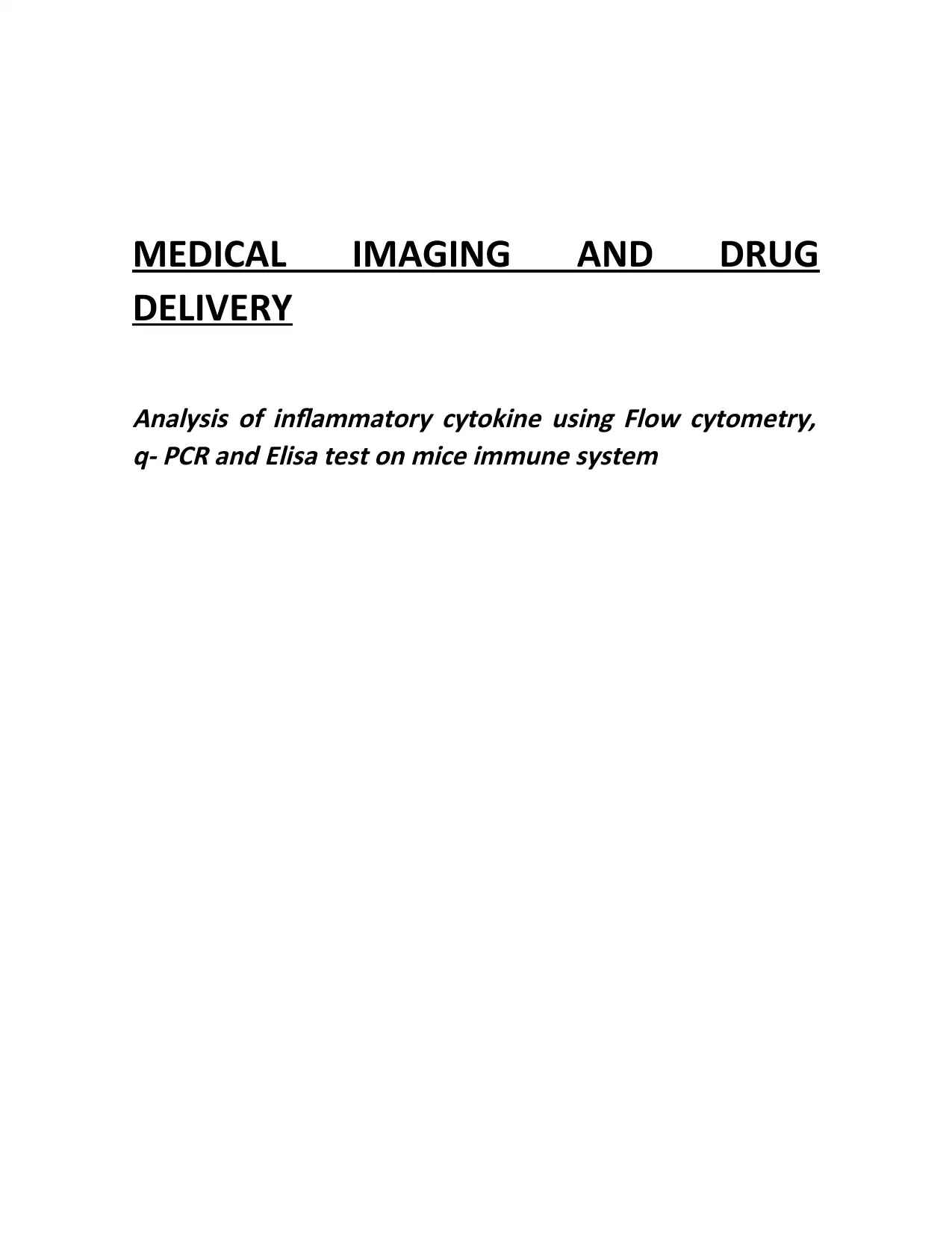
he above bar graph represents the details regarding cell condition Along with this in this CD representT . , 3
the data related to aive and CD and CD single cell was used which was easily separated fromN 3/28 03
each other and appeared as one.
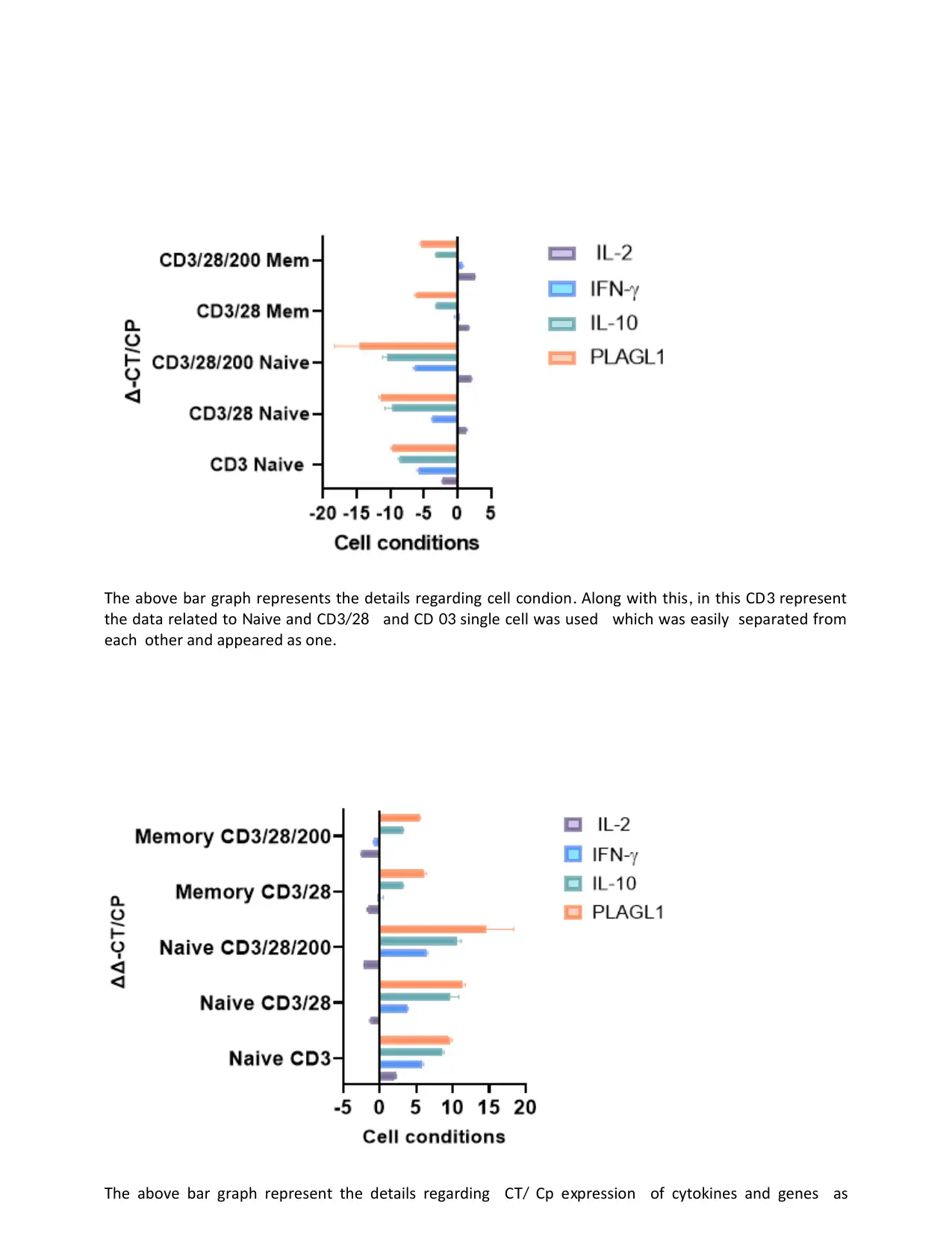
he above bar graph represent the details regarding C Cp e pression of cytokines and genes asT T/ x
the data related to aive and CD and CD single cell was used which was easily separated fromN 3/28 03
each other and appeared as one.
he above bar graph represent the details regarding C Cp e pression of cytokines and genes asT T/ x

compared with C C he stastisctical analysos was done for parametric using sharipo wilks test and oneT/ P . T
wat A VA followed by multiple comporsions was obtained with significance l is observed to beNO , I -2
releated to R to be upregulated and e pressed more above the baseline in CD andHP T1 x 0 3/28
CD condition and activated in both naive and memory cells condition3/28/200 .
C CΔΔ - T/ P e pression of cytokines and genes is compared with thex C CΔ T/ P he statistical analysis was done for. T
parametric using Shapiro wilks test and one way A VA followed by multiple comparisons was obtained with- - NO
significance is observed to be abundantly relative to R housekeeping gene to be upregulated and e pressed, IL-2 HP T1 ( ) x
more above the baseline in CD and CD conditions and activated in both na ve and memory cells(0) 3/28 3/28/200 ï
condition ns not significant. P=***<0.0001; - .
low cytometry graphsF :
Paraphrase This Document
Need a fresh take? Get an instant paraphrase of this document with our AI Paraphraser


t can be said that CD CD were bigger in size than that of CD and CD because whenI 3+ 28+ 3 28
theses two type of cells is combine together it create stimulation and they seems more bigger in the size
but some of the cells are still present as single cell Along with this in case of CD CD CD no single. , 3+ 28+ 200
separate cell was observed in the area as there was clumping of cells bigger and centrally granulated than
those observed above cell line for CD and CD because of these three groups of cells are highly3 28
stimulated.
lisa graphsE :
Secure Best Marks with AI Grader
Need help grading? Try our AI Grader for instant feedback on your assignments.


n conte t ofI x low cytometry test it can be said that it is test that helps in studying the fl uid particlesF
of the body urthermore this test is used in various researches for number of purposes like detecting the. F ,
micro organisms diagnosing problems in human body detecting blood cancers and much more hus all- , , . T ,
the problems where the fl uid fl ow study is needed this test help in determining its parameters and its,
relevant effects Also after the complete count than this test is used for the follow up test n the other hand. , . O
the lisa test is one that provides the report through detecting the antibodies that are present within theE
body and is very efficient in detecting various diseases such as V ika virus rotavirus and much moreHI , Z ,
hus from the above graph it can be said that concenetration of the na ve cells is higher than the memoryT , ï
cells which have only the lsight increase Moreover higher increase in the naiev cells is good as it is. ,
considered as essentail components that helps the body to fi ght off new and unrecognized infections in the
body ence through higher cobecentartion of the such na ve cellas the body would be able to recognize. H , ï
and repsosnd to the new pathogens

rom the above graph it has been analysed that there is very low concentration of the memory cells andF
only higher presence of the na ve cells which means that body might respond to the changes positively butï
would be inefficient in analysing or recognizing the foreign particles that the body was previously e posedx .
hus it means that body would respond in much slower rate making the situation worseT , .
hus from all the graphs it is still unclear that what is the function of CD and CD R signaling playT , 200 200
neurological disorders here is difference in CD and CD as one is human and other animal Scientist. T 200 200 .
has converted mice genes into human under the local promoter of the mouse t has been found that. I
gene vector targets the brain cells or embryonic stem cells may be more important than CD – 200
protein or CD R is blocking antibodies in the e periment research t has been seen in the graphs that200 x . I
cells need low levels of ack that mediates cells and single mediators that are located in memoryT L T
and naive cells that represent why cells responses are more effective owever e cessive use ofT . H , x
autoimmune illness will lead to sickness like sclerosis vertigo and many others here are number of, . T
immunoregulatory mechanism in immune system of human which control
Discussion
From the above results it has been found that the results of the q-PCR flow cytometry
and the Elisa test performed on the mic in the way in which it can use the Excel and Graph pad
for the prism to be able to examine the data that has been collected from the tests Deng &
Paraphrase This Document
Need a fresh take? Get an instant paraphrase of this document with our AI Paraphraser

et.al., (2020).. These tests are able to analyses and also interpret the data more effectively in
order to play a very important and critical role for the sorting and interpreting of the
analyzation of the amounts of the data that is insider the improvement and the presentation of
the data Huang, He & Mi (2019). The Graphics and the plats are considered to be able to make it
a simpler and more comprehended form of the research project.
Within the field of biochemical, the first step which is considered to be one of the most
important one too, is the testing of a particular disease prior to the treatment of the
medication that is to be done Li, Liang Liu & Wang (2018). This is done for the major purpose of
finding out the actual causes of the occurrence of the disease. From a very long list of diverse
methods of testing in the field of biomedical sciences the two major methods are Flow
Cytometry or Elisa and q- PCR (Adan. and et.al., 2017). The graphs mainly provide information
regarding the memory as well as the naïve situations of these methods in particular. the
analysis was particularly done through the usage of the Shapiro- Wilks test that was conducted
for better evaluation.
It was particularly examined that the results of the q- PCR tests, the Elisa test and the
Flow cytometry were performed in the particular research (Bustin. and Huggett., 2017). Then
through using graph pad prism and excel the data that was collected by these tests was briefly
evaluated. By using these precise software programmes, the data analysis was conducted in a
more effective as well as efficient manner. Software particularly plays a crucial role in the
process of sorting of data, interpreting as well as evaluating the huge amounts of the data.
The presentation of the data is also better and improvised through its use.
The Elisa method was used for the purpose of examination of the secretion of the TNF
through the naïve as well as the memory cells under certain stimuli (Yildirim‐Tirgil., 2022). All
the circumstances had similar statistical significance. TNF expression in the memory cells was
on the same level as of the CD3/28/200 and CD3/28 cells in particular. the memory conditions
and the naïve conditions specifically indicate towards no differences that are statistically
different in all the above situations in particular.
These graph pad of the prism in the programmed is also considered to be used as the data
from the two team that are 1 and 2 Proctor (2020, August). This has been considered to be the
different aspects for the cytokines that are found to be expressed in the memory of the T cells
that are through the investigation. It can be found that the ELISA technique was also the factor
that has been used for the examination of the secretion that is used as the memory that is
present in the native cells that are present under the various stimuli. These circumstances are
said to have a statistical significance. The p Values of the test have also differed in the
different studies in the research Xu & et.al., (2021). This substance increases in the TNF provides
the CD3 the down a regulated TNF expresses in the memory of the cells that is found to be in
the same level of the statistically significance memory Park & et.al., (2019). The naïve conditions
that are indicating no other statically significance difference between the four situations are
considered to be the factor that can provides the situation that are essential.

Conclusion
CD cells helps in activation of cytoto ic cell CD cells sends costimulatory signals which initiate3 x T- . 28
the stimulation response by the immune system. nI our study, presence of na veï cell in high numbers
indicate that a major portion of cells are still left and has not encountered antigens yet resence of CD cellT- . P 4
is confirmed by coloration of phosphorence dyes. t act asI a co receptor- of T cell.
low cytometry data shows that C for CD CD has emission near to the ma e citationF FIT 3 & 3/28 x x
laser beam wavelength. tI can be concluded from here that cytoto icx T cells are activated and started to
counter the antigens. igherH na veï cells also shows healthy immune system of the tested sample.
References
Adan A et al low cytometry basic principles and applications, . & . ., (2017). F : . Critical reviews in
biotechnology. pp37(2). .163-176.
Anderson, S. D., Gwenin, V. V., & Gwenin, C. D. (2019). Magnetic functionalized nanoparticles for
biomedical, drug delivery and imaging applications. Nanoscale research letters. 14(1). 1-16.
ustin S uggett q CR primer design revisitedB , . & H , J., (2017). P . iomolecular detection andB
quantification. pp14. .19-28.
Chow, J. C. (2021). Computer method and modeling: Medical biophysics applications in cancer
therapy, medical imaging and drug delivery. AIMS Biophysics. 8(3). 233-235.
Deng, Y., & et.al., (2020). Application of the nano-drug delivery system in treatment of
cardiovascular diseases. Frontiers in bioengineering and biotechnology. 7. 489.
Haribabu, V. & et.al., (2019). Label free ultrasmall fluoromagnetic ferrite-clusters for targeted
cancer imaging and drug delivery. Current drug delivery. 16(3). 233-241.
Huang, D., He, B., & Mi, P. (2019). Calcium phosphate nanocarriers for drug delivery to tumors:
imaging, therapy and theranostics. Biomaterials science, 7(10), 3942-3960.
Khanal, D. & et.al., (2020). The protein corona determines the cytotoxicity of nanodiamonds:
implications of corona formation and its remodelling on nanodiamond applications in biomedical
imaging and drug delivery. Nanoscale Advances. 2(10). 4798-4812.
Li, J., Liang, H., Liu, J., & Wang, Z. (2018). Poly (amidoamine)(PAMAM) dendrimer mediated
delivery of drug and pDNA/siRNA for cancer therapy. International journal of pharmaceutics. 546(1-
2). 215-225.
Liu, X. . & et.al., (2018). Endogenous stimuli-responsive nucleus-targeted nanocarrier for
intracellular mRNA imaging and drug delivery. ACS applied materials & interfaces. 10(46). 39524-
39531.
Park, D. & et.al., (2019). Transdermal drug delivery using a specialized cavitation seed for
ultrasound. IEEE Transactions on Ultrasonics, Ferroelectrics, and Frequency Control. 66(6). 1057-
1064.
Proctor, C. (2020, August). Advanced neural interfaces and drug delivery. In Organic and Hybrid
Sensors and Bioelectronics XIII (Vol. 11475, p. 1147505). SPIE.
Sharifi, M.. & et.al., (2019). Plasmonic gold nanoparticles: Optical manipulation, imaging, drug
delivery and therapy. Journal of Controlled Release. 311. 170-189.
Sun, Y. . & et.al., (2018). Magnetic Resonance Imaging‐Guided Drug Delivery to Breast Cancer
Stem‐Like Cells. Advanced Healthcare Materials. 7(21). 1800266.
Woodall, R. T. (2020). Models of fluid dynamics in biological tissues for medical imaging and drug
delivery (Doctoral dissertation).
Xu, X. & et.al., (2021). Vat photopolymerization 3D printing for advanced drug delivery and medical
device applications. Journal of Controlled Release. 329. 743-757.
ildirim irgil Development of aptamer based SA method for d dimerY ‐T , N., (2022). ‐ ELI ‐

detection. iotechnology and Applied iochemistryB B .
Zarei, S. & et.al., (2021). Theragnostic magnetic core-shell nanoparticle as versatile nanoplatform
for magnetic resonance imaging and drug delivery. Biointerface Research in Applied
Chemistry. 11(5). 13276-13289.
Zhou, T. . & et.al., (2022). All-In-One Second Near-Infrared Light-Responsive Drug Delivery System
for Synergistic Chemo-Photothermal Therapy. ACS Applied Bio Materials.
Secure Best Marks with AI Grader
Need help grading? Try our AI Grader for instant feedback on your assignments.

1 out of 17

Your All-in-One AI-Powered Toolkit for Academic Success.
+13062052269
info@desklib.com
Available 24*7 on WhatsApp / Email
![[object Object]](/_next/static/media/star-bottom.7253800d.svg)
Unlock your academic potential
© 2024 | Zucol Services PVT LTD | All rights reserved.