The Living Body: Components of a Reflex Arc, Endocrine Glands, Nervous and Endocrine System Coordination, Male and Female Reproductive System
VerifiedAdded on 2023/06/13
|10
|1680
|312
AI Summary
This article discusses the components of a reflex arc, function of endocrine glands, nervous and endocrine system coordination, and structure and functions of male and female reproductive system.
Contribute Materials
Your contribution can guide someone’s learning journey. Share your
documents today.

Running head: THE LIVING BODY
The Living Body
Name of student
Name of university
Author note
The Living Body
Name of student
Name of university
Author note
Secure Best Marks with AI Grader
Need help grading? Try our AI Grader for instant feedback on your assignments.
1THE LIVING BODY
3.1 Describe the components of a simple reflex arc
The neurological mechanism, which is responsible for the conduction of nerve impulse in
the autonomous nervous system, that occurs in response to the environmental stimuli (Saladin
2015). It is called an arc because of the shape of pathway it travels with the nerve impulse. There
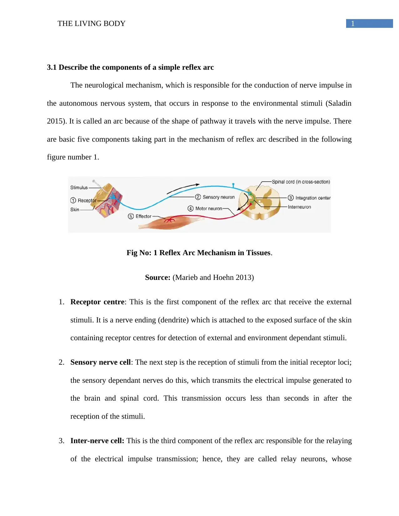
are basic five components taking part in the mechanism of reflex arc described in the following
figure number 1.
Fig No: 1 Reflex Arc Mechanism in Tissues.
Source: (Marieb and Hoehn 2013)
1. Receptor centre: This is the first component of the reflex arc that receive the external
stimuli. It is a nerve ending (dendrite) which is attached to the exposed surface of the skin
containing receptor centres for detection of external and environment dependant stimuli.
2. Sensory nerve cell: The next step is the reception of stimuli from the initial receptor loci;
the sensory dependant nerves do this, which transmits the electrical impulse generated to
the brain and spinal cord. This transmission occurs less than seconds in after the
reception of the stimuli.
3. Inter-nerve cell: This is the third component of the reflex arc responsible for the relaying
of the electrical impulse transmission; hence, they are called relay neurons, whose
2THE LIVING BODY
function is to process and conduct impulse and relaying reflex to the motor nerves from
the grey matter.
4. Motor nerve cell: The fourth component of the reflex arc, carrying the outward impulse
of action to the effecter cells from brain or spinal cord.
5. Effecter nerve cell: The last and fifth component of the reflex arc, giving the reflex
response against the stimuli, which may or may not be at the same position of the
reception centre.
3.2 Identify the function of the main endocrine glands
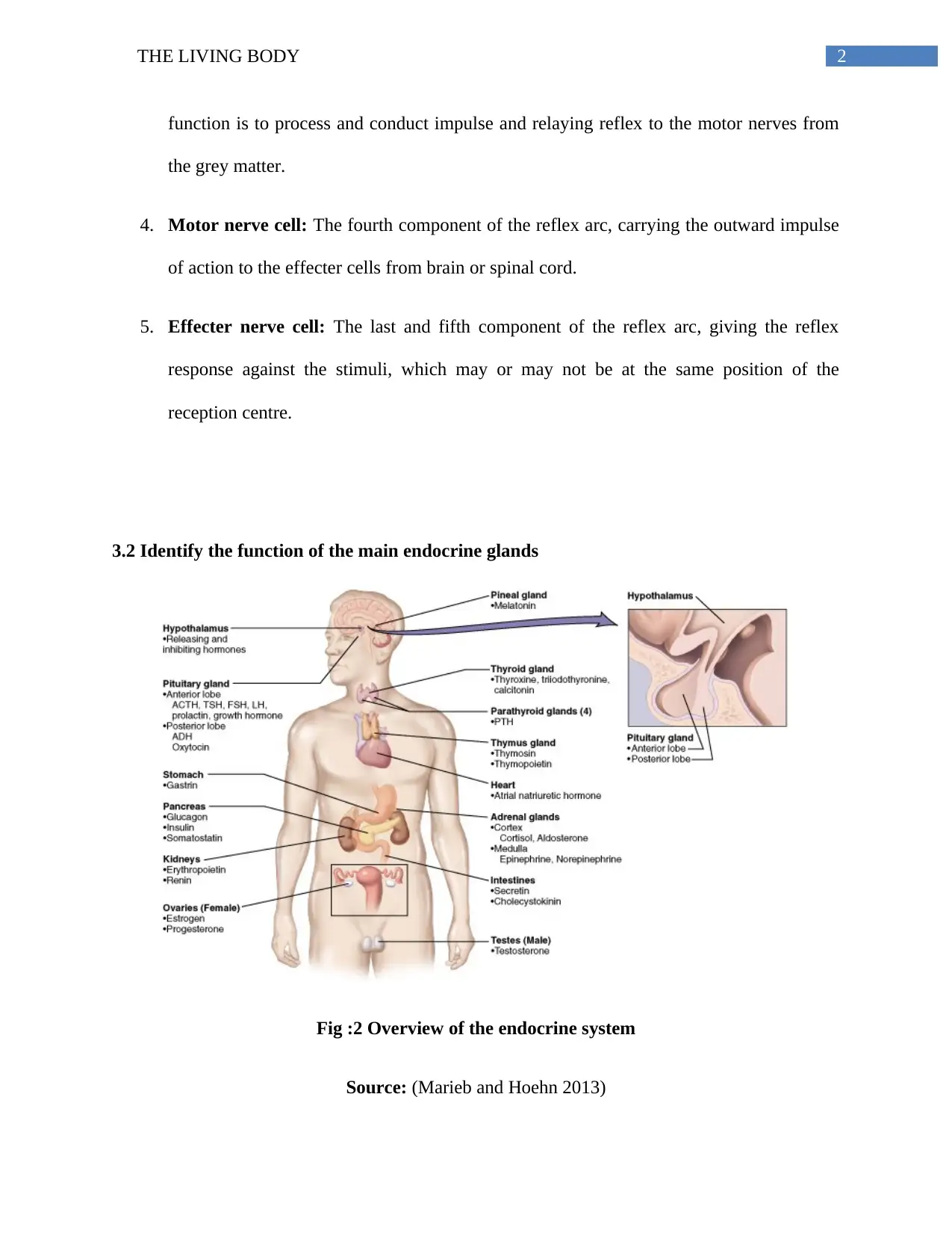
Fig :2 Overview of the endocrine system
Source: (Marieb and Hoehn 2013)
3THE LIVING BODY
Hypothalamus: activation, control and integration of the autonomic peripheral nervous system
and other bodily functions like biological clock, temperature and appetite.
Pituitary Gland: master gland controlling majority of the secretory hormones, body growth,
tissue development, excretion from kidney and sexual maturity.
Adrenal gland: secretion of glucocoticoids, small amount of androgen and oestrogen from
adrenal cortex. Adrenaline hormone secretion. The medulla secretes controls the sympathetic
nervous system and secretion of epinephrine and nor-epinephrine.
Thyroid gland: Controls body metabolic activity and thyroxin production.
Parathyroid gland: Ca2+ and K+ balance as well as parathyroid hormone secretion.
Pancreas: control of carbohydrate metabolism, fat metabolism influence and secretion of
glucagon and insulin.
Ovary and Testes:Production of progesterone and oestrogen. Testes produce testosterone,
which control the sperm development.
3.3 Describe how the nervous and endocrine systems work to coordinate the body systems
Maintenance of homeostasis, growth and reproduction is together controlled by both nervous and
the endocrine system. The hypothalamus is responsible for maintaining the homeostasis as it
regulates majority of the body functions, thermal regulation, body weight and other glandular
secretions, which is interact with the target areas via neuronal network, receive external stimuli
which in turn activates the endocrine systems. Responds to thermal changes in the environment
Secure Best Marks with AI Grader
Need help grading? Try our AI Grader for instant feedback on your assignments.
4THE LIVING BODY
is detected by the nervous system, which stimulates thyroid hormone secretion for controlling the
body temperature and maintaining homeostasis (McAninch and Bianco 2014). Lactation in a
female can be taken as a good example to explain the scenario. When a baby sucks on a mother’s
nipple the sensory nerves in that area receives an external secretory stimulus, which it transmits
into the hypothalamus for inducing the secretion of the hormone oxytocin. Blood vessels carry
the oxytocin hormone to the mammary glands, where the glands receive the secretory stimulus
and milk production is facilitated (Crowley 2014). In a similar case, as seen in pubation period,
the following hormones; testosterone and oestrogen regulate the control of one’s sexual maturity.
The nervous system detects sexual stimulus, transmits those signals into the hypothalamus, and
secretes testosterone, similarly in females the onset of puberty and those bodily stimuli is
detected by the nervous system that stimulates the hypothalamus for oestrogen production and
that causes ovulation followed by menstrual cycle (Plant, 2015). It is observed in case of higher
organisms that the nervous and the endocrine system successfully regulate billions of cells and
maintain the functions of the body. The nervous system detects these stimuli and forms
communications with the target cells with the help of neurotransmitters. The hormones secreted
by the endocrine system travels through the blood vessels into the target area and the target cells
take up these signalling molecules through the extracellular fluid and the nervous system utilize
paracrine signalling which interconnects the target cells with neuronal systems for the
transmission of nerve impulses by the process of neurotransmission (Alberts, 2015).
5THE LIVING BODY
4.1 Outline the structure and functions of the male and female human reproductive system
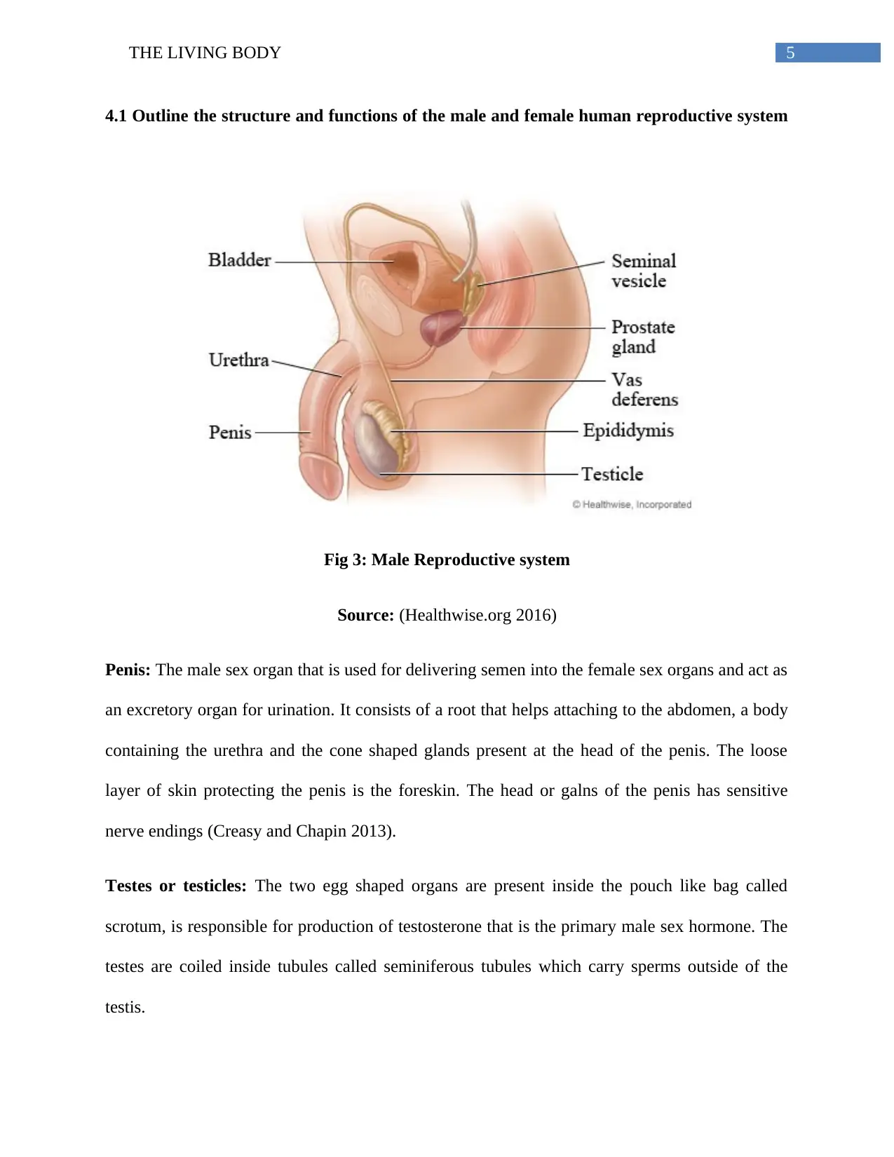
Fig 3: Male Reproductive system
Source: (Healthwise.org 2016)
Penis: The male sex organ that is used for delivering semen into the female sex organs and act as
an excretory organ for urination. It consists of a root that helps attaching to the abdomen, a body
containing the urethra and the cone shaped glands present at the head of the penis. The loose
layer of skin protecting the penis is the foreskin. The head or galns of the penis has sensitive
nerve endings (Creasy and Chapin 2013).
Testes or testicles: The two egg shaped organs are present inside the pouch like bag called
scrotum, is responsible for production of testosterone that is the primary male sex hormone. The
testes are coiled inside tubules called seminiferous tubules which carry sperms outside of the
testis.
6THE LIVING BODY
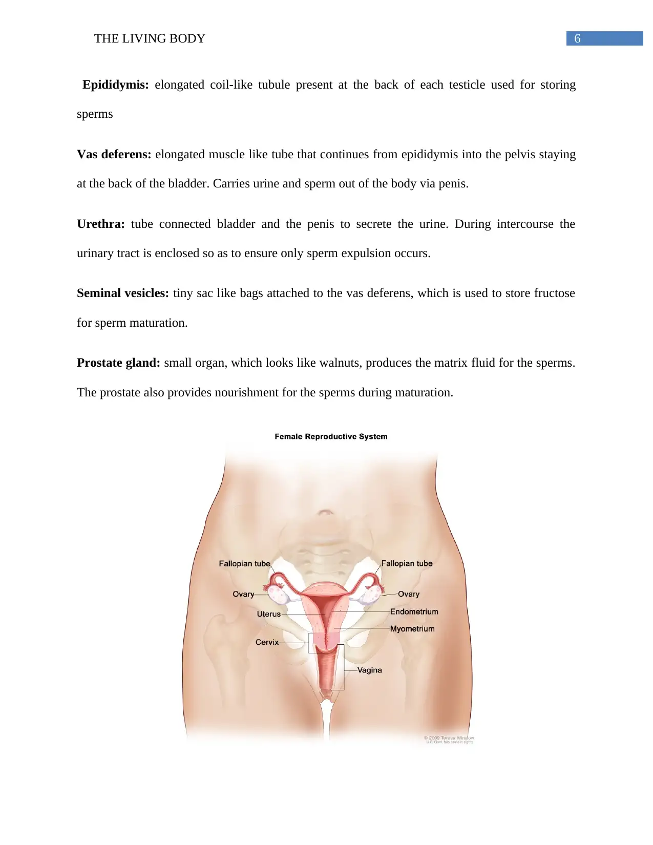
Epididymis: elongated coil-like tubule present at the back of each testicle used for storing
sperms
Vas deferens: elongated muscle like tube that continues from epididymis into the pelvis staying
at the back of the bladder. Carries urine and sperm out of the body via penis.
Urethra: tube connected bladder and the penis to secrete the urine. During intercourse the
urinary tract is enclosed so as to ensure only sperm expulsion occurs.
Seminal vesicles: tiny sac like bags attached to the vas deferens, which is used to store fructose
for sperm maturation.
Prostate gland: small organ, which looks like walnuts, produces the matrix fluid for the sperms.
The prostate also provides nourishment for the sperms during maturation.
Paraphrase This Document
Need a fresh take? Get an instant paraphrase of this document with our AI Paraphraser
7THE LIVING BODY
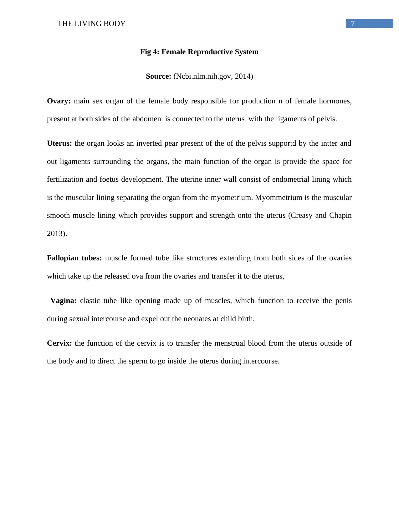
Fig 4: Female Reproductive System
Source: (Ncbi.nlm.nih.gov, 2014)
Ovary: main sex organ of the female body responsible for production n of female hormones,
present at both sides of the abdomen is connected to the uterus with the ligaments of pelvis.
Uterus: the organ looks an inverted pear present of the of the pelvis supportd by the intter and
out ligaments surrounding the organs, the main function of the organ is provide the space for
fertilization and foetus development. The uterine inner wall consist of endometrial lining which
is the muscular lining separating the organ from the myometrium. Myommetrium is the muscular
smooth muscle lining which provides support and strength onto the uterus (Creasy and Chapin
2013).
Fallopian tubes: muscle formed tube like structures extending from both sides of the ovaries
which take up the released ova from the ovaries and transfer it to the uterus,
Vagina: elastic tube like opening made up of muscles, which function to receive the penis
during sexual intercourse and expel out the neonates at child birth.
Cervix: the function of the cervix is to transfer the menstrual blood from the uterus outside of
the body and to direct the sperm to go inside the uterus during intercourse.
8THE LIVING BODY
References:
Alberts, B., 2015. Molecular biology of the cell. New York, NY [u.a.]: Garland Science
Creasy, D.M. and Chapin, R.E., 2013. Male reproductive system. In Haschek and Rousseaux's
Toxicologic Pathology (Third Edition) (pp. 2493-2598).
Crowley, W., 2014. Neuroendocrine Regulation of Lactation and Milk
Production. Comprehensive Physiology, pp.255-291.
Healthwise.org. 2016. Health Content and Patient Education Solutions - Healthwise. [online]
Available at: https://www.healthwise.org/ [Accessed 8 Apr. 2018].
Marieb, E., Hoehn, K. and Hutchinson, M., 2013. Human anatomy & physiology. [San
Francisco, Calif.]: Pearson Education/Benjamin Cummings.
McAninch, E.A. and Bianco, A.C., 2014. Thyroid hormone signaling in energy homeostasis and
energy metabolism. Annals of the New York Academy of Sciences, 1311(1), pp.77-87.
Ncbi.nlm.nih.gov. 2014. [Figure, Anatomy of the female reproductive...] - PDQ Cancer
Information Summaries - NCBI Bookshelf. [online] Available at:
https://www.ncbi.nlm.nih.gov/books/NBK65941/figure/CDR0000257805__158/ [Accessed 8
Apr. 2018].
Saladin, K., 2015. Anatomy & physiology. New York: McGraw-Hill.

9THE LIVING BODY
1 out of 10
Related Documents
Your All-in-One AI-Powered Toolkit for Academic Success.
+13062052269
info@desklib.com
Available 24*7 on WhatsApp / Email
![[object Object]](/_next/static/media/star-bottom.7253800d.svg)
Unlock your academic potential
© 2024 | Zucol Services PVT LTD | All rights reserved.


