Reflective Case Study: Sepsis Management and Care in Intensive Care
VerifiedAdded on 2023/06/13
|14
|3573
|231
Case Study
AI Summary
This nursing case study details the care provided to a 69-year-old male patient, Mr. X, admitted to the hospital with a history of mild COPD and presenting symptoms indicative of sepsis, including pyrexia, poor appetite, breathing difficulties, and chest pain. The study covers the patient's medical history, initial observations, and investigations leading to a diagnosis of sepsis complicated by right middle lobe streptococcus pneumonia. It delves into the pathophysiology of sepsis, focusing on the inflammatory process, acute stress response, and cytokine storm, explaining how these elements manifested in Mr. X's condition. The nursing physical assessment, problems identified, and goals of nursing care are outlined, along with the mechanical ventilation management employed. The case study critically analyzes the nursing interventions and multidisciplinary team efforts over a twelve-hour period, linking clinical observations with theoretical deductions to enhance understanding of the nursing process. Access more solved assignments on Desklib.

Running head: SEPSIS 1
Nursing Case Study
Student’s Name
University Affiliation
Course
Date
Nursing Case Study
Student’s Name
University Affiliation
Course
Date
Paraphrase This Document
Need a fresh take? Get an instant paraphrase of this document with our AI Paraphraser
SEPSIS 2
Introduction
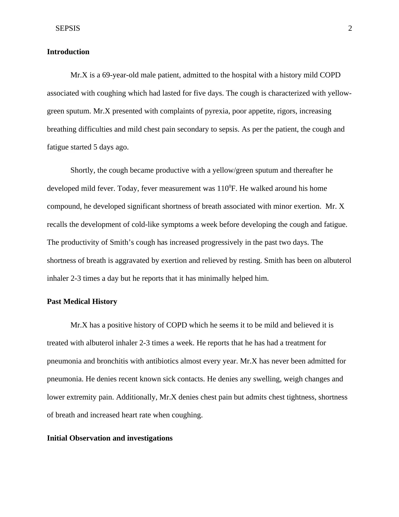
Mr.X is a 69-year-old male patient, admitted to the hospital with a history mild COPD
associated with coughing which had lasted for five days. The cough is characterized with yellow-
green sputum. Mr.X presented with complaints of pyrexia, poor appetite, rigors, increasing
breathing difficulties and mild chest pain secondary to sepsis. As per the patient, the cough and
fatigue started 5 days ago.
Shortly, the cough became productive with a yellow/green sputum and thereafter he
developed mild fever. Today, fever measurement was 1100F. He walked around his home
compound, he developed significant shortness of breath associated with minor exertion. Mr. X
recalls the development of cold-like symptoms a week before developing the cough and fatigue.
The productivity of Smith’s cough has increased progressively in the past two days. The
shortness of breath is aggravated by exertion and relieved by resting. Smith has been on albuterol
inhaler 2-3 times a day but he reports that it has minimally helped him.
Past Medical History
Mr.X has a positive history of COPD which he seems it to be mild and believed it is
treated with albuterol inhaler 2-3 times a week. He reports that he has had a treatment for
pneumonia and bronchitis with antibiotics almost every year. Mr.X has never been admitted for
pneumonia. He denies recent known sick contacts. He denies any swelling, weigh changes and
lower extremity pain. Additionally, Mr.X denies chest pain but admits chest tightness, shortness
of breath and increased heart rate when coughing.
Initial Observation and investigations
SEPSIS 3
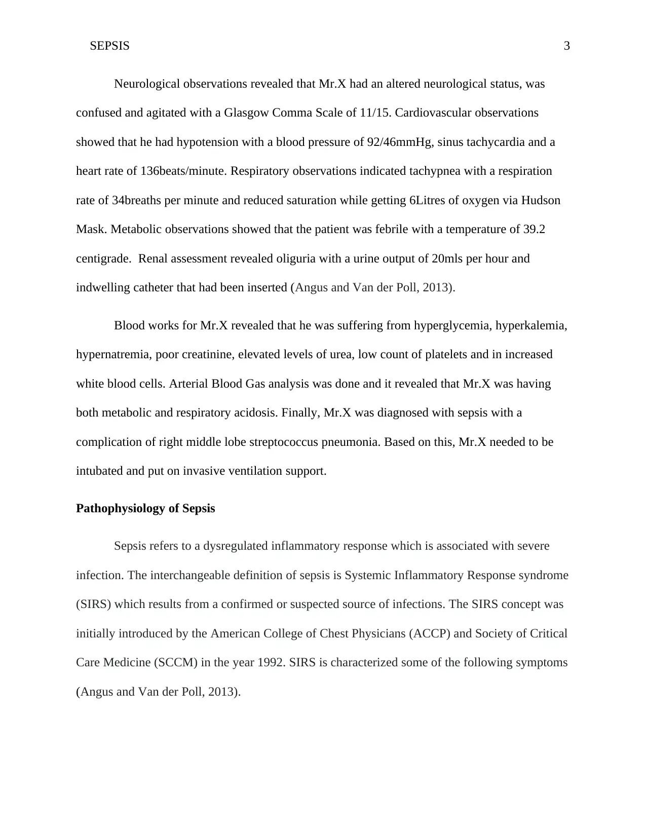
Neurological observations revealed that Mr.X had an altered neurological status, was
confused and agitated with a Glasgow Comma Scale of 11/15. Cardiovascular observations
showed that he had hypotension with a blood pressure of 92/46mmHg, sinus tachycardia and a
heart rate of 136beats/minute. Respiratory observations indicated tachypnea with a respiration
rate of 34breaths per minute and reduced saturation while getting 6Litres of oxygen via Hudson
Mask. Metabolic observations showed that the patient was febrile with a temperature of 39.2
centigrade. Renal assessment revealed oliguria with a urine output of 20mls per hour and
indwelling catheter that had been inserted (Angus and Van der Poll, 2013).
Blood works for Mr.X revealed that he was suffering from hyperglycemia, hyperkalemia,
hypernatremia, poor creatinine, elevated levels of urea, low count of platelets and in increased
white blood cells. Arterial Blood Gas analysis was done and it revealed that Mr.X was having
both metabolic and respiratory acidosis. Finally, Mr.X was diagnosed with sepsis with a
complication of right middle lobe streptococcus pneumonia. Based on this, Mr.X needed to be
intubated and put on invasive ventilation support.
Pathophysiology of Sepsis
Sepsis refers to a dysregulated inflammatory response which is associated with severe
infection. The interchangeable definition of sepsis is Systemic Inflammatory Response syndrome
(SIRS) which results from a confirmed or suspected source of infections. The SIRS concept was
initially introduced by the American College of Chest Physicians (ACCP) and Society of Critical
Care Medicine (SCCM) in the year 1992. SIRS is characterized some of the following symptoms
(Angus and Van der Poll, 2013).
⊘ This is a preview!⊘
Do you want full access?
Subscribe today to unlock all pages.

Trusted by 1+ million students worldwide
SEPSIS 4
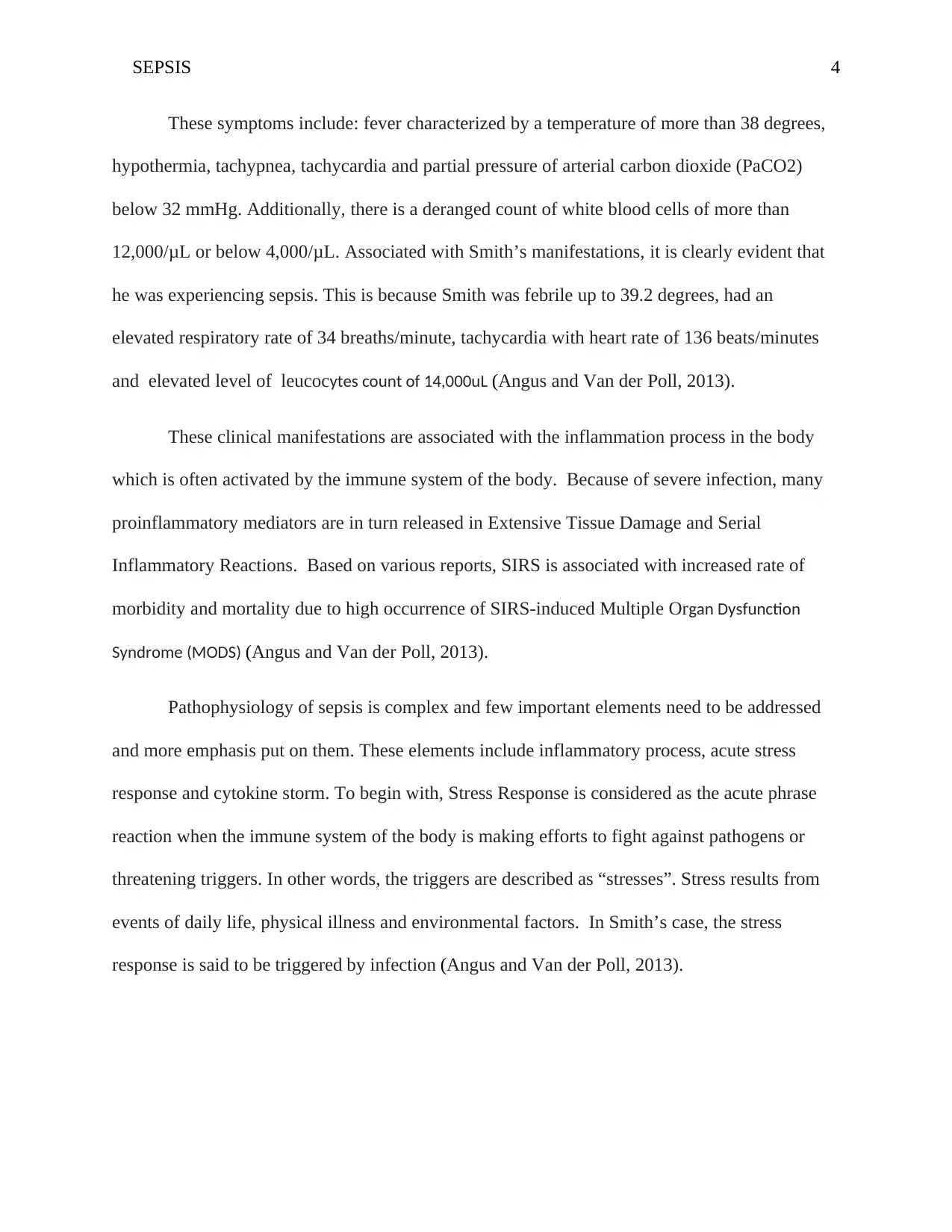
These symptoms include: fever characterized by a temperature of more than 38 degrees,
hypothermia, tachypnea, tachycardia and partial pressure of arterial carbon dioxide (PaCO2)
below 32 mmHg. Additionally, there is a deranged count of white blood cells of more than
12,000/μL or below 4,000/μL. Associated with Smith’s manifestations, it is clearly evident that
he was experiencing sepsis. This is because Smith was febrile up to 39.2 degrees, had an
elevated respiratory rate of 34 breaths/minute, tachycardia with heart rate of 136 beats/minutes
and elevated level of leucocytes count of 14,000uL (Angus and Van der Poll, 2013).
These clinical manifestations are associated with the inflammation process in the body
which is often activated by the immune system of the body. Because of severe infection, many
proinflammatory mediators are in turn released in Extensive Tissue Damage and Serial
Inflammatory Reactions. Based on various reports, SIRS is associated with increased rate of
morbidity and mortality due to high occurrence of SIRS-induced Multiple Organ Dysfunction
Syndrome (MODS) (Angus and Van der Poll, 2013).
Pathophysiology of sepsis is complex and few important elements need to be addressed
and more emphasis put on them. These elements include inflammatory process, acute stress
response and cytokine storm. To begin with, Stress Response is considered as the acute phrase
reaction when the immune system of the body is making efforts to fight against pathogens or
threatening triggers. In other words, the triggers are described as “stresses”. Stress results from
events of daily life, physical illness and environmental factors. In Smith’s case, the stress
response is said to be triggered by infection (Angus and Van der Poll, 2013).
Paraphrase This Document
Need a fresh take? Get an instant paraphrase of this document with our AI Paraphraser
SEPSIS 5
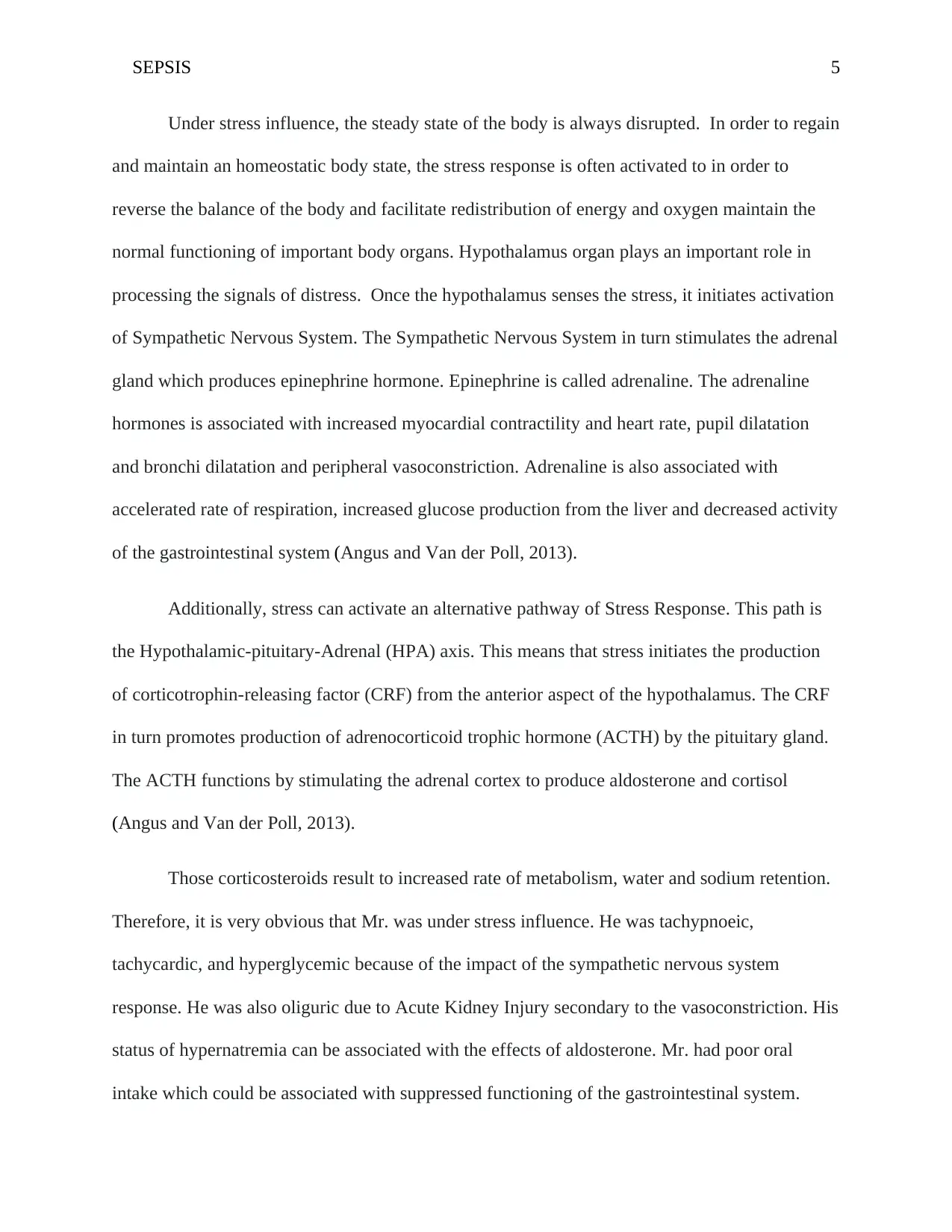
Under stress influence, the steady state of the body is always disrupted. In order to regain
and maintain an homeostatic body state, the stress response is often activated to in order to
reverse the balance of the body and facilitate redistribution of energy and oxygen maintain the
normal functioning of important body organs. Hypothalamus organ plays an important role in
processing the signals of distress. Once the hypothalamus senses the stress, it initiates activation
of Sympathetic Nervous System. The Sympathetic Nervous System in turn stimulates the adrenal
gland which produces epinephrine hormone. Epinephrine is called adrenaline. The adrenaline
hormones is associated with increased myocardial contractility and heart rate, pupil dilatation
and bronchi dilatation and peripheral vasoconstriction. Adrenaline is also associated with
accelerated rate of respiration, increased glucose production from the liver and decreased activity
of the gastrointestinal system (Angus and Van der Poll, 2013).
Additionally, stress can activate an alternative pathway of Stress Response. This path is
the Hypothalamic-pituitary-Adrenal (HPA) axis. This means that stress initiates the production
of corticotrophin-releasing factor (CRF) from the anterior aspect of the hypothalamus. The CRF
in turn promotes production of adrenocorticoid trophic hormone (ACTH) by the pituitary gland.
The ACTH functions by stimulating the adrenal cortex to produce aldosterone and cortisol
(Angus and Van der Poll, 2013).
Those corticosteroids result to increased rate of metabolism, water and sodium retention.
Therefore, it is very obvious that Mr. was under stress influence. He was tachypnoeic,
tachycardic, and hyperglycemic because of the impact of the sympathetic nervous system
response. He was also oliguric due to Acute Kidney Injury secondary to the vasoconstriction. His
status of hypernatremia can be associated with the effects of aldosterone. Mr. had poor oral
intake which could be associated with suppressed functioning of the gastrointestinal system.
SEPSIS 6
The second element of the pathophysiology of sepsis is the, the inflammatory cascade
which plays a crucial role in the pathophysiology of Systemic Inflammatory Response
Syndrome. The mediators of inflammation process are related to the mechanisms involved in
Systemic Inflammatory Response. Excessive production of pro-inflammatory mediators lead to
inflammation and inhibit the functioning of the Compensatory anti-Inflammatory Response. Pro-
inflammatory mediators eventually compromise the body’s immune system.
Cytokines are one of the vital elements of the immune system. Local cytokines are
immediately activated following an insult to repair the wounds and stimulate the innate immune
system. Following release of local cytokines, some of the cytokines get into circulation system.
Presence of cytokines in the circulation promotes release of growth factors and adhesion of
platelets and macrophages which help in local damage recovery. However, severe infection and
homeostasis that cannot be restored lead to occurrence of cytokine storm (Angus and Van der
Poll, 2013).
Cytokine storm specifically results from complex progression. Cytokines are always
made by monocytes, macrophages, platelets, mast cells and endothelial cells which make up the
components of the initial immune defensive system. The multitude of cytokines induces
Interleukin-1 (IL-1) and cytokine Tissue Necrosis Factor-alpha (TNF-a) and interleukin-1 (IL-1).
These elements lead to removal of the Nuclear Factor-KB (NF-KB) Inhibitor which
consequently prompts the release of more proinflammatory mediators like interferon gamma,
Interleukin-8 (IL-8) and interleukin-6 (IL-6). Generally, cytokines do initiate the production of
immune cells, which then induce more cytokines in the circulation system (Angus and Van der
Poll, 2013).
⊘ This is a preview!⊘
Do you want full access?
Subscribe today to unlock all pages.

Trusted by 1+ million students worldwide
SEPSIS 7
The cytokines greatly impact the body. This includes indirect and direct contribution of
morbidity and mortality in SIRS. TNFa is associated with fever clotting problems, abnormal
hemodynamic values, low count of white blood cells and elevated level of liver enzymes. IL-1 is
connected to loss of appetite, fever, haemodynamic abnormalities, general body weakness,
neutrophilia and headache. IL-6 has strong relationship with impaired functioning of the lungs
and fever. It also acts as a severity determinant of mortality rate associated with SIRS (Angus
and Van der Poll, 2013).
The massive cytokine accumulation can lead to wide spreading vasodilatory impacts.
This is because they stimulate the release of some vasodilators like nitric oxide. To add on this,
cytokines are known for promoting adhesion of the endothelial cells and immune cells which
lead to leaky endothelium and fluid loss from intercellular space to the extracellular space. The
cytokines cascade can lead to disorders of clotting due to the high fibrinogen concentration
involved in the process of inflammation. The fibrinogen produced is always converted from
thrombin, generated by tissue factor. Tissue factor refers to the substance expressed by the
surface of white blood cells. The tissue factor can also be induced by endotoxin and TNFa from
the infection. The fibrinogen can be converted into fibrin forming clots. Therefore, excessive
amount of fibrin during inflammation process leads to extensive clotting disorders (Angus and
Van der Poll, 2013).
Based on all these mechanisms, it can be concluded that Mr.X’s fever is highly likely
related to the production of Interleukin-1, TNFa and interleukin-6. Interleukin-1 could be the
likely contributor of his reduced appetite and elevated count of white blood cells. Interleukin-6
could lead to worsening of Mr.X’s already affected lung functioning. Mr.X had increased count
Paraphrase This Document
Need a fresh take? Get an instant paraphrase of this document with our AI Paraphraser
SEPSIS 8
of white blood cells which could have been contributed by the interleukin-1 and immune
response.
The hypotension (92/46mmHg) could be associated with the effects of vasodilation due
to the effect of the Sympathetic nervous system. Hypotension led to reduced kidney perfusion
hence causing acute kidney failure and reduced urinary output (20mls/hour). The acute kidney
injury may consequently affect the patterns of potassium elimination so that Mr.X was
discovered having high levels of potassium. The low count of platelets could be associated with
to the massive cytokine production and endothelium damage.
Primary Nursing Physical Assessment
Upon assessment, Mr.X was agitated, confused with a GCS of 12/15. He was febrile with
a temperature of 39.1 centigrade secondary to the inflammation process and cytokine production
involved in sepsis. He had poor urine output with 20mls/hour following hypotension which led
to reduced kidney perfusion hence low urinary output. Mr.X had an increased heart rate (134
beats/minute) and respiration rate (34 breaths/minute) following stimulation of the sympathetic
nervous system and metabolic and respiratory acidosis. Production of cytokines could also ahve
led to compromised lung functioning hence leading to dyspnea. Additionally, dyspnea was also
related to right lobe streptococcus pneumonia which led to compromised functioning of the lung
leading to shortness of breath and coughing.
The main problems for Mr.X were hyperthermia, hypoxia, impaired gas
exchange/dyspnea and risk of fluid volume deficit. The Based on assessment, the goal of nursing
of nursing care was to ensure that the patient displays respiratory rate and Arterial Blood Gas
Analysis (ABGs) within their normal ranges, with clear breath sounds and he experiences no
SEPSIS 9
dyspnea. The patient will also experience no complications associated with hyperthermia and
demonstrate a temperature within the normal range of values. The problems that were addressed
were impaired gas exchange and hyperthermia.
Mechanical Ventilation Management
Mechanical Ventilation Management of Mr.X involved use of Synchronized Intermittent
Mandatory Ventilation Volume Control. SIMV is used very commonly in Intensive Care Units
(ICU). Using the volume control mode, Mr.X was given ventilation support using a set of tidal
volume in mandatory breaths. For an effective ventilation support, I had to set up some specific
values for the SIMV volume-controlled mode. The values were respiratory and tidal volume.
Tidal volume is an amount of oxygen given by the machine or that amount in the lungs when a
patient breathes without support. The respiratory rate has to be set for mandatory breaths. In the
SIMV volume controlled mode, the ventilator or patient triggered ventilation.
The SIMV mode helped Mr.X reduce the energy required in breathing especially in his
high energy-consuming status of sepsis. The model also allowed him get an extra breath in order
to expel the accumulative level of carbon dioxide which led to respiratory acidosis hence
improving the acidosis. Additionally, due to pneumonia and thee accumulative inflammatory
response syndrome, the lungs could be fragile and stiff secondary to mass accumulation of
cytokines and the inflammation process. Therefore, the volume controlled ventilator acted as
protective strategy to prevent complications related to ventilators such as volutrauma. The tidal
volume was set at 9ml/kg to avoid ventilator-related complications.
⊘ This is a preview!⊘
Do you want full access?
Subscribe today to unlock all pages.

Trusted by 1+ million students worldwide
SEPSIS 10
Profoundly, ventilation can compromise cardiovascular functioning via conflicting,
complex, and opposite processes. The processes give a reflection of the interaction between
ventricular pump function, myocardial reserve, blood flow distribution, circulating blood
volume, autonomic tone, lung volume, endocrinologic responses, intrathoracic pressure (ITP),
and the surrounding pressures for the remaining part of circulation.
Mechanical ventilation generates an increased airway pressure hence increased in
intrathoracic pressure. This may consequently may decrease intra-abdominal organ and systemic
perfusion, which may significantly affect the homeostasis an organ functioning. Therefore,
critically ill patients can develop a Systemic Inflammatory Response hence culminating to
multiple-organ dysfunction syndrome and mortality. Therefore, I closely monitored the patient
while on mechanical ventilation.
Since Mr.X was on intubation and mechanical ventilation, I had to implement some
interventions to prevent aerodigetive tract colonization which result severe complications
associate with ventilation. These interventions included avoidance of unprescrtibed antibiotics,
chlorhexidine mouth rinsing, stress ulcer prophylaxis, selective digestive decontamination and
short-course parenteral prophylactic antibiotics in high-risk patients. I also avoided use of
weaning protocols, semi-recumbent positioning, and sub-glottic suctioning.
Obviously, a very a key element is the haemodynamic monitoring, which has been very
vital in the assessment of the patient. According to literature, dynamic and ongoing
haemodynamic monitoring is vital in judging the response to the treatment modalities and fluid
challenge. Mechanical ventilation induces cyclic variations in blood flow in the vena cava,
pulmonary artery and aorta. At the bedside, changes in respiration in aortic blood flow are
Paraphrase This Document
Need a fresh take? Get an instant paraphrase of this document with our AI Paraphraser
SEPSIS 11
indicated by “swings” in blood pressure whose magnitude is dependent on volume status (Green,
2015).
Nursing interventions
From the assessment the patient had a temperature of 39.20C which hyperthermic. The
aim of nursing care was to ensure that patient demonstrates a normal temperature and prevent
any complications associated with hyperthermia. To achieve this, the following interventions
were implemented; Monitoring his temperature–degree and pattern. A temperature of 38.90C to
41.20C it’s a suggestion of acute infection process. Consistent fever lasting for more than 24 ours
is an indication of pneumococcal pneumonia. Administering antipyretics like acetaminophen.
Antipyretics are useful in reducing fever due to its central action on the hypothalamus (Green,
2015).
Regarding impaired gashouse exchange, the goal of nursing care was to ensure that the
patient displays respiratory rate and Arterial Blood Gas Analysis (ABGs) within normal range,
with clear breath sounds. The patient should demonstrate reduced dyspnea. To achieve this goal,
I monitored rate and depth of respirations noting use of accessory muscles when breathing.
Rapid and shallow respirations often occur due to stress, hypoxemia and circulating endotoxins.
Dyspnea and hypoventilation indicate ineffective compensatory mechanisms hence indicating
the need for ventilator support. I auscultated the patient for breath sounds noting stridor,
crackles, wheezes and areas of absent or decreased ventilation. Presence of respiratory distress
and adventitious sounds indicate lung atelectasis, pulmonary congestion and interstitial edema. I
assessed Mr.X for sensorium changes. Cerebral functioning is sensitive to decreased oxygenation
such as reduced perfusion or hypoxemia (Green, 2015).
SEPSIS 12
I maintained the airway of the patient by positioning him comfortably with head elevated
at 30degrees. Head elevation promotes lung expansion hence reducing respiratory effort. I noted
coughing and production of purulent sputum. Pneumonia is one of the commonest hospital-
acquired infection that occurs via aspiration of oropharyngeal microorganisms. I frequently re-
positioned Mr.X and encouraged him to cough and do breathing exercises. I also suctioned him
as per the indications. Good pulmonary toilet is vital in reducing perfusion/ventilation
imbalance. This also facilitated removal of secretions from the airway to ensure maximum gas
exchange (Green, 2015).
I also monitored pulse oximetry and ABGs for Mr.X. Hypoxemia is associated with
decreased rate of ventilation and changes in the pulmonary system such as interstitial edema,
atelectasis or pulmonary shunting. Hypoxemia is also associated with increased demand for
oxygen which is caused by infection or fever. Respiratory acidosis characterized by a pH of
below 7.35 and PaCO2 more than 40 mm Hg, happens because of ventilation-perfusion
imbalance and hypoventilation. As septic condition deteriorates, metabolic acidosis characterized
by a pH of below 7.35 and HCO3 below 22-24 mEq/L, develops due to accumulation of lactic
acid from anaerobic metabolism (Green, 2015).
Regarding medical treatment, Mr.X had been prescribed intravenous antibiotics by the
physician such as vancomycin to treat pneumonia and infections of the skin and soft tissue.
These antibiotics were administered following the nursing protocol of drug administration. The
patient was also on vasopressin which was meant to constrict hence helping in increasing blood
pressure to increased tissue-perfusion. Management of Mr.X was multidisciplinary whereby the
nurse, doctor, nutritionist and counsellor were involved to give holistic care to the patient
(Green, 2015.
⊘ This is a preview!⊘
Do you want full access?
Subscribe today to unlock all pages.

Trusted by 1+ million students worldwide
1 out of 14
Related Documents
Your All-in-One AI-Powered Toolkit for Academic Success.
+13062052269
info@desklib.com
Available 24*7 on WhatsApp / Email
![[object Object]](/_next/static/media/star-bottom.7253800d.svg)
Unlock your academic potential
Copyright © 2020–2026 A2Z Services. All Rights Reserved. Developed and managed by ZUCOL.


