Perceptions of Indian Radiographers about Patient Care and
VerifiedAdded on 2022/08/12
|57
|13384
|19
AI Summary
Perceptions of Indian radiographers about patient care and safety in MRI: A case study conducted in a medical imaging center, Kerala.
Contribute Materials
Your contribution can guide someone’s learning journey. Share your
documents today.
Perceptions of Indian Radiographers about Patient Care and Safety In MRI
Name
Institution
Tutor
Date
Name
Institution
Tutor
Date
Secure Best Marks with AI Grader
Need help grading? Try our AI Grader for instant feedback on your assignments.
Abstract
Since the 1990s, MRIs have gained significant popularity in healthcare settings and
research. However, is not properly conducted, MRI procedures may have significant
adverse effects on the patients, or the imaging personnel. Therefore, adequate safety
measures must be adhered to. The aim of the study is to explore the perceptions of
Indian radiographers about patient safety and care in MRI. Descriptive and survey
research designs were applied to address the research objectives. Data was collected
from 29 radiographers working with MRIs, who were selected from different health
imaging facilities within India. The findings of the study indicated that radiographers
perceive patient safety and care as a priority in their practice. Also, demographic factors
apart from education did not affect the radiographers’ view of patient health and safety.
Education determined the levels of skills and knowledge regarding MRI safety
procedures and standards. Further, the work environment was a key determinant of
adherence or radiographers to safety guidelines. The radiographers understand and
uphold the importance of safety and care of the patient before, during and after the
imaging procedures.
Table of Contents
Abstract..............................................................................................................................2
CHAPTER ONE: INTRODUCTION...................................................................................5
1.Introduction………………………………......................................................................5
1.1 Preventing Decontamination, Infection and Hospital-Acquired Infections..................6
1.3 Objectives…………………………….........................................................................8
CHAPTER 2: LITERATURE REVIEW.............................................................................10
2.Introduction……………………………........................................................................10
2.1 Risks Associated with MRI.....................................................................................10
2.2 Prevention of MRI ...…………………………………………………………………....11
2.2.1 Prevention by Restriction and Access Control................................................12
2.2.2 Risks Related To the Refrigerant.....................................................................13
2.2.3 Risks Related To Magnetic Field Gradients....................................................14
2.2.4 Risks Related to Antennas...............................................................................15
2.3 Responsibilities of the Radiologist.........................................................................17
CHAPTER 3: RESEARCH METHODOLOGY.................................................................20
3.Introduction………………………………....................................................................20
3.1 Research Design………………………...................................................................21
3.2 Population of the Study…………............................................................................22
3.3 Sampling Technique……………….........................................................................22
3.4 Instruments…………………………........................................................................23
3.4.1 Questionnaire...................................................................................................23
3.4.2 Interviews.........................................................................................................24
3.5 The validity of Instruments (Pilot study).................................................................25
3.6 Data Collection Procedures...................................................................................25
3.7 Methods of Data Analysis......................................................................................26
3.7.1 Quantitative Data Analysis...............................................................................26
3.7.2 Qualitative Data Analysis.................................................................................27
3.8 Ethical Consideration.....…………………………………………………………….....28
CHAPTER 4. RESULTS AND FINDINGS.......................................................................28
4.0 Demographic Characteristics.................................................................................28
4.1 Findings from the Questionnaire............................................................................31
4.1.2 Part A...............................................................................................................31
4.1.3 Part B...............................................................................................................32
4.1.4 Part C...............................................................................................................32
4.2 Interview Findings……………….............................................................................32
4.2.1 Phase 1: Planning the MRI examination.........................................................33
4.2.2 Phase 2: Producing Correct Images................................................................35
4.2.3 Phase 3: Evaluating the MRI examination.......................................................36
CHAPTER 5. DISCUSSION............................................................................................38
CHAPTER 6. CONCLUSION..........................................................................................39
References.......................................................................................................................41
Secure Best Marks with AI Grader
Need help grading? Try our AI Grader for instant feedback on your assignments.
CHAPTER ONE: INTRODUCTION
1.Introduction
In the recent two decades, the safety and care of patients have managed to garner a lot
of attention. The Institute of Medicine report published in 1999, "To Err is Human",
indicated that between 44,000 and 98000 patients die in American Hospitals as a result
of preventable errors (Lark, Kirkpatrick, and Chung, 2018). Notably, many broad
epidemiological types of research have claimed that a made or the increased level of
risk is mainly because of mistakes that occur within the current medical environment
(Hartwig et al. 2009; Ko and Yu, 2015; Mello and Hemenway, 2004 and; Hiivala et al.,
2016). Improving the safety level of patients has become paramount within all
healthcare services and procedures. A recent case study revelation of medical
misconduct involving MRI, in a Taiwan hospital, clearly states that the risk sources tend
to lack adequate clinical training and inaccurate and insufficient communication
amongst the MRI staff and clinical team (Lee, Lin and Chan, 2015).
There is a rising consideration of MRI by practitioners due to the exquisite soft-tissue
delineation provided by the imaging modality. The MRI process involves exposure to
powerful static magnetic fields and gradient fields and radiofrequency energy. Without
going into the details and technical aspects of its operation, an MRI machine generates
magnetic fields to provide high definition images of all parts of the human body (Kanal
et al. 2007; Sammet 2016). The advantage compared to other imaging devices, it emits
no radiation that could harm patients or professional users. Its major drawback is that it
is a very powerful electromagnet which necessarily requires perfect control of the
environment near the installation. It is essential for staff, visitors and patients to
understand the primary hazards associated with the environment and select the safest
and rigorously protection procedures to follow (Durbridge 2011). Within the Indian
context, key safety considerations that are related to the patients are associated with
magnetic resonance imaging (MRI) should always consider the following aspects
(Durbridge 2011).
The associated behaviour of ferromagnetic objects when they are exposed to a
robust magnetic field as the forces can react over the ferromagnetic implant
thereby, resulting in it to transit in a different direction causing injury or potential
death. Additionally, many external ferromagnetic objects might also get
influenced by the strong magnetic fields and tend to become airborne and will
transmit rapidly towards the isocenter of the magnetic. This situation might also
cause injury or death (Durbridge, 2011).
Gradient or static magnetic fields might also have an impact over other medical
devices, whether external or implanted and can cause them to malfunction
(Durbridge 2011).
Acoustic sounds that are associated with frequently switching gradient coils can
also be a severe risk for the patients. This can be avoidable through
appropriately using noise reduction and hearing protection technologies
(Durbridge 2011).
The risks associated with radiofrequency (RF) include heating up of tissues
because of RF energy deposition, which can be measured as the specific
absorption rate (SAR). Resultantly, this is more evident because of the
increasing field strengths mainly because of the increased frequency of the RF
pulses. Additionally, the energy of RF can also be deposited within wires, cables,
tattoos and skin patches causing these elements to get heated and potentially
result in burns to the patient (Callaghan et al. 2019; Durbridge 2011; Takahashi,
Fujimoto, Hamada, Tezuka, and Tanaka, 2016).
1.1 Preventing Decontamination, Infection and Hospital-Acquired Infections
As per a study undertaken by the Centres for Disease Control and Prevention (CDC),
basic control over infections within hospitals must include all the general principles to
avoid transmission within all patient care, as well as specific ways for preventing that,
are suspected or known to be infected within a transmittable microorganism (Centers for
Disease Control and Prevention, 2017). General principles to be applied by healthcare
professionals include:
Using personal and protective equipment at the time of exposure to that of
infectious material (Centers for Disease Control and Prevention, 2017);
Performing hand hygiene and using all five moments (World Health Organization,
2018) before and after touching the patient and appropriately using the solutions
for washing hands or disinfection (Centers for Disease Control and Prevention,
2017);
Assure staff awareness about issues that are related to patients in isolation
particularly, the contact and respiratory types of isolation (Centers for Disease
Control and Prevention, 2017);
Paraphrase This Document
Need a fresh take? Get an instant paraphrase of this document with our AI Paraphraser
Assuring safer procedures for injections and follow the rule 'one syringe, one
needle and one use' (Centers for Disease Control and Prevention, 2017).
Handling laundry garments and textiles very carefully and dispose of them off per
the hospital policy (Centers for Disease Control and Prevention, 2017);
Ensuring cleanliness of the materials related to sterilization and disinfection as
well as the environment of the patient (Centers for Disease Control and
Prevention, 2017).
Transmission prevention processes should also be used within patients who have been
infected or occupied with particular infectious agents and which include (Centers for
Disease Control and Prevention, 2017):
Precautions undertaken during contact in case of contact transmission occur in a
patient. This is achieved through isolating the patient, making use of personal
protective equipment, making use of disposable equipment and assuring
surfaces are appropriately cleaned.
Airborne precautionary measures in case infectious microorganisms transmit
through airborne routes like measles, tuberculosis or chickenpox. Droplet
precaution can be used along with restricting susceptible healthcare
professionals to access the room by immunizing all susceptible people.
Droplet precautions can be used for agents that are transmitted by respiratory
droplets such as talking, coughing or sneezing. This can be attained through the
continuous usage of facemasks, using personal protective equipment as well as
limiting patient transport and movement.
1.2 Aim
Patient care and safety is a priority in healthcare settings (Aspden Corrigan and
Wolcott, 2004; Levine et al. 2007). Radiologists with healthcare units all over India that
are involved in MRI needs to be aware of these considerations and should, as such,
undertake appropriate safety procedures that must be in place. With millions of MRI
scans being performed Indian hospitals coupled with growth within the MRI activity
between 2011 and 2015 (Arora, 2014), the need for ensuring best practice for the care
and safety of patients across the country has become empirical. The research study
aims to determine the perception of Indian Radiographers for MRI patient safety and
care.
1.3 Objectives
The survey aims to meet the following objectives:
1. To assess the knowledge of Indian radiographers regarding patient care and
safety in MRI procedures.
2. To investigate the perceptions of Indian Radiographers for regarding patient care
and safety in MRI procedures.
CHAPTER 2: LITERATURE REVIEW
2.Introduction
There have been continuous and rapid developments in MRI techniques that have
widely been applied in the past two decades. The technical developments are
demonstrated by the optimization and improvements in conventional MRI techniques
such as MR fingerprinting and compressed sensing MRI and the new pulse sequences
such as DWIBS (Diffusion-weighted Whole-body Imaging with Background body signal
Suppression) and CEST-MRI (Chemical Exchange Saturation Transfer MRI). The rapid
development in MRI techniques has resulted in an increased application of MRI
techniques and the emergence of new knowledge in biomedical research and clinical
sciences (Dong, Andrews, Xie, and Yokoo, 2015). With the wide adoption and utilization
of MRI, patient safety and risk associated with the modality has been a significant issue
of concern for practitioners and researchers (Andersen, 2007; Lee, Lin and Chan,
2015).
2.1 Risks Associated with MRI
Any human endeavour creates risks. This is particularly the case for health-related
activities and medical imagery. The proper functioning of an imaging firm or service is
closely linked to controlling the risks that its activity generates. As patient and
professional safety is a fundamental issue in imaging procedures, it is, therefore,
essential to continually comply with safety rules and to prevent risks (European
Medicine Agency, 2017)
Secure Best Marks with AI Grader
Need help grading? Try our AI Grader for instant feedback on your assignments.
The projectile effect is certainly one of the most important risks in the MRI. The list of
objects responsible for accidents associated with MRI is long. It involves retaining
oxygen shells, helium cans, equipment for monitoring and assistance in resuscitation,
scissors, reflex hammers, stethoscopes, mobile phones, barrettes, keys, wheelchairs.
When they enter the magnetic field, these objects undergo a violent attraction and reach
speeds of several meters per second (Cross, Hoff, and Kanal, 2018).). The main
accidents reported in the literature concern projectiles with a large mass, such as
oxygen shells. In 2001, a 6-year-old boy died in the United States from head trauma
following a shock from a ferromagnetic oxygen shell that penetrated the magnet at high
speed (Bellin et al., 2002). Other cases of oxygen shells projected in an MRI, with
material damage, but without victims have been reported in the literature. If the danger
increases with the mass of the projectile, even small objects such as coins can be
thrown into the magnet and injure the patient.
Another risk concerns metallic implants as well as implantable medical devices or even
intracorporeal metallic foreign bodies. In such a case, the associated threat comes from
displacements or rotational movements of these foreign bodies or implants.
Haemorrhages caused by the displacement of intracranial ferromagnetic vascular clips,
intraocular metallic foreign bodies, bullets or fragments of shells resulting from to
exposure electromagnetic field causes significant harm. Malfunctions of certain
implanted devices (pacemakers, neurostimulators, cochlear implants, insulin pumps can
cause considerable injury (International Organization for Standardization, 2012).
2.2 Prevention of MRI Risks'
The time-varying gradient fields, main magnetic fields involved, and radiofrequency
energy involved in MRIs require a variety of unique safety considerations. The
practitioner who works in the MRI environment needs to understand the
translation/torque, projectile effect, human health effects and personal items required for
the MRI environment (Durbridge, 2011). There are various strategies for achieving
safety in the MRI environment, such as restricting access to the area, taking precaution
when handling equipment and providing all safety information before entering into the
area.
2.2.1 Prevention by Restriction and Access Control
Accessing the room where the examination will take place should be restricted to
people who are not aware of the relative constraints associated with intense magnetic
fields. The danger it represents is always materialized by signs placed over the
examination room door, or even of the surrounding premises. The magnetic field limit of
0.5 Millitesla (mT) is considered to be an "exclusion zone" prohibited for people with
particular active medical devices (pacemaker for example), ferromagnetic objects and
certain electronic equipment. Such a limitation is generally a part of the magnet room
because of the progress of the shielding: "active" shielding at the level of the magnet
(additional magnetic field coils) and "passive" shielding fixed on the walls, ceilings or
floor if necessary (metal plates). If this might not be the case with higher fields, this limit
might be marked over the ground (Crook and Robinson, 2009; Expert Panel on MR
Safety et al. 2013).
Access control is generally provided by the radiography experts working in the MRI
environment (Consiglio 2006). It concerns both personnel (paramedical, medical,
maintenance personnel, maintenance personnel, etc.) and patients. Anyone entering
the examination room must have all ferromagnetic metallic objects they carry removed.
It is essential to check that they are not carrying a prohibited medical device in the 0
zones, 5 mT and, finally, have him deposit his goods (watch, credit card, mobile phone,
etc.). They must also have them remove jewellery, removable dentures and any object
harmful for the quality of exploration (artifacts). A meticulous search for
contraindications by a questionnaire, oral or written, is an essential prerequisite for
carrying out the examination. All MR personnel must go through an MR-screening
process as an important component of their employment interview to ensure the safety
of MR environments (Expert Panel on MR Safety et al. 2013; Sawyer‐Glover and
Shellock, 2000).
In the field of implanted materials, a compatibility check with the maker is many times
required. Certain devices usually contraindicated (for example, pacemakers) can cause
possible adverse effects for the staff and the patient when used in MRI. For instance, for
implants that have strong ferromagnetism, the rotational and translational forces in the
MRI procedure may reach the implant and dislodge or move the device from its position
(Expert Panel on MR Safety et al. 2013). If there is any doubt about the possible
presence of an implant, an active implantable medical device or metal shards which
would constitute contraindications, an x-ray of the region can be performed. Finally, for
accompanying persons who may attend the examination in the room (which is frequent
Paraphrase This Document
Need a fresh take? Get an instant paraphrase of this document with our AI Paraphraser
in pediatric MRI), the same precautions should be taken as for the personnel (Mayeda-
Letourneau, 2014).
2.2.2 Risks Related To the Refrigerant
Prevention within this aspect consists essentially of periodically checking the existing
installation to check that there is no leakage or obstruction at the end of the tube.
Besides, staff training within emergency processes is very important. Indeed, in the
event of incomplete or total failure of the quench tube, and, consequently, of the release
of gaseous helium in the examination room, the patient must first be evacuated, then
operate the ventilation in extraction mode to expel the gas from the room. Lastly, the
quench is voluntarily provoked by pressing the "magnet stop" switch in the event of an
accident with the risk of projection of metallic elements into the magnet, in the event of
injury or blockage of the patient or personnel, but also the event of a fire in the room.
However, this procedure which brutally "stops" the magnetic field should only be used
as a last resort (The Society and College of Radiographers, 2018).
2.2.3 Risks Related To Magnetic Field Gradients
Magnetic field gradients have two effects for the patient:
Peripheral Nerve Stimulation. They manifest as tingling or a slight muscle twitch in
certain areas of the body. This effect is the result of certain arrangements (in particular
echo-planar or PPE) during which the switching of the gradients is exceedingly fast,
which eventually results in low-frequency electromagnetic fields which can produce
currents in the tissues (hence neurostimulations) (Woodward, 2001).
Preventing this phenomenon requires compliance with the patient's installation
procedures. In particular, contact with the hands, knees or feet should be avoided as it
leads towards a closed and conductive loop favouring the instruction of an electric
current within the nerve fibres (and therefore stimulation). It should also be assured that
the patient does not wear any metallic object or conductive material favouring the
induction of electric current.
Also, the devices generally offer power supervision of the gradients at two levels, the
reduced level reducing the risk of generating neurostimulations. In either case, the
radiologist should pay close attention to all the sensations that are felt by the patient in
order, if necessary, to stop the acquisition sequence (Gilk et al., 2006).
Acoustic Noise. Principles of the operation of the gradient coils, such a noise arises
because of the vibration of gradient coils following the injection of electric current. The
noise emitted is comparative to the strength of the electric current and the intensity of
the magnetic field, ensuring high noise level whenever the major magnetic field
increases.
Here too, prevention comes first and foremost through proper care of the patient with
the installation of earplugs or earmuffs, or both. In general, reduces noise by around 30
to 35 dB. From a technological point of view, might also include different methods to
reduce noise, either software or hardware sequences.
2.2.4 Risks Related to Antennas
The application of radiofrequency pulses during the acquisition sequences, i.e. high-
frequency electromagnetic fields produced by the transmitting antenna, causes energy
to be transmitted into the tissues the result of which can be overheating. Subsequently,
this disposition of energy is measured by the SAR (Specific Absorption Rate), which is
calculated in W / kg. The international standard IEC 60601-2-33 of July 1995 sets the
energy transmission limits aimed at not exceeding a body temperature increase of 1 °
C. In practice, the SAR depends on the parameters of the sequences used, in particular
the number of radiofrequency pulses (in sequences of rapid spin echo type and
derivatives) or the tilt angle of these pulses (Gupta et al., 2007).
The transmitting antenna can be the source of another risk: as a matter of fact, the
meditation of the radiofrequency field in certain skin areas can result in second as well
as third-degree burns. Such a danger is magnified by the presence of electric cables
which forms the shape of loops over the skin (ECG cables, antenna cables, etc.), by
contact between a conductive metallic element and the skin (for example patches on
skin containing a metallic foil or piercings) and by areas of contact with the skin forming
a conductive loop (for example at the level of the calves, thighs, or crossed arms)
(Viscuse, Khasawneh, and Constantinou, 2015; Takahashi et al. 2016).
The prevention of tissue overheating is mainly ensured by software fitted to each
device, which continuously calculates the SAR according to the parameters of the
sequences used. The operator is thus alerted to the modifications to be implemented in
the event of the threshold being exceeded. Furthermore, avoid switching to high energy
transmission mode for young children as well as for patients with hyperthermia or
whose thermoregulatory capacities are impaired. Care should also be taken not to cover
patients too much and to ensure good ventilation of the tunnel (Expert Panel on MR
Safety 2013).
Secure Best Marks with AI Grader
Need help grading? Try our AI Grader for instant feedback on your assignments.
To avoid the risk of skin burns, never leave an antenna cable in contact with the
patient's skin. The operator must also take care to ensure a minimum distance of 5 mm
between the patient and the lining of the tunnel, confirm the absence of metallic
conductive element in contact with the skin and avoid contact with skin areas (risk of
burns at the points of contact). As always, the operator must remain attentive to the
patient (call bulb) by asking him to report any sensation of abnormal heat (Expert Panel
on MR Safety 2013).
The American College of Radiology (ACR) report released in 2013 provided simple and
reasonable steps that can be applied in the reducing injury during MRI. The steps
include removing all unnecessary electric conductive material external to the patient and
placing thermal insulation between the patient and any electrically conductive material.
Other measures include setting wires and leads far from the RF coils within the inner
wall, ensuring that the tissues of the patient do not form conductive loops by reducing
contact between parts of the skin in areas imaged. Radiographers should also use ice
packs and cold compresses for leads that are electrically conductive and need to be
patient's contact during imaging, in unresponsive or unconscious patients, and concern
areas such as tattoos. Also, it is crucial to continually monitor the patient during the MRI
procedure for any discomfort or overheating sensation (Jankharia, 2008; Viscuse et al.,
2015).
2.3 Responsibilities of the Radiologist
The pre-examination interview is the ideal time to provide information to the patient, in a
personal way and according to the type of examination prescribed for it. It is important
to check if the patient is wearing makeup, hairstyling devices (e.g. wig), jewellery or
clothing that is too thick, which may affect its security or cause artifacts in the images.
The heating of metals, in contact with the patient's skin, is to be prevented (Elliot, 2006).
Radiologists have a unique role in ensuring the safety of their patients. According to
Srinivas (2004) and kalyanpur (2008) a radiologist in India must:
Explain the nature of examination in details including duration, goals, positioning,
noises and vibration heard during the examination and respiratory phases linked
to the examination apnea to such the patient or, if applicable, the companion.
If necessary, consult the patient's radiological file, prior to the examination;
If the patient is unable to communicate or is not accompanied, carry out a visual
examination of it, in order to detect any scars (signs of surgery not mentioned or
born which could involve a risk linked to metal);
Must provide for the cancellation of the examination or screening for foreign
bodies by radiography
Notify if there is risk material present in the patient;
Warn the patient of the risks linked to metal and the possibility of overheating
(during examination sequences) metal objects that cannot be removed from the
body. In that case, prevent external metallic objects from coming into direct
contact with the patient's skin;
Inform patients with tattoos or permanent makeup of the risk of burns associated
with these elements;
As a preventive measure, a cold water compress can be applied to the tattooed
area during the examination to reduce the risk of burns;
Ask the patient to mention (by the warning button) if they feel a burning
sensation. If such is the case, the exam should be stopped. The radiologist will
conduct the test after thorough examination;
The radiologist also assures that all transdermal patches (e.g. nicotine,
nitroglycerin) are removed from the patient's skin before the examination, even if
these patches are not ferromagnetic (a metallic component is found in some
stamps and may heat up during the exam);
The radiologist will also avoid covering the patient too much during the
examination, given the rise in body temperature caused by radiofrequency waves
emitted during the sequences;
Maintain visual and auditory contact with the patient, especially during the
acquisition of images and when moving the table;
Suggest to the patient, or to any other person who must remain in the magnet
room during the sequences, hearing protection offering a reduction of at least
twenty decibels, except in the case where the service can prove that the noise
level does not exceed 85 dB.
The radiologist also assures that hearing protectors are used correctly. Anyone
refusing hearing protection should sign a refusal form;
If necessary, remove the baby's identification bracelet to proceed with the MRI
examination and act in accordance with the rules in force in the establishment.
Paraphrase This Document
Need a fresh take? Get an instant paraphrase of this document with our AI Paraphraser

CHAPTER 3: RESEARCH METHODOLOGY
3.Introduction
The research methodology chapter discusses the methodology, research design,
instruments and procedures used by the researcher in data collection and analysis in
the study in order to achieve the desired objective of the study. The main aim of the
study is to investigate the perceptions of Indian radiographers about patient safety in the
MRI. Research methodology refers to a systematic approach used by a researcher in
the evaluation of a given research problem, find solutions and obtain relevant answers
regarding the given investigation (Chakravarthy, Kumar, and BIST, 2018). This entire
procedure is considered as rigorous and systematic and will eventually lead toward
acquiring new knowledge (Jamshed, 2014). Basic objectives of a research methodology
are to explain, assess, control as well as forecast facts, behaviour and phenomena.
It is very important to understand the perceptions of Indian radiographers in a reliable
way so that the research is valid and adds in value to the particular research aspect.
Throughout this chapter, the methodology and procedures that have been used for the
exploration of the underlying phenomenon will be discussed in detail. The chapter will
focus on research design, selecting and profiling the respondents or participants of the
research, instrumentation, data collections and analysis.
In such cases, qualitative methods are considered to be very helpful to create or
develop reasonable structures so to provide an alternate route for the development of
theories by exploring phenomena in their natural settings (Creswell and Creswell, 2017;
Gunnell, 2016; Rahman, 2016; Yilmaz, 2013). These approaches can be used as a part
of refining existing literature and theories (Thompson, Gamble and Strickland, 2004).
3.1 Research Design
Aggarwal and Ranganathan (2019) described research design as the collective method
and procedures for gathering and analyzing specified variable data in a particular study
question. The researcher will use a descriptive research design to investigate the
perceptions of Indian radiographers' on patient safety in MRI. Descriptive research
design describes the demographics of participants or phenomena in the research
(Atmowardoyo, 2018). Descriptive research designs discuss the distribution of different
variables in a given study without focusing on the causes of the phenomena (Aggarwal
and Ranganathan (2019). Descriptive research design enables the researcher to
investigate the relationship between radiographers' demographics such as age and the
perceptions of radiographers on the safety of patient undergoing MRI. Many times the
research design also includes using visual aids such as charts and graphs so as
provide ease to the reader in the understanding of data distribution. Descriptive
research designs are cost-effective, quick and have lenient ethical consideration except
for studies seeking to collect information of confidential nature such as substance use
and sexual practices (Aggarwal and Ranganathan, 2019). Descriptive research design
is suitable in this research since the study that will seek only to understand
radiographers' perception of the safety of patient undergoing MRI used and is not be
meant to cause any prejudice to the participants, and the identity of the participants will
not be disclosed.
The study incorporated both qualitative and quantitative user research design using
structured interviews and descriptive surveys. A structured interview is a process in
which the interviewee is guided throughout the interview in a specific pattern and in
Secure Best Marks with AI Grader
Need help grading? Try our AI Grader for instant feedback on your assignments.
such a way that the information received is in alignment with the research objectives
(Pontin, 2000). In addition to this, a descriptive study provides a meaningful way of
assessing and investigating the behaviour and attitude of people when they are a part of
a specific situation (Carter, 2000 and; Gray, 2004).
3.2 Population of the Study
The population in the study refers to all the elements or individuals for which the
researcher seek generalization. Researchers need to draw a small sample of fairly
eligible participants from the target population (Asiamah et al., 2017; Martínez-Mesa et
al., 2016; Martirosyan et al. 2010). The population in the study included radiographers in
India. Purposive sampling was used to detect eligible participants from the entire
population of Indian radiographers.
3.3 Sampling Technique
When a research study makes use of a smaller data set, the inconveniences can be
overcome rather quickly and effectively which occurs because of homogeneity, lack of
time as well as improving the quality and accuracy of the data (Atkinson, 2000; Aderet
al., 2008). The sampling technique chosen for the study was purposive sampling.
Purposive sampling is a technique used to select information-rich cases that can
provide required information regarding the phenomena under investigation. The cases
identified through purposive sampling are available, experienced, knowledgeable and
willing to participate in the study (Moser and Korstjens, 2018; Palinkas et al., 2015;
Sargeant, 2012). The advantage of using purposive sampling is that researchers gain
an in-depth understanding of the phenomena (Gentles et al., 2015). Purposive sampling
was essential for selecting participants from a population of Indian radiographers.
The partner organization contact method was essential for the identification and
recruitment of participants (McCullagh, Sanon, and Cohen, 2014). The researcher
contacted five healthcare organizations to seek permission to conduct the research
within their premises. The partner organization contact method is important for
identifying participants that are difficult to reach and involves facilitation by
organizational management or leadership (Ellard-Gray et al. 2015; Alice, Palakiko,
Daniggelis, and Makahi, 2015). The researcher engaged with the managers in five
imaging centres within India who assisted in the identification of information-rich
radiographers who have specialized in MRI. The working relationship established
between the managers and the organizational leaders helped in building trust and
confirming issues of confidentiality and avoiding information misrepresentation.
3.4 Instruments
A self-administered survey questionnaire were used for attaining data from that of the
participants. The purpose was to achieve additional data, verify as well as validate
results obtained from the research (Polgar and Thomas, 2000). Formation and
supervision of policy implementation appear to be the responsibility of the hospital and
its management (Beddoe et al., 2004). As a result, conducting interviews with the
management members was rendered to be necessary. It was considered to be an
effective way of obtaining comprehensive and detailed information related to safety
management.
3.4.1 Questionnaire
The research questionnaire was filled up by the radiographers and was categorized into
three parts. Part A included different questions related to the personal information of the
research participants like their age, sex, education level and their knowledge about the
safety and care of the patients along with the effects of the magnetic field. The
questions in part A were essential for identifying the relationship between demographic
characteristics and the perceptions of radiographers regarding patient safety.
In Part B, radiographers were required to fill up their qualifications, frequency and
reasons for repeated radiologic examinations over patients as well as make patients
alert about the dangers associated with the application and exposure towards
established safety rules. The questions in Part B were important in understanding the
actual perceptions of radiographers regarding patient safety, and the actions they take
towards the realization of patient safety.
Part C fielded questions related to the characterization of the radiographers' workplace.
This included determining safety measures related to work accidents and errors while
concluding this section with patient safety rules. The questions triggered responses to
gain information regarding the perception of radiographers on how the work
environment affects their adherence to patient safety rules and over-all patient safety.
3.4.2 Interviews
Studies on the uses of individual research interviews in the research studies have
revealed a wide variety of research practices and ways of reporting these practices in
empirical articles (Royer, Baribeau and Duchesne, 2009). Semi-structured interviews
Paraphrase This Document
Need a fresh take? Get an instant paraphrase of this document with our AI Paraphraser
are flexible as they contain open-ended questions that help to provide to gather in-depth
information regarding the study phenomena (Alshenqeeti, 2014; Casey and Houghton,
2010; Walshe, Ewing, and Griffiths, 2011). The analysis indicates that researchers use
more than thirty interview techniques that they use to know and understand phenomena
and, even, to demonstrate the existence of some of their aspects. Among other results,
it is also noted that several sampling techniques constituting samples of extremely
varied sizes (from three to more than 150 participants) are used. While they allow a
better understanding of the outline of research practices by interview, these
observations also raise questions about the use of research interviews in the
disciplines. To see more clearly, it has been studied more specifically as to how
interviews occupy a central place and play a preponderant role in terms of
instrumentation in a research study (Tierney and Dilley, 2002). The research conducted
semi-structured interviews to gain an in-depth understanding of the perceptions of
radiographers regarding patient safety in MRI procedures in India.
3.5 The validity of Instruments (Pilot study)
Within this research report, the pilot testing of research instruments has been employed
so as to determine all misunderstandings and uncertainties, if any within every item.
Pilot studies help in the identification of unclear questions and wrongly worded
statements to improve research validity (Bolderston, 2012; In, 2017; Vogel and Draper-
Rodi. 2017). Also, it helps the researcher to understand the feasibility of the interview.
The process of pilot testing was to pay attention to instances when participants
hesitated in answering or were willing to ask clarification about the questions. This can
serve as an indication that questions are vague, are difficult for the participants to
understand or can generate more than one meaning. Noting down such instances also
took place.
Three participants were selected from the sample and issued with questionnaires. They
were taken through the interview process. The pilot results study were examined and
thoroughly checked as to whether there is any need for modification. The improvement
was made accordingly. Also, the items on the questionnaire were assessed carefully
after which vague and incorrect statements will be rectified before the actual study takes
place. The pilot results were not used as part of the final research.
3.6 Data Collection Procedures
When the validity and reliability of the data collection instruments were done and
considered to be valid, the process of orientation was undertaken for administering the
research instrument - questionnaires. The researcher was responsible for administering
the research instruments. During the administration of research instruments, the period
of orientation was extended within which the actual reason of conducting this research,
and ethical considerations were explained so that the respondents could gain thorough
knowledge and information either collectively or over an individual basis within their
respective roles.
To achieve successful interviews, excellent rapport development practices were
undertaken with the participants so that valuable information and experience sharing
can take place (Bell, Fahmy, and Gordon, 2014; Elmir et al. 2011; McCullagh et al.
2014; Sanjari et al., 2014; Sivell et al., 2019). Additionally, research protocol and ethical
considerations have also been discussed and explained to the interviewee before
proceeding with the interview. Furthermore, the research also closely considers the
ethical priority of this research report and for this purpose; all the interviewees that were
a part of this study were presented anonymous names. Moreover, every interview was
conducted over an irregular basis, including some separate interview sessions as well,
which can have a duration of at least about 20 minutes at an average. Document
analysis was also done to link the research findings with existing literature.
3.7 Methods of Data Analysis
The data of this research has been attained from several types of research instruments,
and the same has been examined through implementing various methods that were
purely based upon the specific nature of the data.
3.7.1 Quantitative Data Analysis
The different types of percentages are used to explain the underlying characteristics of
the respondents of the study as well as determining the difference between the
responses that come from a different group of participants as well as highlighting
significant differences amongst the responses of the research participants of the other
group. On the other side, the types of means are used to calculate the average of the
class size.
3.7.2 Qualitative Data Analysis
Document analysis involves the use of secondary data, which appropriate as it
uncovers unexpected situations that occur in the real world, and contains detailed
documentation (Barnes, Dang, Leavitt, Guarana, and Uhlmann, 2015; Cheng and
Phillips, 2014; Dunn, Arslanian-Engoren, DeKoekkoek, Jadack, and Scott, 2015).
Secure Best Marks with AI Grader
Need help grading? Try our AI Grader for instant feedback on your assignments.
Thematic analysis is an easy and suitable data analysis method. The data attained from
the questionnaires were rearranged in an ordered way to work upon improving its
processing in the Statistical Package for Social Sciences (SPSS) version 16.0. Ordinal
and nominal levels of measurement were made part of the study mainly because the
research involves descriptive survey (Burns and Grove, 2001). The results were
presented through descriptive statistics in the form of percentages, frequency tables
and charts. To cross-check and supplement the data attained through reviewing all the
above-mentioned research instruments, current along with other related documents
such as the MRI patient history record, policy documents, records that pertain the safety
guidelines needs to be implemented in the MRI unit Codes are created for categorizing
collected data and different elements like behaviours, attitude towards different
situations and activities were coded accordingly.
3.8 Ethical Considerations
According to Polgar and Thomas (2000), within any type of scientific research, a
primary factor that needs to be carefully considered is the protection of the rights and
welfare of the participants. Human participants in qualitative research are the sources of
data and researchers have the responsibility for protecting them from potential harm
(Cosac, 2017; McKenna and Gray, 2018; Sanjari et al., 2014; Yip, Han, and Sng, 2016).
The ethical considerations are presented in the Belmont report, which asserts that
ethical research is based on three principles namely autonomy, respect for persons and
beneficence (Parker, 2016; Nijhawan et al., 2013; Sanjari et al., 2014; Tajir, 2018). As a
result, ethical approval was sought from the Institutions Review Board (IRB). All
participants were approached to provide informed consent in the form of written forms of

consent after the research objective has been clearly explained to them. Moreover, the
participants of the study were assured of the confidentiality of the identity, anonymity, as
well as the information provided.
participants of the study were assured of the confidentiality of the identity, anonymity, as
well as the information provided.
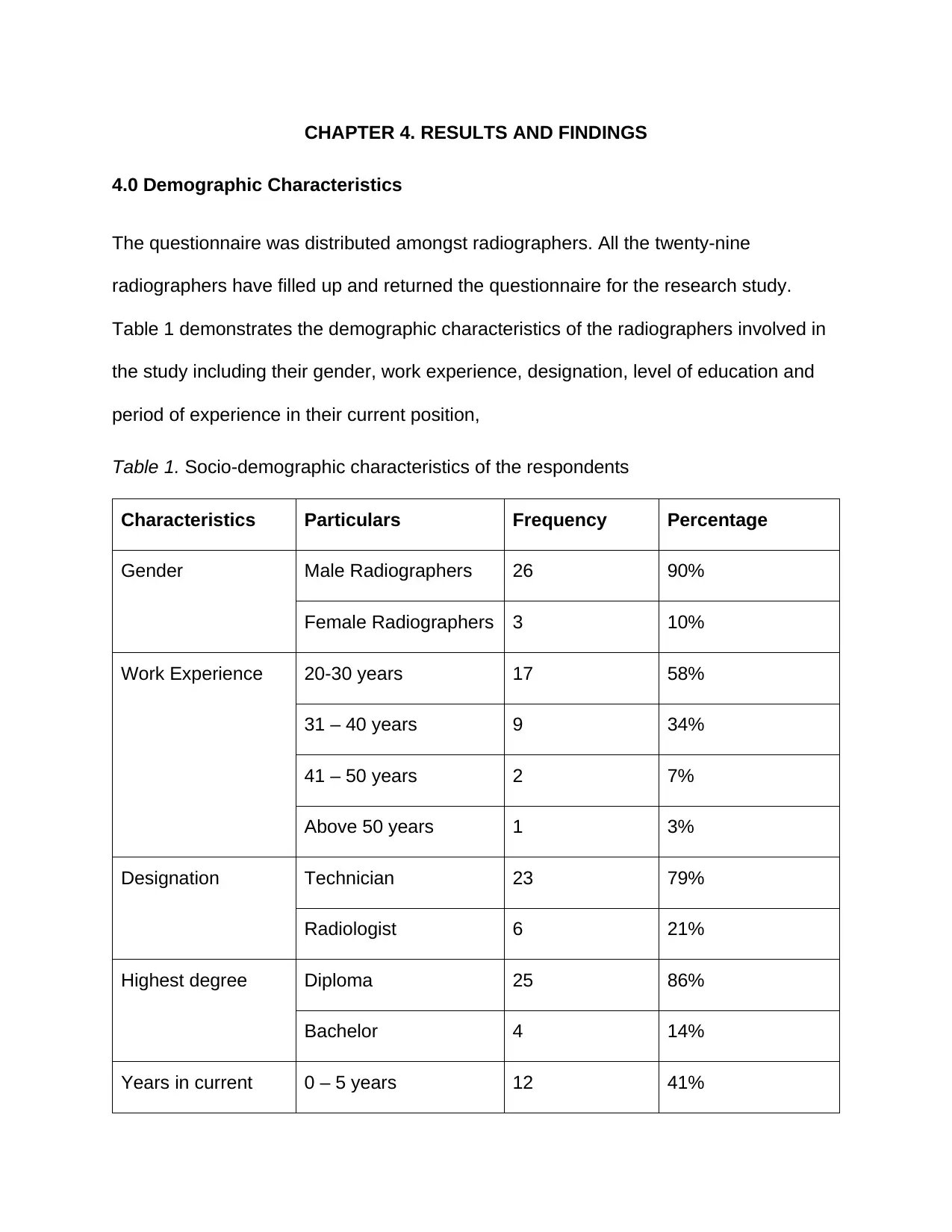
CHAPTER 4. RESULTS AND FINDINGS
4.0 Demographic Characteristics
The questionnaire was distributed amongst radiographers. All the twenty-nine
radiographers have filled up and returned the questionnaire for the research study.
Table 1 demonstrates the demographic characteristics of the radiographers involved in
the study including their gender, work experience, designation, level of education and
period of experience in their current position,
Table 1. Socio-demographic characteristics of the respondents
Characteristics Particulars Frequency Percentage
Gender Male Radiographers 26 90%
Female Radiographers 3 10%
Work Experience 20-30 years 17 58%
31 – 40 years 9 34%
41 – 50 years 2 7%
Above 50 years 1 3%
Designation Technician 23 79%
Radiologist 6 21%
Highest degree Diploma 25 86%
Bachelor 4 14%
Years in current 0 – 5 years 12 41%
Paraphrase This Document
Need a fresh take? Get an instant paraphrase of this document with our AI Paraphraser
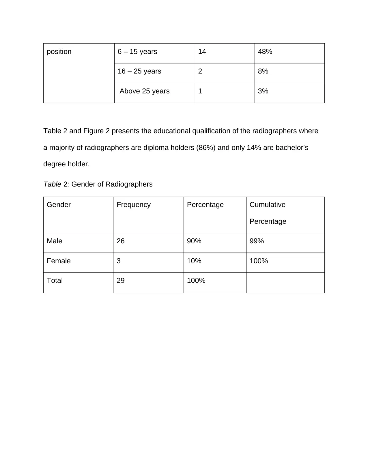
position 6 – 15 years 14 48%
16 – 25 years 2 8%
Above 25 years 1 3%
Table 2 and Figure 2 presents the educational qualification of the radiographers where
a majority of radiographers are diploma holders (86%) and only 14% are bachelor’s
degree holder.
Table 2: Gender of Radiographers
Gender Frequency Percentage Cumulative
Percentage
Male 26 90% 99%
Female 3 10% 100%
Total 29 100%
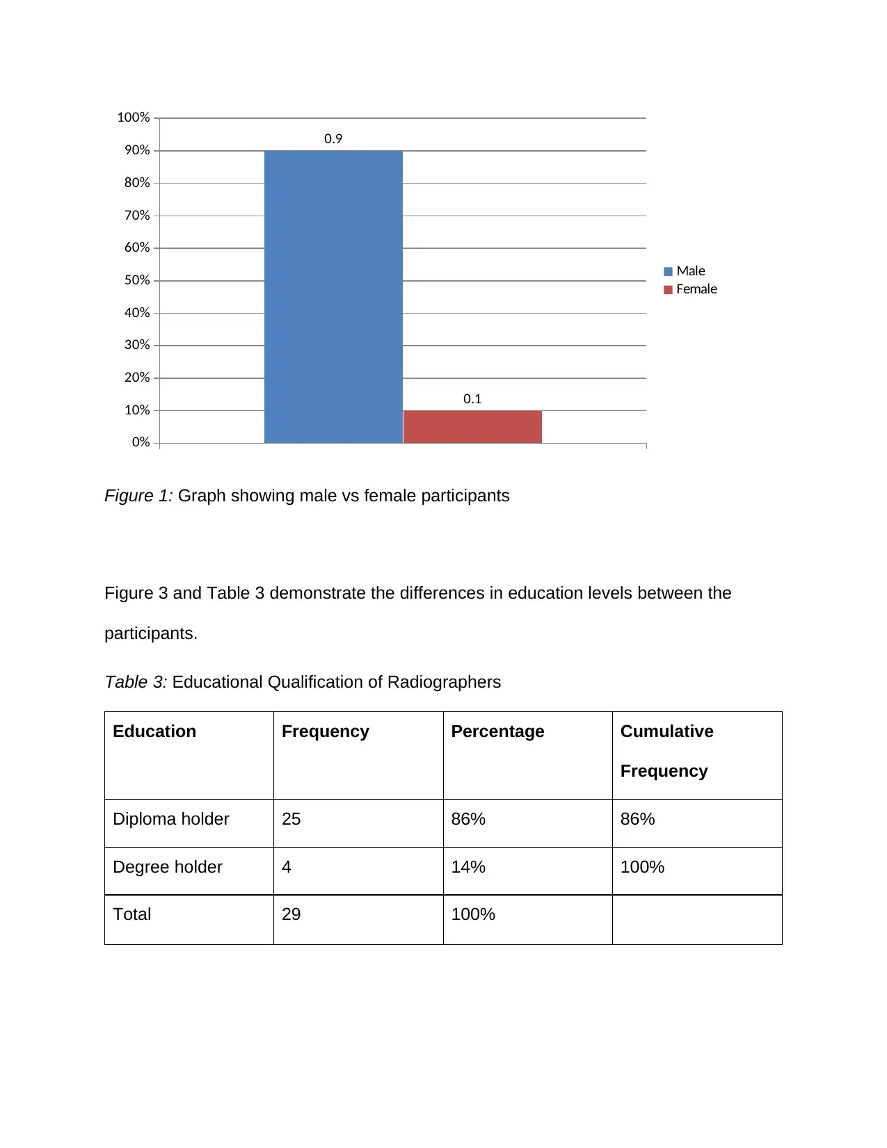
0%
10%
20%
30%
40%
50%
60%
70%
80%
90%
100%
0.9
0.1
Male
Female
Figure 1: Graph showing male vs female participants
Figure 3 and Table 3 demonstrate the differences in education levels between the
participants.
Table 3: Educational Qualification of Radiographers
Education Frequency Percentage Cumulative
Frequency
Diploma holder 25 86% 86%
Degree holder 4 14% 100%
Total 29 100%
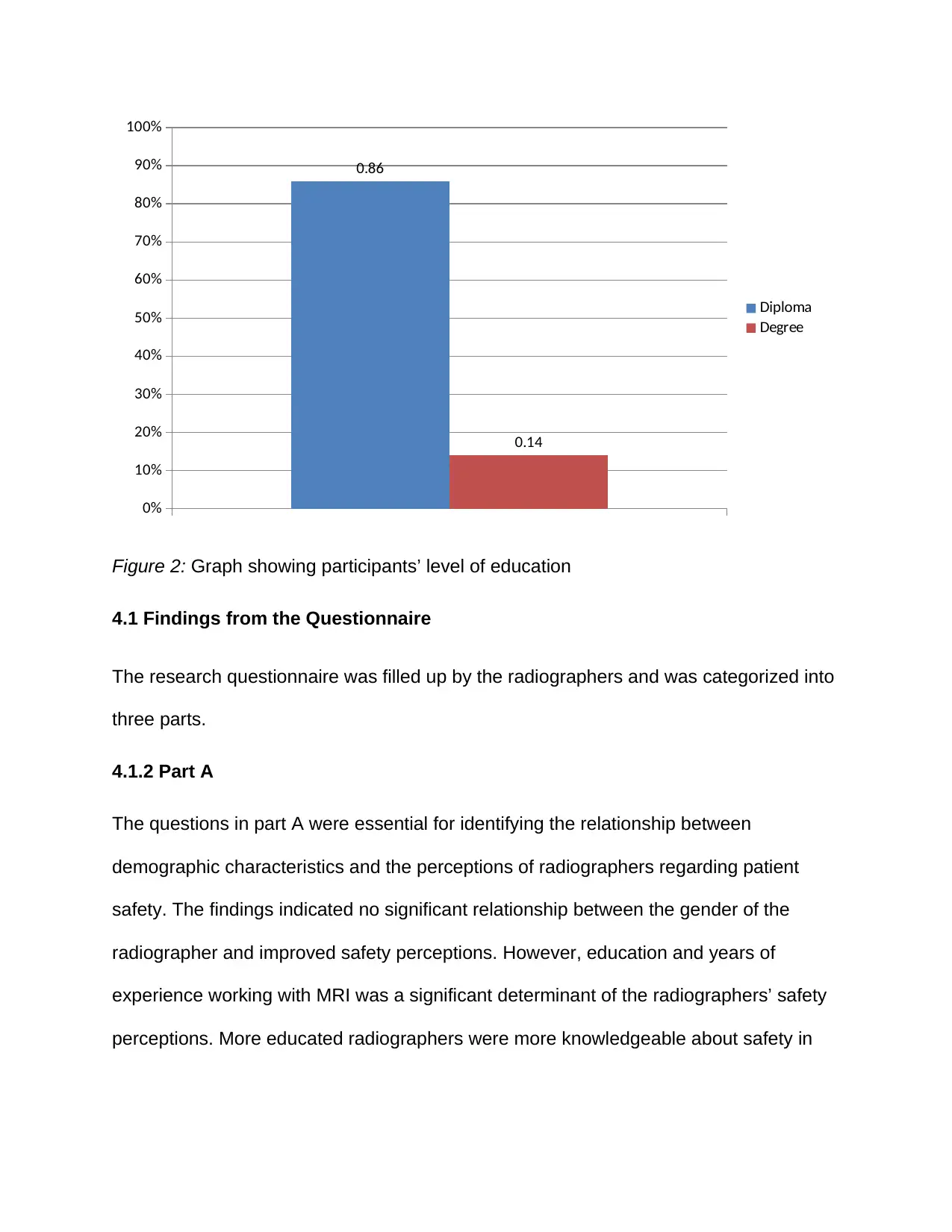
0%
10%
20%
30%
40%
50%
60%
70%
80%
90%
100%
0.86
0.14
Diploma
Degree
Figure 2: Graph showing participants’ level of education
4.1 Findings from the Questionnaire
The research questionnaire was filled up by the radiographers and was categorized into
three parts.
4.1.2 Part A
The questions in part A were essential for identifying the relationship between
demographic characteristics and the perceptions of radiographers regarding patient
safety. The findings indicated no significant relationship between the gender of the
radiographer and improved safety perceptions. However, education and years of
experience working with MRI was a significant determinant of the radiographers’ safety
perceptions. More educated radiographers were more knowledgeable about safety in
Secure Best Marks with AI Grader
Need help grading? Try our AI Grader for instant feedback on your assignments.
MRI environments and were kore positive about safety. Radiographers with degrees
prioritized safety as a component of their work.
4.1.3 Part B
The questions in Part B were important in understanding the actual perceptions of
radiographers regarding patient safety, and the actions they take towards the realization
of patient safety. The findings indicated that most radiographers perceive patient safety
as a priority in their work. They maintained that effective communication, and prompt
action in emergency situations is an important priority. Most participants agreed that
they were keen to follow the safety guidelines provided by regulatory bodies and the
healthcare organizations in which they work as a strategy of ensuring safety of
themselves and their patients.
4.1.4 Part C
Part C fielded triggered responses to gain information regarding the perception of
radiographers on how the work environment affects their adherence to patient safety
rules and over-all patient safety. The findings indicated that the characteristics of work
environments such as availability of equipment, frequent trainings, managerial
behaviour and a safety culture determine the motivation of the radiographers to ensure
patient safety.
4.2 Interview Findings
All the interviews were taken within the regular settings of organizational environments.
The participants were willing to share their knowledge and information over the study
area. According to Lichtman (2010), creating a rapport of some level is rendered as a
very important aspect for conducting an interview which will be useful for obtaining
meaningful data.
Questions related to the framework of operational safety of the MRI unit, practical
safety-related issues that are being faced by the MRI unit and related training
programmes were some major questions that were interviewed by the managers of the
imaging medical center. The entire interview was recorded electronically after which it
was transcribed and data grouped into different themes and then analysed. Electronic
recording ensured that all details in the interview process are captured; hence ensuring
validity and reliability of data collection (Al-Yateem, 2012; Markle, West, and Rich, 2011;
Tessier, 2012)
The perception of radiographers in patient safety and care is explained as a process
which involved a chain of judgement related to patient safety and care. The interviews
revealed that there are different phases involved which illustrate how radiographers will
conduct MRI which suits individual patient’s requirements. The interview questions
focused on investigating the perceptions of radiographers regarding safety in the
planning phase of the MRI, during the procedure itself and during evaluation of the
images.
4.2.1 Phase 1: Planning the MRI examination
The findings indicated adequate knowledge among the radiographers regarding safety
considerations while planning MRI procedures for their patients. They indicated that the
initial phases comprises of (a) critically assessing referrals; (b) interviewing and
observing patients for gathering missing information; (c) judging the communicative
capacity and (d) identification of safety aspects. For instance, one participants stated,
“We tend to emphasize over the importance of assessing information provided in
the referral prior to meeting the patient. In the process, of MRI, we select the
method of examination in advance or if conventional methods of examining takes
place, we will decide which method is appropriate with respect to the description
of the medical history of the patient. While outlining the procedure to undertake,
we put into consideration the important factors such as whether the patient has
internal or external implants, the mobility of patints to determine the possibility of
skin-to-skin contact.”
The findings also indicated that patient safety is a priority in MRI procedures and must
be considered during planning. Another pasrticipant asserted that “We take safety as
the first priority, not only because the rules and regulations demand so, but because it
is part of our job to protect patients and fellow personnel from harm or danger”
The radiographers also considered interviews as an important data collection tool for
assessing the suitability of patients for MRI and the safety precautions required. A fifth
participant mentioned that
“By interviewing the patient or observing about any symptoms, information will
be gathered with respect to the medical status of the patient. In case, it is
different from the information provided in the referral, it will be judged whether
the issue in question is appropriate. We will engage a multidisciplinary team in
case of doubt to ascertain the procedure and priorities of MRI to undertake. In
addition, the psychological condition of the patient is also considered closely.
There may be patients which have undergone a serious disease or trauma
whereas, there may be some patients who may undergo anxiety related to the
Paraphrase This Document
Need a fresh take? Get an instant paraphrase of this document with our AI Paraphraser
examination or are worried about the diagnosis. We emphasize over the
importance of this aspect as it will be useful for the safety and care of the patient
during the process of examination. An established rapport amongst the
radiographer and the patient tends to ease up cooperation level and as such
prevents motion artefacts.”
Another participant added that:
“We cannot underscore the importance of patient examination prior to an MRI
procedure. The process may be long and tiresome, but patient safety is of more
importance. For instance, judging the communicative capacity of the patient is
important as low communication capacity due to functional impairment or
language difficulties serves to be a challenge as the examination time Is limited.
If these factors are not considered closely, it can result in motion artefacts.”
Checking and examination of equipment before conducting any MRI procedure also
emerged as an important consideration for radiographers. One radiographer stated that
“Another safety aspect is related to the method selected for conducting MRI on a
patient. These safety aspects includes checking the level of radiation for fertile
women and children along with understanding counter indications implants within
the body or allergies as these aspects can pose a safety risk for MRI patients.”
4.2.2 Phase 2: Producing Correct Images
The findings indicated that producing the correct MRI images is a complex task that
required high level of safety. The phase involves various activities including (a)
positioning the patient correctly within the machine; (b) verifying with the patient so as to
establish cooperation and; (c) producing images. The radiographers must ensure that
the patient is safe and they perceive patient safety to be an essential part of their
decision-making. For instance, one radiographer mentioned that:
“We have to ensure that the patient is correctly, safely and comfortably
positioned in the machine. This is a safety guideline which prevents movement
during the examination so that accurate results can be obtained. Many times,
patients will be uncomfortable in a position however; it is the responsibility of the
radiographer to explain its importance so that adequate images can be attained.”
In addition, a second radiographer stated that:
“Positioning the patient accurately is very important when working with MRI as
position is important for image production. In comparison to the conventional
technique, the radiographer will be working more with the machine by selecting
the correct parameters and protocols, timing contrast injections as well as
instructing patients about the best breathing procedures for visualizing for
visualizing appropriate pathology and anatomy.”
Communication is also another important safety measure in MRI environments. Another
participant added that “Radiographers also constantly communicate with the patients
during the examination so that their current condition can be analysed. Radiographers
also inform about special breathing processes as well as body sensations which may
occur in the use of intravenous contrast.”
4.2.3 Phase 3: Evaluating the MRI examination
There was a consensus that final assessment of the MRI images is essential to ensure
that the correct parts were imaged. The accurate images preceded appropriate safe and
quality treatment. For example, a participant mentioned that: “After producing images,
the radiographers will evaluate the image quality and assess as to whether the correct
anatomical area has been covered and whether the image quality is sufficient enough
for pathology visualization.” A second participant asserted that: “An experienced
radiographer is aware when the quality of the image is sufficient with respect to the
conditions during the process of examination.”
Secure Best Marks with AI Grader
Need help grading? Try our AI Grader for instant feedback on your assignments.
CHAPTER 5. DISCUSSION
The aim of the research study is to determine the perception of Indian Radiographers
for MRI patient safety and care. Main findings of the study indicated that radiographers
consider patient safety and care to be their responsibility along with obtaining optimal
quality of the image by considering the individual needs of the patient and the need to
cooperate (Pooley et al. 2008). Safety aspects are related with the usage of medicine
distribution and technology with particular reference to contrast media and radiation
level (Tsai et al., 2015).The results present that the actions and judgement of a
radiographer starts from the production phase where the radiographer takes into
account safety and care aspects like contrast allergy as mentioned by Mangahy et al.
2018). Moreover, positioning the patient for correct examination is also very important.
The ability of the radiographer to visualize the image in advance helps their decision as
to how to place the patient correctly and safely and is also a major feature which
explains their professional skills. Similarly, Expert Panel on MR Safety et al. (2013)
reported that radiographers should assess the MRI room and only allow access by
restricted people who understand the constraints associated with magnetic fields.
Additionally, Elliot (2006) and Srinivas (2004) mentioned that radiologists have an
important role to play in ensuring that safety of patients. They can make safety
decisions by conducting pre-examination interviews with the focus of providing
information to the patients and their families. A safety culture was mentioned as a vital
component in MRI, similar to the findings of Crisp and Dawdy (2018).
In addition to this, establishing cooperation between the radiographer and patient before
the examination is very important for the safety and well-being of the patient (Mello and
Hemenway, 2004). How this is done by radiographers is briefly explained however, it is
not clarified in these results. Hospitals that have diagnostic radiological equipment’s are
under the requirement to undertake measures that assure the safety of both the patient
and the staff.
The findings of the study indicated that radiographers understand the safety precautions
and procedures involved in MRI operations. More than 70% of the radiographers
reported that they completely explain the processes prior to carrying out exposures over
the patients. , radiographers, as a matter of fact, tend to explain the process of
examination to the patients and get compete cooperation which helps in reducing
repeated investigations by failure to keep on with the instructions. In addition to this,
approximately, 20% of the radiographers reported that were forced to repeat imaging
procedures mainly because of technical faults or poor processing of the investigation.
No matter what are the reasons, repetition of MRI procedures should always be avoided
mainly because of the cumulative impact of the patients. Reducing the number of
repeated investigations and this situation can be effectively attained if the machines are
inspected on a regular basis for optimal performance and the patients are informed
about careful handling of the examination. When these two elements are under control
repeated investigations so as to confirm previous results might not be necessary as the
radiographers will be legible and clear thereby, reducing risks (Chang et al., 2008).
Additionally, radiographers also reported that protected devices were used at the time of
investigation. This aspect is grossly is unacceptable and inadequate. It is recommended
that protective devices should always be used. Alongside being a fundamental right,
patients should be active participants in the investigation process so as to keep
radiation exposure to minimum.
radiation exposure to minimum.
Paraphrase This Document
Need a fresh take? Get an instant paraphrase of this document with our AI Paraphraser
CHAPTER 6. CONCLUSION
From the research findings, it is possible to conclude that Indian radiographers perceive
patient care and safety as a priority in MRI. They also have the requisite knowledge and
skills regarding patient care and safety in MRI, but would prefer continuous trainings to
enhance their knowledge. Level of education is a significant determinant of the
perception and knowledge of radiographers regarding safety.
There are various limitations associated with the study. For instance, a sample of 29
radiographers may be small and limit generalizability. The sample may not reflect the
view of the entire population of radiographers in MRI within India. Also, the findings on
gender may be biased as only a few women were involved in the study compared to
their male counterparts
References
Aggarwal, R., and Ranganathan, P. (2019) Study designs: Part 2–Descriptive
studies. Perspectives in clinical research, 10(1), 34. doi:10.4103/picr.PICR_154_18
Alice, M. T., Palakiko, M. D. M., Daniggelis, E., and Makahi, M. E. (2015) Facilitating
community participants’ research engagement: community members’ perceptions of
community-based research. International journal of nursing and clinical practices, 2.
doi:10.15344/2394-4978/2015/142
Alshenqeeti, H. (2014) Interviewing as a data collection method: A critical
review. English linguistics research, 3(1), 39-45. doi:10.5430/elr.v3n1p39
Andersen, E. (2007) Magnetic resonance imaging—safety and health issues. Aaohn
Journal, 55(4), 137-139.
Arora, R. (2014) The training and practice of radiology in India: Current
trends. Quantitative imaging in medicine and surgery, 4(6), 449. doi:
10.3978/j.issn.2223-4292.2014.11.04
Asiamah, N., Mensah, H. K., and Oteng-Abayie, E. F. (2017) General, target, and
accessible population: Demystifying the concepts for effective sampling. The
Qualitative Report, 22(6), 1607. Available at https://nsuworks.nova.edu/tqr/
Aspden P., Corrigan J., and Wolcott J. (2004). Patient safety: achieving a new
standard of care. Washington, DC: National Academies Press.
Atmowardoyo, H. (2018). Research methods in TEFL studies: Descriptive research,
case study, error analysis, and R and D. Journal of Language Teaching and
Research, 9(1), 197-204.
Barnes, C. M., Dang, C. T., Leavitt, K., Guarana, C. L., and Uhlmann, E. L. (2018)
Archival data in micro-organizational research: A toolkit for moving to a broader set
of topics. Journal of Management, 44(4), 1453-1478.
doi:10.1177/0149206315604188
Beddoe, A., Temperton, D., Rafiqi, E., Larkin, E., and Jones, T. (2004)
Implementation of IRR99 and IRMER2000 in UK hospitals. Radiation Protection in
Australasia, 21(2), 59.
Bell, K., Fahmy, E., and Gordon, D. (2016) Quantitative conversations: the
importance of developing rapport in standardised interviewing. Quality and
quantity, 50(1), 193-212. doi:10.1007%2Fs11135-014-0144-2
Bellin, M. F., Jakobsen, J. Å., Tomassin, I., Thomsen, H. S., and Morcos, S. K.
(2002) Contrast medium extravasation injury: guidelines for prevention and
management. European radiology, 12(11), 2807-2812. doi:
10.1007/s003300100933.
Bolderston, A. (2012). Conducting a research interview. Journal of Medical Imaging
and Radiation Sciences, 43(1), 66-76. doi:10.1016/j.jmir.2011.12.002
Burns, N., and Grove, S. (2009) The practice of nursing research conduct, critique
and utilization.. St Louis, Mo.: Elsevier, 122e123, 176e210, 341, pp.352-353.
Available at
https://www.scirp.org/(S(vtj3fa45qm1ean45vvffcz55))/reference/ReferencesPapers.a
spx?..
Secure Best Marks with AI Grader
Need help grading? Try our AI Grader for instant feedback on your assignments.
Callaghan, M. F., Negus, C., Leff, A. P., Creasey, M., Burns, S., Glensman, J., ...
and Weiskopf, N. (2019). Safety of tattoos in persons undergoing MRI. New England
Journal of Medicine, 380(5), 495-496.
Casey, D., and Houghton, C. (2010) Clarifying case study research: Examples from
practice. Nurse researcher, 17(3). doi:10.7748/nr2010.04.17.3.41.c7745
Centers for Disease Control and Prevention (2017) ‘Infection control basics’.
Available at https://www.cdc.gov/infectioncontrol/basics/index.html
Chang, P. Y., Hsieh, M. C., La, Y. J., and Chan, W. P. (2008) Analysis of revised
imaging reports in diagnostic radiology: an example from a medical center in
Taiwan. Chinese Journal of Radiology, 33(1), 23-29.
Cheng, H. G., and Phillips, M. R. (2014) Secondary analysis of existing data:
opportunities and implementation. Shanghai archives of psychiatry, 26(6), 371.
Available at http://www.shanghaiarchivesofpsychiatry.org/
Consiglio, N. O. (2006) MRI and patient safety. Canadian Journal of Medical
Radiation Technology, 37(2), 5-9.
Cormack, D. (2000). The Research Process in Nursing, 4th edn. Backwell Science.
Cosac, D. C. D. S. (2017) Autonomy, consent and vulnerability of clinical research
participants. Revista Bioética, 25(1), 19-29. doi:10.1590/1983-80422017251162
Creswell, J. W., and Creswell, J. D. (2017). Research design: Qualitative,
quantitative, and mixed methods approaches. Thousand Oaks, CA: Sage
publications.
Crisp, S., and Dawdy, K. (2018) Building a magnetic resonance imaging safety
culture from the ground up. Journal of medical imaging and radiation
sciences, 49(1), 18-22.
Crook, N., and Robinson, L. (2009) A review of the safety implications of magnetic
resonance imaging at field strengths of 3 Tesla and above. Radiography, 15(4), 351-
356.
Cross, N. M., Hoff, M. N., and Kanal, K. M. (2018) Avoiding MRI-related accidents: a
practical approach to implementing MR safety. Journal of the American College of
Radiology, 15(12), 1738-1744.
Dong, Z., Andrews, T., Xie, C., and Yokoo, T. (2015). Advances in MRI techniques
and applications. BioMed research international, 2015.. doi:10.1155/015/139043
Dunn, S. L., Arslanian-Engoren, C., DeKoekkoek, T., Jadack, R., and Scott, L. D.
(2015) Secondary data analysis as an efficient and effective approach to nursing
research. Western journal of nursing research, 37(10), 1295-1307.
doi:10.1177/0193945915570042
Durbridge, G. (2011) Magnetic resonance imaging: fundamental safety
issues. journal of orthopaedic and sports physical therapy, 41(11), 820-828.
Ellard-Gray, A., Jeffrey, N. K., Choubak, M., and Crann, S. E. (2015) Finding the
hidden participant: Solutions for recruiting hidden, hard-to-reach, and vulnerable
populations. International Journal of Qualitative
Methods, 14(5).doi:10.1177/1609406915621420
Elliot, (2006). ‘India Awakens’, Time Magazine ‘India, Inc.’ edition, p. 38
Elmir, R., Schmied, V., Jackson, D., and Wilkes, L. (2011). Interviewing people
about potentially sensitive topics. Nurse researcher, 19(1),12-16.
doi:10.7748/nr2011.10.19.1.12.c8766
European Medicines Agency, 2017, ‘EMA's final opinion confirms restrictions on use
of linear gadolinium agents in body scans’, EMA website. Available from
https://www.ema.europa.eu/medicines/human/referrals/gadolinium-containing-
contrast-agents
European Society of Radiology (2018). ESR EuroSafe imaging. Available at
http://www.eurosafeimaging.org/
Expert Panel on MR Safety:, Kanal, E., Barkovich, A. J., Bell, C., Borgstede, J. P.,
Bradley Jr, W. G., ... and Larson, P. A. (2013) ACR guidance document on MR safe
practices: 2013. Journal of Magnetic Resonance Imaging, 37(3), 501-530.
Gentles, S. J., Charles, C., Ploeg, J., and McKibbon, K. A. (2015). Sampling in
qualitative research: Insights from an overview of the methods literature. The
qualitative report, 20(11), 1772-1789. Available at https://nsuworks.nova.edu/tqr/
Gilk, T., and Kanal, E. (2006) Integrating MRI facility design and risk
management. Journal of Healthcare Risk Management, 26(3), 29-37.
Gillies, A., (2002). Using research in Nursing: a workbook for practitioners. Radcliffe
Publishing.
Gray, D.E. (2019). Doing research in the real world. Sage Publications Limited.
Gunnell, M. (2016). Research Methodologies: A comparison of quantitative,
qualitative, and mixed methods. Available at https://www.linkedin.com
Paraphrase This Document
Need a fresh take? Get an instant paraphrase of this document with our AI Paraphraser
Gupta, A. K. (2007) Modern trends in planning and designing of hospitals: Principles
and practice. Indian Journal of Medical Research, 126(2), 167-169.
Hartwig, V., Giovannetti, G., Vanello, N., Lombardi, M., Landini, L., and Simi, S.
(2009) Biological effects and safety in magnetic resonance imaging: a
review. International journal of environmental research and public health, 6(6), 1778-
1798.
In, J. (2017) Introduction of a pilot study. Korean journal of anesthesiology, 70(6),
601. doi:10.4097%2Fkjae.2017.70.6.601
International Commission on Radiological Protection (2007). The 2007
recommendations of the International commission on radiological protection. ICRP
Publication 103 Ann. ICRP 37 (2–4). Available at
http://www.icrp.org/publication.asp?id=ICRP%20Publication%20103
International Organization for Standardization (2012). ISO/TR 12296:2012.
Ergonomics - manual handing of people in the healthcare sector. International
Organization for Standardization, Geneva. Availabel
athttps://www.iso.org/standard/51310.html
Jamshed, S. (2014) Qualitative research method-interviewing and
observation. Journal of basic and clinical pharmacy, 5(4), 87. doi:10.4103%2F0976-
0105.141942
Jankharia, G. R. (2008) Commentary-radiology in India: the next decade. The Indian
journal of radiology and imaging, 18(3), 189.
Kalyanpur, A. (2008) Commentary-radiology in India: the next decade. The Indian
journal of radiology and imaging, 18(3), 191.
Kanal, E., Barkovich, A. J., Bell, C., Borgstede, J. P., Bradley Jr, W. G., Froelich, J.
W., ... and Lester Jr, J. W. (2007). ACR guidance document for safe MR practices:
2007. American Journal of Roentgenology, 188(6), 1447-1474.
Ko, Y., and Yu, S. (2017) The relationships among perceived patients' safety culture,
intention to report errors, and leader coaching behavior of nurses in Korea: A pilot
study. Journal of patient safety, 13(3), 175-183.
doi.10.1097/PTS.0000000000000224
Lark, M. E., Kirkpatrick, K., and Chung, K. C. (2018) Patient safety movement:
history and future directions. The Journal of hand surgery, 43(2), 174-178.
doi:10.1016/j.jhsa.2017.11.006
Lee, C. H., Lin, M. F., and Chan, W. P. (2015) Management of a sandbag accident
in an MRI unit. Magnetic resonance imaging, 33(9), 1187-1189.
Levine, G. N., Gomes, A. S., Arai, A. E., Bluemke, D. A., Flamm, S. D., Kanal, E., ...
and Shellock, F. S. (2007) Safety of magnetic resonance imaging in patients with
cardiovascular devices: an American Heart Association scientific statement from the
Committee on Diagnostic and Interventional Cardiac Catheterization, Council on
Clinical Cardiology, and the Council on Cardiovascular Radiology and Intervention:
endorsed by the American College of Cardiology Foundation, the North American
Society for Cardiac Imaging, and the Society for Cardiovascular Magnetic
Resonance. Circulation, 116(24), 2878-2891.
Madhok, R., Roy, N., and Panesar, S. (2012) Patient safety in India: time to speed
up our efforts to reduce avoidable harm. The National medical journal of India, 25(3),
129.
Mangahy, A., Altolub, M., Batarfi, M., and Alshulayyil, M. (2018) Assessment of
Clinical Competences and Self-confidence of Radiologist and Technologist at Asir
Region Hospitals, Saudi Arabia. The Egyptian Journal of Hospital Medicine, 71(2),
2544-2552.
Martínez-Mesa, J., González-Chica, D. A., Duquia, R. P., Bonamigo, R. R., and
Bastos, J. L. (2016) Sampling: how to select participants in my research
study?. Anais brasileiros de dermatologia, 91(3), 326-330.
Martirosyan, L., Arah, O. A., Haaijer-Ruskamp, F. M., Braspenning, J., and Denig, P.
(2010) Methods to identify the target population: implications for prescribing quality
indicators. BMC health services research, 10(1), 137. doi:10.1186/1472-6963-10-
137
Mayeda‐Letourneau, J. (2014) Safe patient handling and movement: a literature
review. Rehabilitation Nursing, 39(3), 123-129. doi: 10.1002/rnj.133
McCullagh, M. C., Sanon, M. A., and Cohen, M. A. (2014) Strategies to enhance
participant recruitment and retention in research involving a community-based
population. Applied Nursing Research, 27(4), 249-253.
doi:10.1016/j.apnr.2014.02.007
McKenna, L., and Gray, R. (2018) The importance of ethics in research
publications. Collegian, 25(2), 147-148. doi:10.1016/j.colegn.2018.02.006
Secure Best Marks with AI Grader
Need help grading? Try our AI Grader for instant feedback on your assignments.
Mello, M. M., and Hemenway, D. (2004). Medical malpractice as an epidemiological
problem. Social science and medicine, 59(1), 39-46.
Moser, A., and Korstjens, I. (2018) Series: Practical guidance to qualitative research.
Part 3: Sampling, data collection and analysis. European Journal of General
Practice, 24(1), 9-18. doi:10.1080/13814788.2017.1375091
Nijhawan, L. P., Janodia, M. D., Muddukrishna, B. S., Bhat, K. M., Bairy, K. L.,
Udupa, N., and Musmade, P. B. (2013) Informed consent: Issues and
challenges. Journal of advanced pharmaceutical technology and research, 4(3), 134.
doi:10.4103%2F2231-4040.116779
Palinkas, L. A., Horwitz, S. M., Green, C. A., Wisdom, J. P., Duan, N., and
Hoagwood, K. (2015) Purposeful sampling for qualitative data collection and
analysis in mixed method implementation research. Administration and policy in
mental health and mental health services research, 42(5), 533-544.
doi:10.1007/s10488-013-0528-y
Parker, G. E. (2016). A framework for navigating institutional review board (IRB)
oversight in the complicated zone of research. Cureus, 8(10)..
doi:10.7759/cureus.844
Polgar, S., and Thomas, S. A. (2011) Introduction to Research in the Health
Sciences E-Book. Elsevier Health Sciences.
Pontin, D. (2000) Descriptive research. The Research Process in Nursing (4thedn),
Oxford: Blackwell Science.
Pooley, R. A., Felmlee, J. P., and Gorny, K. R. (2013) MR siting: improving efficiency
and safety. Journal of the American College of Radiology, 10(9), 721-722.
Pullen Jr, R. L. (2008) Preparing a patient for magnetic resonance
imaging. Nursing2019, 38(10), 22.
Rahman, M. S. (2017) The Advantages and Disadvantages of Using Qualitative and
Quantitative Approaches and Methods in Language" Testing and Assessment"
Research: A Literature Review. Journal of Education and Learning, 6(1), 102-112.
doi:10.5539/jel.v6n1p102
Sage, W. M., Jablonski, J. S., and Thomas, E. J. (2015) Use of nondisclosure
agreements in medical malpractice settlements by a large academic health care
system. JAMA internal medicine, 175(7), 1130-1135.
Sammet, S. (2016) Magnetic resonance safety. Abdominal Radiology, 41(3), 444-
451. doi:10.1007/s00261-016-0680-4
Sanjari, M., Bahramnezhad, F., Fomani, F. K., Shoghi, M., and Cheraghi, M. A.
(2014) Ethical challenges of researchers in qualitative studies: The necessity to
develop a specific guideline. Journal of medical ethics and history of medicine, 7.
Available at http://jmehm.tums.ac.ir/index.php/jmehm
Sargeant, J. (2012). Qualitative research part II: Participants, analysis, and quality
assurance. Journal of Graduate Medical Education, 4(1)1-3. doi:10.4300%2FJGME-
D-11-00307.1
Sawyer‐Glover, A. M., and Shellock, F. G. (2000) Pre‐MRI procedure screening:
recommendations and safety considerations for biomedical implants and
devices. Journal of Magnetic Resonance Imaging, 12(1), 92-106.
Sileyew, K. J. (2019). Research Design and Methodology. In Text Mining-Analysis,
Programming and Application. IntechOpen.
Sivell, S., Prout, H., Hopewell-Kelly, N., Baillie, J., Byrne, A., Edwards, M., ... and
Nelson, A. (2019). Considerations and recommendations for conducting qualitative
research interviews with palliative and end-of-life care patients in the home setting: a
consensus paper. BMJ supportive and palliative care, 9(1), e14-e14.
doi:10.1136/bmjspcare-2015-000892
Srinivas, S. (2004). Technological learning and the evolution of the Indian
pharmaceutical and biopharmaceutical sectors (Doctoral dissertation,
Massachusetts Institute of Technology).
Tajir, G. K. A. (2018). Ethical treatment of participants in public health
research”, Journal of Public Health and Emergency, 2(1)1-10. Available at
http://jphe.amegroups.com/
Takahashi, T., Fujimoto, N., Hamada, Y., Tezuka, N., and Tanaka, T. (2016) MRI-
related thermal injury due to skin-to-skin contact. European Journal of
Dermatology, 26(3), 296-298.
Tang, A., Tam, R., Cadrin-Chênevert, A., Guest, W., Chong, J., Barfett, J., ... and
Poudrette, M. G. (2018) Canadian Association of Radiologists white paper on
artificial intelligence in radiology. Canadian Association of Radiologists'
Journal, 69(2), 120-135. doi:10.1148/radiol.2018182404
The Society and College of Radiographers (2018). Obtaining consent: a clinical
guideline for the diagnostic imaging and radiotherapy workforce”. Available at
Paraphrase This Document
Need a fresh take? Get an instant paraphrase of this document with our AI Paraphraser
https://www.sor.org/sites/default/files/document-versions/obtaining_consent_170118
.pdf
Tsai, L. L., Grant, A. K., Mortele, K. J., Kung, J. W., and Smith, M. P. (2015) A
practical guide to MR imaging safety: what radiologists need to
know. Radiographics, 35(6), 1722-1737.
Viscuse, P. V., Khasawneh, M., and Constantinou, C. (2015). MRI-Induced Second
and Third Degree Burns to Hands Bilaterally: A Case Report. Annals of Hematology
and Oncology, 2(7). Available at
https://austinpublishinggroup.com/hematology/fulltext/hematology-v2-id1053.php
Vogel, S., and Draper-Rodi, J. (2017). The importance of pilot studies, how to write
them and what they mean. International Journal of Osteopathic Medicine, 23, 2-3.
doi:10.1016/j.ijosm.2017.02.001
World Health Organization (2018). Clean Care is Safer Care. About SAVE LIVES:
Clean your hands. World Health Organization, Geneva. Available at
http://www.who.int/gpsc/5may/background/5moments/en/
Yilmaz, K. (2013) Comparison of quantitative and qualitative research traditions:
Epistemological, theoretical, and methodological differences. European journal of
education, 48(2), 311-325. Available at
https://onlinelibrary.wiley.com/journal/14653435
Yip, C., Han, N. L. R., and Sng, B. L. (2016) Legal and ethical issues in
research. Indian journal of anaesthesia, 60(9), 684. doi:10.4103%2F0019-
5049.190627

1 out of 57
Related Documents


Your All-in-One AI-Powered Toolkit for Academic Success.
+13062052269
info@desklib.com
Available 24*7 on WhatsApp / Email
![[object Object]](/_next/static/media/star-bottom.7253800d.svg)
Unlock your academic potential
© 2024 | Zucol Services PVT LTD | All rights reserved.