Pathophysiology and Treatment of Ischemic Stroke
VerifiedAdded on 2023/01/19
|4
|1442
|21
Presentation
AI Summary
This presentation discusses the pathophysiology, clinical manifestations, and treatment of ischemic stroke. It highlights the risk factors such as hypertension and atrial fibrillation, and explores the interventions for stroke rehabilitation. The case study of a 66-year-old man with extra-cranial embolism is used to illustrate the concepts.
Contribute Materials
Your contribution can guide someone’s learning journey. Share your
documents today.
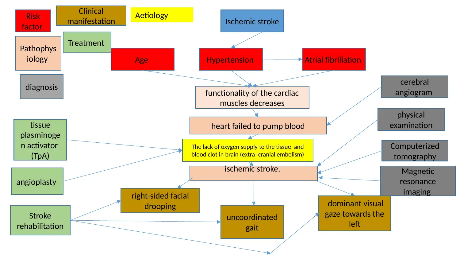
Ischemic stroke
Hypertension
functionality of the cardiac
muscles decreases
Atrial fibrillationAge
heart failed to pump blood
ischemic stroke.
The lack of oxygen supply to the tissue and
blood clot in brain (extra=cranial embolism)
right-sided facial
drooping
uncoordinated
gait
dominant visual
gaze towards the
left
physical
examination
Computerized
tomography
Magnetic
resonance
imaging
cerebral
angiogram
tissue
plasminoge
n activator
(TpA)
angioplasty
Stroke
rehabilitation
Risk
factor
Pathophys
iology
diagnosis
Clinical
manifestation
Treatment
Aetiology
Secure Best Marks with AI Grader
Need help grading? Try our AI Grader for instant feedback on your assignments.
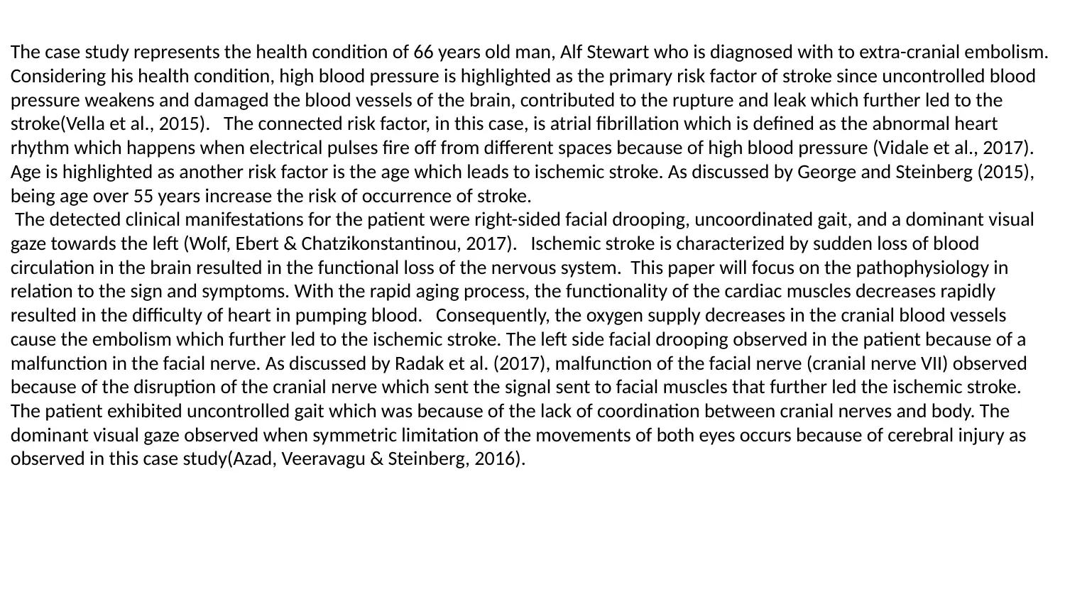
The case study represents the health condition of 66 years old man, Alf Stewart who is diagnosed with to extra-cranial embolism.
Considering his health condition, high blood pressure is highlighted as the primary risk factor of stroke since uncontrolled blood
pressure weakens and damaged the blood vessels of the brain, contributed to the rupture and leak which further led to the
stroke(Vella et al., 2015). The connected risk factor, in this case, is atrial fibrillation which is defined as the abnormal heart
rhythm which happens when electrical pulses fire off from different spaces because of high blood pressure (Vidale et al., 2017).
Age is highlighted as another risk factor is the age which leads to ischemic stroke. As discussed by George and Steinberg (2015),
being age over 55 years increase the risk of occurrence of stroke.
The detected clinical manifestations for the patient were right-sided facial drooping, uncoordinated gait, and a dominant visual
gaze towards the left (Wolf, Ebert & Chatzikonstantinou, 2017). Ischemic stroke is characterized by sudden loss of blood
circulation in the brain resulted in the functional loss of the nervous system. This paper will focus on the pathophysiology in
relation to the sign and symptoms. With the rapid aging process, the functionality of the cardiac muscles decreases rapidly
resulted in the difficulty of heart in pumping blood. Consequently, the oxygen supply decreases in the cranial blood vessels
cause the embolism which further led to the ischemic stroke. The left side facial drooping observed in the patient because of a
malfunction in the facial nerve. As discussed by Radak et al. (2017), malfunction of the facial nerve (cranial nerve VII) observed
because of the disruption of the cranial nerve which sent the signal sent to facial muscles that further led the ischemic stroke.
The patient exhibited uncontrolled gait which was because of the lack of coordination between cranial nerves and body. The
dominant visual gaze observed when symmetric limitation of the movements of both eyes occurs because of cerebral injury as
observed in this case study(Azad, Veeravagu & Steinberg, 2016).
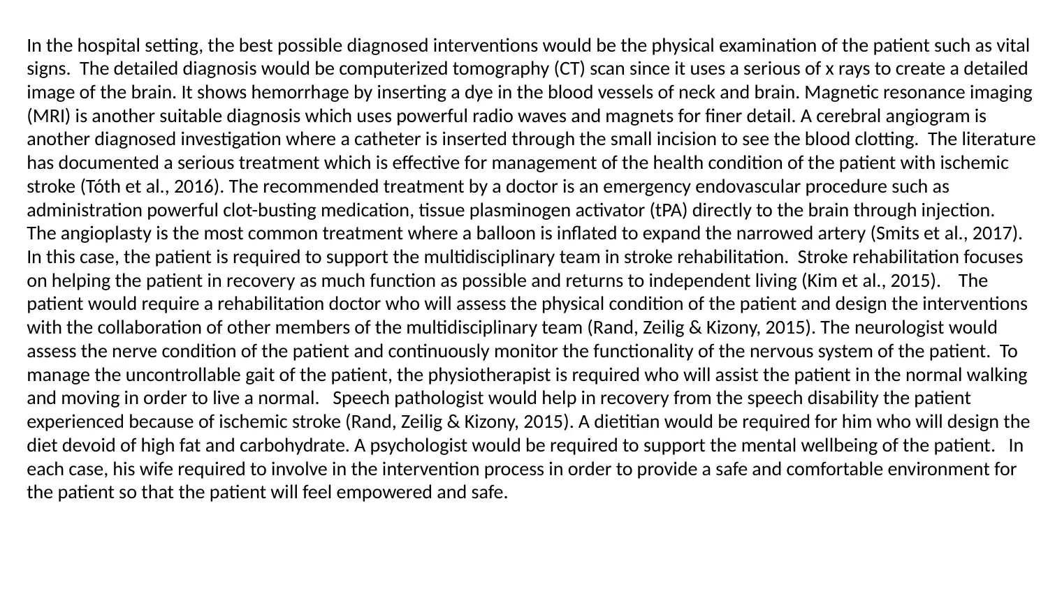
In the hospital setting, the best possible diagnosed interventions would be the physical examination of the patient such as vital
signs. The detailed diagnosis would be computerized tomography (CT) scan since it uses a serious of x rays to create a detailed
image of the brain. It shows hemorrhage by inserting a dye in the blood vessels of neck and brain. Magnetic resonance imaging
(MRI) is another suitable diagnosis which uses powerful radio waves and magnets for finer detail. A cerebral angiogram is
another diagnosed investigation where a catheter is inserted through the small incision to see the blood clotting. The literature
has documented a serious treatment which is effective for management of the health condition of the patient with ischemic
stroke (Tóth et al., 2016). The recommended treatment by a doctor is an emergency endovascular procedure such as
administration powerful clot-busting medication, tissue plasminogen activator (tPA) directly to the brain through injection.
The angioplasty is the most common treatment where a balloon is inflated to expand the narrowed artery (Smits et al., 2017).
In this case, the patient is required to support the multidisciplinary team in stroke rehabilitation. Stroke rehabilitation focuses
on helping the patient in recovery as much function as possible and returns to independent living (Kim et al., 2015). The
patient would require a rehabilitation doctor who will assess the physical condition of the patient and design the interventions
with the collaboration of other members of the multidisciplinary team (Rand, Zeilig & Kizony, 2015). The neurologist would
assess the nerve condition of the patient and continuously monitor the functionality of the nervous system of the patient. To
manage the uncontrollable gait of the patient, the physiotherapist is required who will assist the patient in the normal walking
and moving in order to live a normal. Speech pathologist would help in recovery from the speech disability the patient
experienced because of ischemic stroke (Rand, Zeilig & Kizony, 2015). A dietitian would be required for him who will design the
diet devoid of high fat and carbohydrate. A psychologist would be required to support the mental wellbeing of the patient. In
each case, his wife required to involve in the intervention process in order to provide a safe and comfortable environment for
the patient so that the patient will feel empowered and safe.
Azad, T. D., Veeravagu, A., & Steinberg, G. K. (2016). Neurorestoration after stroke. Neurosurgical focus, 40(5), E2.
doi.org/10.3171/2016.2.FOCUS15637
George, P. M., & Steinberg, G. K. (2015). Novel stroke therapeutics: unraveling stroke pathophysiology and its impact on
clinical treatments. Neuron, 87(2), 297-309. doi.org/10.1016/j.neuron.2015.05.041
Joundi, R. A., Martino, R., Saposnik, G., Giannakeas, V., Fang, J., & Kapral, M. K. (2017). Predictors and outcomes of
dysphagia screening after acute ischemic stroke. Stroke, 48(4), 900-906. doi.org/10.1161/STROKEAHA.116.01532
Radak, D., Katsiki, N., Resanovic, I., Jovanovic, A., Sudar-Milo Kim, E. H., Tolhurst, A. T., Szeto, H. H., & Cho, S. H. (2015).
Targeting CD 36‐Mediated Inflammation Reduces Acute Brain Injury in Transient, but not Permanent, Ischemic Stroke. CNS
neuroscience & therapeutics, 21(4), 385-391. doi.org/10.1111/cns.12326
Ivanovic, E., Zafirovic, S., ... & R Isenovic, E. (2017). Apoptosis and acute brain ischemia in ischemic stroke. Current
vascular pharmacology, 15(2), 115-122. doi.org/10.1111/cns.12326
Rand, D., Zeilig, G., & Kizony, R. (2015). Rehab-let: touchscreen tablet for self-training impaired dexterity post stroke:
study protocol for a pilot randomized controlled trial. Trials, 16(1), 277. Doi.org/10.1186/s13063-015-0796-9
Smits, P. C., Abdel-Wahab, M., Neumann, F. J., Boxma-de Klerk, B. M., Lunde, K., Schotborgh, C. E., ... & Hambrecht, R.
(2017). Fractional flow reserve–guidóed multivessel angioplasty in myocardial infarction. New England Journal of
Medicine, 376(13), 1234-1244. DOI: 10.1056/NEJMoa1701067
Vella, J., Zammit, C., Di Giovanni, G., Muscat, R., & Valentino, M. (2015). The central role of aquaporins in the
pathophysiology of ischemic stroke. Frontiers in cellular neuroscience, 9, 108. doi.org/10.3389/fncel.2015.00108
Vidale, S., Consoli, A., Arnaboldi, M., & Consoli, D. (2017). Postischemic inflammation in acute stroke. Journal of Clinical
Neurology, 13(1), 1-9. doi.org/10.3988/jcn.2017.13.1.1
Wolf, M. E., Ebert, AToth, M., Little, P., Arnberg, F., Häggkvist, J., Mulder, J., Halldin, C., ... & Holmin, S. (2016). Acute
neuroinflammation in a clinically relevant focal cortical ischemic stroke model in rat: longitudinal positron emission
tomography and immunofluorescent tracking. Brain Structure and Function, 221(3), 1279-1290.
doi: 10.1586/14737175.4.2.255
. D., & Chatzikonstantinou, A. (2017). The use of routine EEG in acute ischemic stroke patients without seizures:
generalized but not focal EEG pathology is associated with clinical deterioration. International Journal of
Neuroscience, 127(5), 421-426. doi.org/10.1080/00207454.2016.1189913
1 out of 4
Your All-in-One AI-Powered Toolkit for Academic Success.
+13062052269
info@desklib.com
Available 24*7 on WhatsApp / Email
Unlock your academic potential
© 2024 | Zucol Services PVT LTD | All rights reserved.