Understanding Glyceryl Trinitrate Action
VerifiedAdded on 2020/03/13
|8
|1779
|33
AI Summary
This assignment delves into the pharmacological action of glyceryl trinitrate, a potent vasodilator drug. It explains how glyceryl trinitrate works by releasing nitric oxide (NO), leading to relaxation and dilation of blood vessels. The assignment highlights its impact on systemic vascular resistance (SVR), cardiac preload, and afterload. It also discusses common adverse effects like facial flushing, hypotension, and headaches, emphasizing the importance of monitoring blood pressure during administration.
Contribute Materials
Your contribution can guide someone’s learning journey. Share your
documents today.
Running head: ACUTE HEART FAILURE
ACUTE HEART FAILURE
Name of the University
Name of the Student
Author Note
ACUTE HEART FAILURE
Name of the University
Name of the Student
Author Note
Secure Best Marks with AI Grader
Need help grading? Try our AI Grader for instant feedback on your assignments.
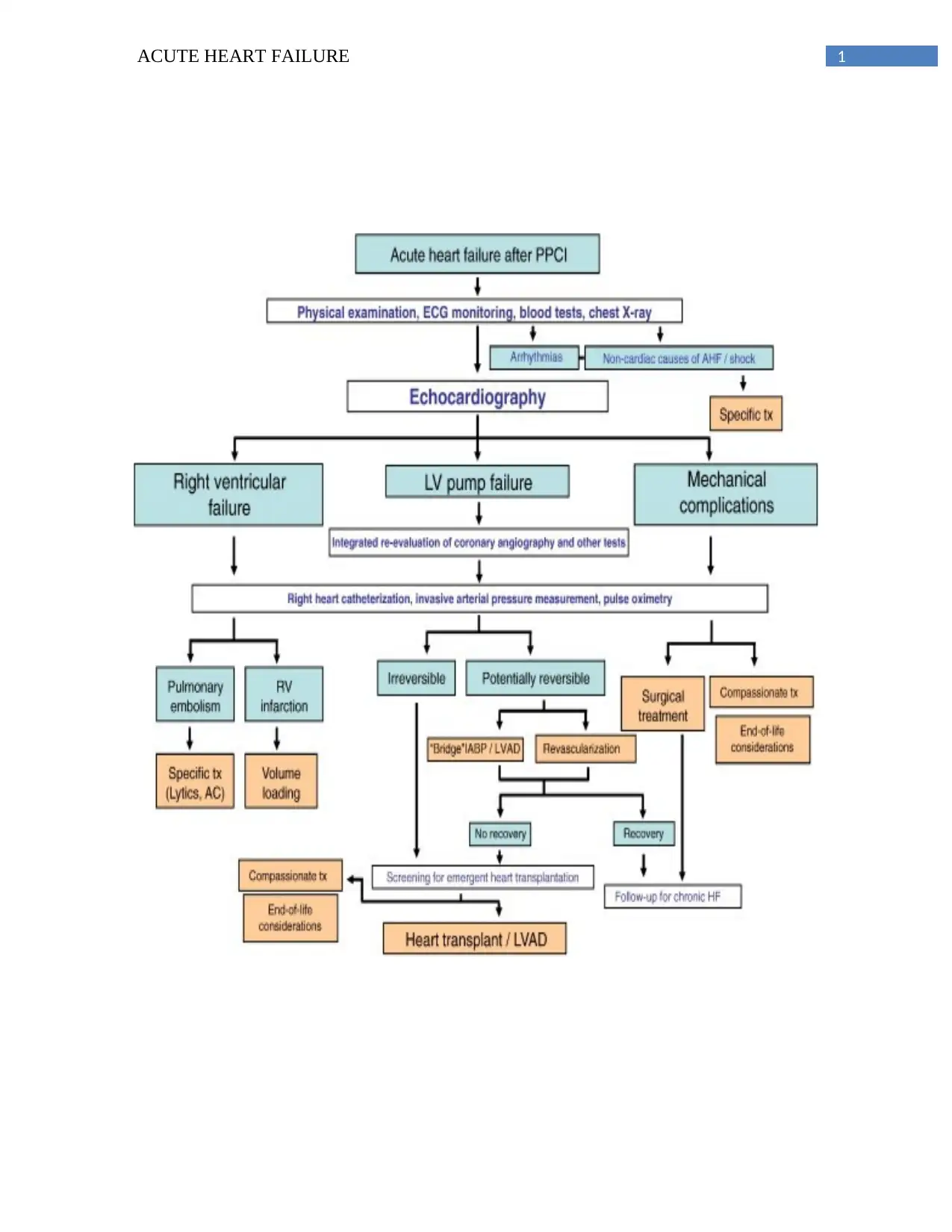
1ACUTE HEART FAILURE
2ACUTE HEART FAILURE
Q1.
A diagnosis has been done on Mrs. Brown with acute exacerbation of heart failure
(AIHF). The symptoms of AIHF are initially caused by the pulmonary oedema due to an
increased filling pressure in the left ventricle (LV) (Copstead & Banasik, 2013). To meet the
metabolic demand of the body, the LV is responsible for pumping an sufficient amount of
oxygenated rich blood to the body. The LV becomes impaired to perform this activity, which
leads to increase the blood volume and blood pressure in the LV (Gallagher, 2012). This
situation results to the flowing of the blood forwarded into the body, which can cause the
increased blood in the LV to revert through the pulmonary veins and left atrium, resulting to an
increment in the capillary pressure (Craft & Gordon, 2015). The fluid more possibly becomes to
penetrate into the interstitial spaces and the lung’s alveoli from the capillary walls causing a lift
in the capillary hydrostatic pressure leading to pulmonary oedema, as a result of increase of the
pulmonary circulation hydrostatic pressure (Fenwick, 2015). The pulmonary gas exchange
function can be significantly impaired due to the increased fluid and in the alveoli and airway as
the interference of the gas exchange with the lung’s ventilation process. Hence, severe dyspnoea
would be experienced by Mrs. Browns. Due to the movement of the air that passes through the
alveolar flied, sounds of the crackles are heard from the patient having pulmonary oedema
(Fenwick, 2015). It is indicated from the oxygen saturation reading that Mrs. Brown is having
inadequate supply of blood in her body. The situation is resulted by the incapability of the lungs
for not effective oxygenation of the blood. Therefore, while leaving the pulmonary circulation,
the blood gets poorly oxygenated (Gallagher, 2012). The failing heart is responsible for not
delivering the adequate oxygenated blood to reach the tissue oxygen needs, resulting into the
hypoxia and reduced tissue perfusion, as stated by Fenwick (2015). AHF is connected with a
3ACUTE HEART FAILURE
remarkable reduction in the cardiac output (CO), myocardial contractility and stroke volume.
Accumulated blood pressure and tachycardia has been experienced by Mrs. Brown due to the
activation o compensatory mechanism responding to the reduced CO. The activation of the
symoathetic nervous system is stimulated by the decreased cardiac output to discharge the no
noradrenalin and adrenaline that can be the reason for increment in vasoconstriction (McCance
& Huether, 2014). With the activation of the vasoconstriction, the further elevation happens in
the myocardial contractility, peripheral vascular constriction and the HR. A decreased blood flow
to the kidneys is caused by the decreased CO, resulting to a step down in the glomerular filtration
rate, additionally (McCance & Huether, 2014). Chopsted and Banasik (2013) has showed in
response to the above stated situation, that, to release rennin, which transforms the
angiotensinogen to angiotensin I from angiotensin II the kidneys are stimulated by the rennin-
angiotensin-aldosteron. A lift in the arterial blood pressure is caused by the increase of the
peripheral vasoconstriction from such situation. In addition, Gordon and Craft (2015) stated that,
the release of antidiuretic hormone (ADH) is stimulated by the posterior pituitary, in response to
the reduction of the cerebral perfusion pressure caused by the low CO. An important role is
played by the ADH in raising the reabsorption of the water of the renal tubules, which results in
increased volume of blood and the water retention.
Q2.
If the prescribed oxygen therapy is provided to Mrs. Brown, it will relieve the symptoms
of her connected with acute hypoxia and dyspnoea. The density of oxygen in airway and the
alveolar space is increased and the levels of carbon dioxide are decreased by the disposal of
oxygen. Therefore, this helps the gases to spread out into the capillaries of pulmonary by
crossing the membrane of the alveolar capillary (Wagner & Hardin-Pierce, 2014). As the result,
Secure Best Marks with AI Grader
Need help grading? Try our AI Grader for instant feedback on your assignments.
4ACUTE HEART FAILURE
the function of the pulmonary gas exchange improves and the symptoms of the dyspnoea
decreases.the oxygen level in the blood is increased and the requirement for the tissue perfusion
is improved by the oxygen therapy (Powell, Graham, O’Reilly & Punton, 2016). Optimally, a
pulse oximetry has been used for monitoring the effectiveness of oxygen therapy disposal
provided to Mrs. Brown.
Mrs. Brown has been placed in the high Fowler position while her feet is dangling at the
bedside, which improves the gas exchange function and the ventilation by enhancing her thoracic
capacity (Gallagher, 2012). Moreover, the cardiac preload caused by the ineffective systematic
circulation is decreased by this particular position. During diastole, the blood amount returning
to the left ventricle gets less overfilled, as a result of the decreased return of venous (Wagner &
Hardin-Pierce, 2014). Consequently, this improves the LV performance.
Q3.
Furosemide is one of the loop diuretic drugs. The reabsorption of the chloride and the
sodium ions into the interstitial fluid from the loops inhibited by the direct working of the
ascending loop’s medullary part of Henle, which result into a hypotonic interstitial fluid
environment (Bullock & Manias, 2013). The reduction of the pulmonary venous pressure is
helped by the Furosemide, by circulating the oedemous fluid, which can be responsible for
resulting to the optimal exchange of gas. The intravascular volume is decreased by this which
can lead to a reduction of return of venous to the preload and LV. This can benefit the
improvement of the cardiac output by allowing the overfilled LV to constrict more effectively by
the reduced venous return (Gallagher, 2012). The electrolyte imbalances and the dehydration are
affected by the common adverse. The main affected electrolyte is the potassium ions and
5ACUTE HEART FAILURE
imbalances in the potassium level can lead to cardiac dysrhythmias, hypokalaemia and
confusions in aged patients (Riley, 2013). Thus, the nurses are suggested to monitor and
document the fluid and electrolyte status of the patient, prior to begin the therapy of intravenous
(IV) furosemide. The IV furosemide usage on an aged patient needs to be as low dose as
possible, and generally not more than 4mg per minute not to cause ototoxicity. The patient needs
to be monitored frequently for dizziness, headache, dry mouth and loss of skin turgor as the signs
of dehydration. For further advice, these symptoms are needed to be documented and reported to
the doctors (Bullock & Manias, 2013).
Glyceryl trinitrate is one of the peripheral vasodilator drugs, which is absorbed by the endothelial
cells of the wall of the blood vessel and transformed into nitric oxide (NO) in the vascular
muscle. The activation of the second manager system depended on calcium is stimulated by the
NO for releasing cyclic guanosine monophosphate thatalerts the myosin’s activity resulted into
the dilating of the blood vessels (Gallagher, 2012). The NO levels in the vascular smooth muscle,
which is responsible for the activation of the vasodilatation, is increased by the glyceryl
trinitrate. This results into the systematic vascular response (SVR) and reduction of venous
return that further results into decreased cardiac preload and cardiac after load. Dilating the
pulmonary vasculature is acted by this which is responsible for the result of the increase in
venous capacitance (Gallagher, 2012). The facial flushing, hypotension and the headache is
caused by the common adverse. The blood pressure that is resulted from the reduced SVR is
decreased by glyceryl trinitrate. Thus, is considered to be important to observe the blood pressure
of the patient frequently, every 5 to 10 minutes for avoiding the systematic hypotension. The
nurses are required to document and report to the doctors immediately, if a large reduction in the
systolic blood pressure can be seen in the patient (Riley, 2013).
6ACUTE HEART FAILURE
Reference:
Bullock, S., & Manias, E. (2013). Fundamental of Pharmacology (7th ed.). Pearson Australia.
Copstead, L., & Banasik, J. (2013). Pathophysiology (5th ed.). Elsevier Astralia.
Craft, J., & Gordon, C. (2015). Understanding Pathophysiology (2nd ed.). Chatswood, Australia:
Elsevier Australia.
Fenwick, R. (2015). Mnagaement of acute heart failure in the emergency department. Emergency
Nurse. 23(8), 26-35. Retrieved from
http://search.proquest.com/docview/1784630412/fulltextPDF/A56CA91C5E14460PQ/1?
accountid=36155
Gallagher, R. (2012). Problems of oxygenation: perfusion. In Brown, D., & Edwards, H. (3rd
edition.). Lewis’s medical-surgical nursing: assessment and management of clinical
problems. (pp. 883-898). Chatswood, NSW: Elsevier Australia.
McCance, K., & Huether, S. (2014). Pathophysiology: the biologic basis for disease in adults and
children (7th edi.). Elsevier Australia.
Powell, J., Graham, D., o’Reilly, S., & Punton, G. (2016). Acute pulmonary oedema. Nursing
Standard. 30(23),51. Retrieved from
http://search.proquest.comezproxy.uws.edu.au/docview/1784938311.fulltext/
71A552B44E73PQ/1?accountid=36155
Riley, J. (2013). Acute decompensate heart failure: diagnosis and management. British Journal of
Nursing. 22(22), 1290-1295. Retrieved from
Paraphrase This Document
Need a fresh take? Get an instant paraphrase of this document with our AI Paraphraser
7ACUTE HEART FAILURE
http://web.b.ebscohost.com.ezproxy.uws.edu.au/ehost/pdfviewer?sid=c609c3a5-1919-
41fc-8ff8-eac9facfea9f%40sessionmgr4009&vid=5&hid=4201
Wagner, K., & Hardin-Pierce, M. (2014). High-acuity nursing (6th ed.). Upper saddle river, New
Jersey: Pearson.
http://web.b.ebscohost.com.ezproxy.uws.edu.au/ehost/pdfviewer?sid=c609c3a5-1919-
41fc-8ff8-eac9facfea9f%40sessionmgr4009&vid=5&hid=4201
Wagner, K., & Hardin-Pierce, M. (2014). High-acuity nursing (6th ed.). Upper saddle river, New
Jersey: Pearson.
1 out of 8
Related Documents
Your All-in-One AI-Powered Toolkit for Academic Success.
+13062052269
info@desklib.com
Available 24*7 on WhatsApp / Email
Unlock your academic potential
© 2024 | Zucol Services PVT LTD | All rights reserved.