ProductsLogo
LogoStudy Documents
LogoAI Grader
LogoAI Answer
LogoAI Code Checker
LogoPlagiarism Checker
LogoAI Paraphraser
LogoAI Quiz
LogoAI Detector
PricingBlogAbout Us
logo

Anatomy and Physiology of Human Skeleton - Desklib

Verified

Added on  2023/06/08

|8
|1560
|262
AI Summary
This article discusses the anatomy and physiology of the human skeleton, including the types of bones, their functions, and the different types of joints. It also covers the muscles and joints involved in running and computer use. The article cites references from Seeley's Anatomy & Physiology and Ross & Wilson Anatomy and Physiology in Health and Illness.

Contribute Materials

Your contribution can guide someone’s learning journey. Share your documents today.
Document Page
Running head:ANATOMY AND PHYSIOLOGY OF HUMAN SKELETON
Anatomy and physiology of human skeleton
Name of student:
Name of university:
Author note:

Secure Best Marks with AI Grader

Need help grading? Try our AI Grader for instant feedback on your assignments.
Document Page
1
ANATOMY AND PHYSIOLOGY OF HUMAN SKELETON
TAQ 1
1.1
Bones are living growing tissue that comprises important part of the skeleton. Bones
consist of three types of cells namely osteoblasts, osteocytes and osteoclasts. Bones are
composed of two types of tissues such as compact bone and cancellous bone. Concellous bones
are the network of trabeculae or rod-like structures. In addition, bones have osteoid which is a
mix of collagen and proteins, inorganic mineral salts, blood vessels, nerve, cartilage and
membranes. The endosteum is the membrane lining the cavity of the bones. The peristeum is the
tough and fibrous membrane surrounding the outer side of the bones at places where the
articulatory cartilage do not cover them.
The primary functions of the bones, making up the skeletal system are to store minerals,
assist in movement, provide protection and support to vital organs and aid in movement by
working with the ligament, tendons, skeletal muscles and joints . Bones act as the framework for
the body, and provide mechanical support for the internal organs of the body. Further, assistance
is provided by the bones in movement as the muscles attached to the bones take part in
movement. Bones further store important minerals such as phosphorous and calcium. Red bone
marrow present within the larger bones is known to produce red blood cells. Yellow bone
marrow within long bones act as chemical energy reserve(VanPutte, Regan, and Russo 2017).
1.2
The functions of the bones are primarily determined by their structures. Such can be
observed through the variety in the sizes and shapes of bones and joints, which allows only
Document Page
2
ANATOMY AND PHYSIOLOGY OF HUMAN SKELETON
certain types of movements, which in turn allows it to perform specific tasks. For example, the
socket type joint of the femur bone allows greater angle of movement of the legs, but the joint at
the knee only allows movement in one direction, like a hinge, which allows the ability to walk.
Similarly, the structure of the vertebra and the ribs allows it to provide a protective covering for
the spinal cord and the thoracic organs and the hollow chamber of the cranium allows the
protection of the brain, while the perforations at the base of the cranium providing path for the
passage of the cranial nerves. The relation between structure and function of the bones also can
be found in the different types of ridges on the bones (such as the deltoid) which provides point
of attachments for the muscles, ligaments and tendons (VanPutte, Regan, and Russo 2017).
TAQ 2
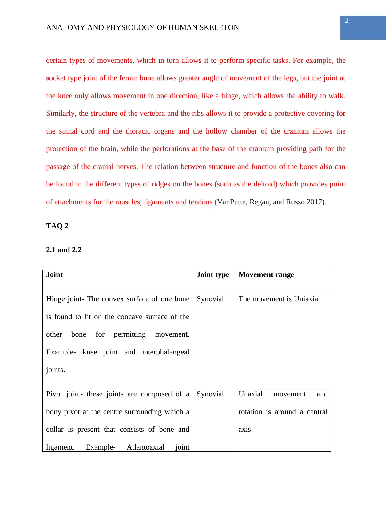
2.1 and 2.2
Joint Joint type Movement range
Hinge joint- The convex surface of one bone
is found to fit on the concave surface of the
other bone for permitting movement.
Example- knee joint and interphalangeal
joints.
Synovial The movement is Uniaxial
Pivot joint- these joints are composed of a
bony pivot at the centre surrounding which a
collar is present that consists of bone and
ligament. Example- Atlantoaxial joint
Synovial Unaxial movement and
rotation is around a central
axis
Document Page
3
ANATOMY AND PHYSIOLOGY OF HUMAN SKELETON
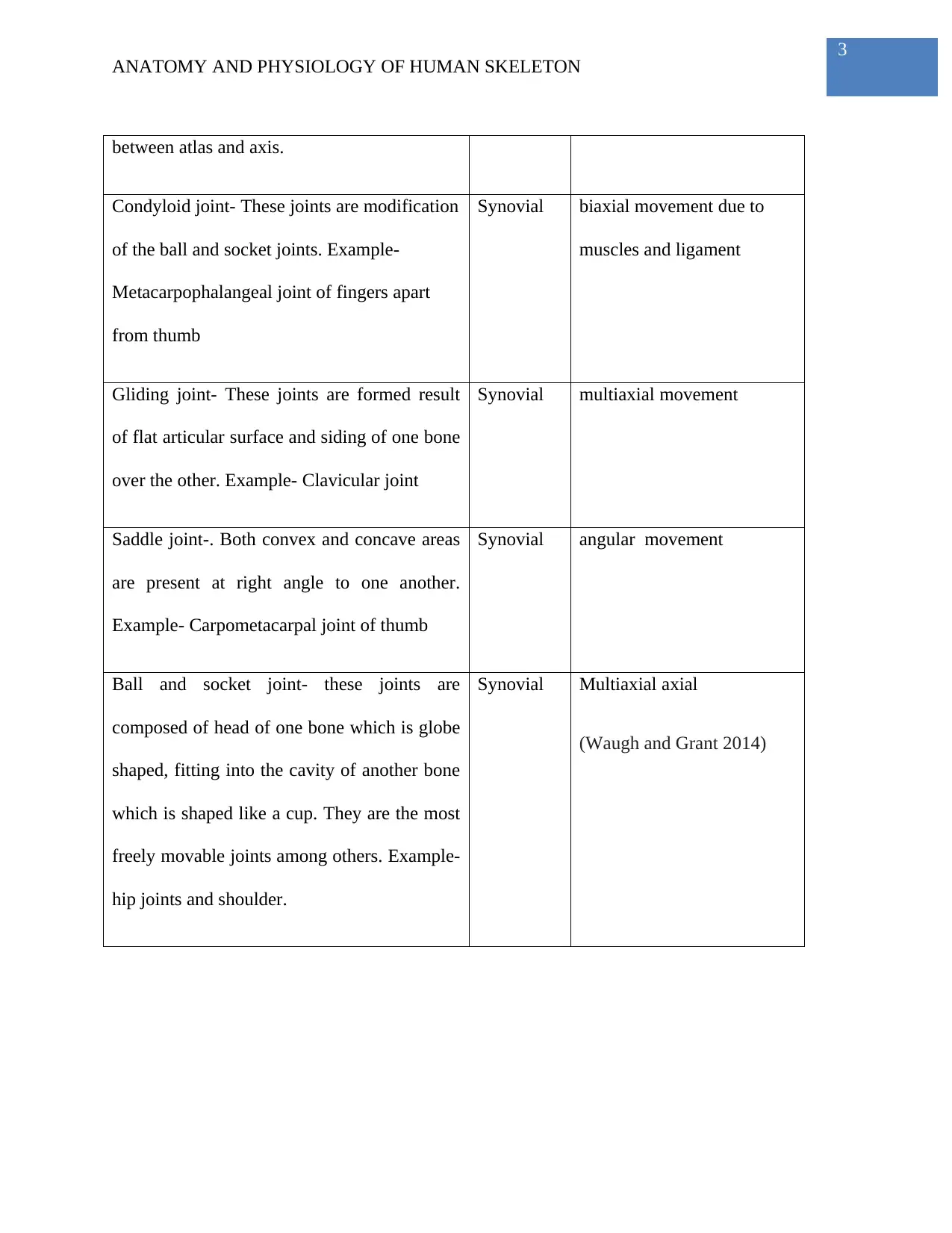
between atlas and axis.
Condyloid joint- These joints are modification
of the ball and socket joints. Example-
Metacarpophalangeal joint of fingers apart
from thumb
Synovial biaxial movement due to
muscles and ligament
Gliding joint- These joints are formed result
of flat articular surface and siding of one bone
over the other. Example- Clavicular joint
Synovial multiaxial movement
Saddle joint-. Both convex and concave areas
are present at right angle to one another.
Example- Carpometacarpal joint of thumb
Synovial angular movement
Ball and socket joint- these joints are
composed of head of one bone which is globe
shaped, fitting into the cavity of another bone
which is shaped like a cup. They are the most
freely movable joints among others. Example-
hip joints and shoulder.
Synovial Multiaxial axial
(Waugh and Grant 2014)

Paraphrase This Document

Need a fresh take? Get an instant paraphrase of this document with our AI Paraphraser
Document Page
4
ANATOMY AND PHYSIOLOGY OF HUMAN SKELETON
TAQ 3
2.3
The action of the leg in running movement takes place in a sagittal plane about a frontal
axis and the hip, knee and ankle joints is involved in it. In this regard, the bones of the hip that
have an involvement are the femur and pelvic girdle which form a ball and socket joint. The knee
bones such as the tibia and femur a hinge joint. The ankle bones that are involved are the
calcaneus and tibia which form a modified joint. These mentioned joints are known to be
producing two distinct actions. The first phase is the driving phase when there is contact of the
leg with the ground and recovery phase is when there is no contact of the leg with the ground.
The action carried out by the knee is extension. The quadriceps group of muscles is the agonist
muscles working in this regard. The action carried out by the ankle is plantar flexion.
. The arms and legs are swing in opposite pairs in sync at the time of running. The arms
are pumped forward and backward in line with the movement direction. The swing of the arms is
not across the body and the elbows point backwards. Further, the arms are in swing from the
shoulder and not the elbows. The elbows are held at a 90 degree angle, and the angle of the
elbow might be fluctuating in a slight manner at the time of the swing of the arm(VanPutte,
Regan, and Russo 2017).
3.4
The main muscles and joints that are used while using a computer are the vertebrae for
maintaining the posture. The main body movement takes place in the shoulder girdle, wrists and
elbows. During sitting, the majority of the muscles and joints of the body are in an inactive state
Document Page
5
ANATOMY AND PHYSIOLOGY OF HUMAN SKELETON
apart from the triceps and biceps. All muscles are in antagonistic pairs such as the bicep and
tricep. For the movement of the arms, the tricep is relaxed and the bicep is contracted. As a result
the arm is allowed to move in a free manner. The elbow is in flexion so that the hands can type in
a comfortable manner. Wrist, fingers and knuckles are involved when the person is using a
keyboard of the computer. The fingers are the metacarpus bones extending from the second row
of carpal bones. A bone of such characteristics that is responsible for combining with the
proximal phalanx for making up the carpometacarpal joint in the thumb, further contributing to
the thumb movement. The fingers have three individual bones and joints that are capable of
flexion and extension. Further, the movements primarily originate in the forearm (Waugh and
Grant 2014).
TAQ 5
3.2
Muscle contraction is a complex process that involves the activation of specific sites in a
muscle fiber, as a result of which the length or the tension within the muscle changes. The
contraction of certain muscles such as the skeletal muscles helps in locomotion, since these
muscles are connected to the bones, and the muscular contraction leads to these bones being
moved, thereby allowing locomotion. For example, contraction of the bicep muscle of the arm
helps in the movement of the forearm towards the body, while contraction of the triceps helps to
stretch the arm. Thus the bicep and triceps muscles work antagonistically to allow the
movements of the hand or forearm (VanPutte, Regan, and Russo 2017.
3.3
Document Page
6
ANATOMY AND PHYSIOLOGY OF HUMAN SKELETON
The antagonist muscles are the one that are responsible for producing an opposing joint
torque to the agonist muscles. The torque aids in the control of motion of the part of the body.
The terms flexion refers to the bending movement in case of which the angle between the two
parts is decreased. The muscles are the flexor muscles and the opposing muscle of a flexor is
termed as extensor muscle. For each of these pairs, one muscle has the role of agonist while the
other has the role of antagonist. The agonist undergoes contraction for causing the movement,
while the antagonist relaxes to stretch (VanPutte, Regan, and Russo 2017).

Secure Best Marks with AI Grader

Need help grading? Try our AI Grader for instant feedback on your assignments.
Document Page
7
ANATOMY AND PHYSIOLOGY OF HUMAN SKELETON
References
VanPutte, C.L., Regan, J.L. and Russo, A., 2017. Seeley's anatomy & physiology. McGraw-Hill
Education.
Waugh, A. and Grant, A., 2014. Ross & Wilson Anatomy and Physiology in Health and Illness
E-Book. Elsevier Health Sciences.
1 out of 8
[object Object]

Your All-in-One AI-Powered Toolkit for Academic Success.

Available 24*7 on WhatsApp / Email

[object Object]