logo

Techniques Used to Study Structures of Animal Cell

Describe techniques used to observe and study structures in an animal cell, including specimen preparation, cell structure and function, and modern biology techniques.

6 Pages1101 Words340 Views
   

Added on  2023-06-03

About This Document

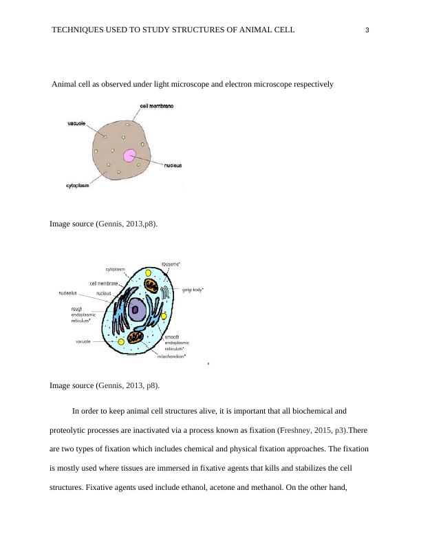
This article discusses the various techniques used to study animal cell structures, including microscopy, staining techniques, and cell fractionation. It covers the differences between light and electron microscopes, the process of fixation, and the steps involved in cell fractionation. The article also provides an overview of staining techniques and their applications.

Techniques Used to Study Structures of Animal Cell

Describe techniques used to observe and study structures in an animal cell, including specimen preparation, cell structure and function, and modern biology techniques.

   Added on 2023-06-03

ShareRelated Documents
TECHNIQUES USED TO STUDY STRUCTURES OF ANIMAL CELL 1
TECHNIQUES USED TO STUDY STRUCTURES OF ANIMAL CELL
Course name
Professor’s name
University name
City, State
Date of submission
Techniques Used to Study Structures of Animal Cell_1
TECHNIQUES USED TO STUDY STRUCTURES OF ANIMAL CELL 2
Introduction
The animal cell is the smallest unit of an organism. Animals have different types of cell
structures and cell components. These cell structures are observed and studied using cell biology
techniques. These techniques include microscope, staining techniques and Cell fractionation
(Gennis, 2013, p6).
A microscope varies from each other with different sizes and it’s appropriate. The two
common microscopes utilized in the observation of cell structures such as mitochondria are light
and electron microscope. Both of these two types have different features which are suitable for
different functions. Their similarities are that they both visualize small objects that cannot be
seen by naked eye, both are utilized in research applications and both involve specimen
preparations like staining, sectioning, and mounting. Besides, both light and electron microscope
follow Abbe’s law while visualizing small objects (Pawley, 2010, p10).
Some of the differences between the two are that a light microscope is used to view both
live and dead specimens whereas electron microscope is utilized to view only dead specimens as
electrons that it uses can destruct living cells. Secondly, the resolving power for light microscope
very low and is usually below 3 micrometers whereas electron microscope has a high-resolution
power of up to 0.01 nanometers. The other difference between the two is that a light microscope
utilizes light and glass lenses to view images whereas the electron microscope utilizes beam of
electrons and magnetic lenses to view images (Gennis, 2013, p8).
Techniques Used to Study Structures of Animal Cell_2
TECHNIQUES USED TO STUDY STRUCTURES OF ANIMAL CELL 3
Animal cell as observed under light microscope and electron microscope respectively
Image source (Gennis, 2013,p8).
Image source (Gennis, 2013, p8).
In order to keep animal cell structures alive, it is important that all biochemical and
proteolytic processes are inactivated via a process known as fixation (Freshney, 2015, p3).There
are two types of fixation which includes chemical and physical fixation approaches. The fixation
is mostly used where tissues are immersed in fixative agents that kills and stabilizes the cell
structures. Fixative agents used include ethanol, acetone and methanol. On the other hand,
Techniques Used to Study Structures of Animal Cell_3

End of preview

Want to access all the pages? Upload your documents or become a member.

Related Documents
Biochemistry Name of the University Author
|18
|2748
|113

Tissue and Organ Staining Assessment Practical
|11
|2350
|261

Cell Biology: Structure and Function of Cells
|10
|1391
|93

Electron Microscopy Of Subcellular Structure
|9
|2408
|169

Study on Massons Trichrome (MT) Procedure
|5
|1040
|94

Assignment on Lab Report - 1
|7
|1422
|19