ProductsLogo
LogoStudy Documents
LogoAI Grader
LogoAI Answer
LogoAI Code Checker
LogoPlagiarism Checker
LogoAI Paraphraser
LogoAI Quiz
LogoAI Detector
PricingBlogAbout Us
logo

Biology: Nucleus, Rough Endoplasmic Reticulum, Cell Membrane, Glycolipids, Cholesterol, Membrane Proteins, Transport Proteins, Carbohydrate Group, Bulk Transport

Verified

Added on  2023/05/31

|10
|2140
|257
AI Summary
This article discusses the structure and function of the nucleus, rough endoplasmic reticulum, and cell membrane. It also covers glycolipids, cholesterol, membrane proteins, transport proteins, carbohydrate group, and bulk transport.

Contribute Materials

Your contribution can guide someone’s learning journey. Share your documents today.
Document Page
Running head: BIOLOGY
BIOLOGY
Name of the Student:
Name of the University:
Author note:

Secure Best Marks with AI Grader

Need help grading? Try our AI Grader for instant feedback on your assignments.
Document Page
1BIOLOGY
Nucleus
The nucleus of the cell forms one of its most essential organelles, and is commonly
prevalent in the cells of eukaryotes. Due to its high level of specialization, the nucleus functions
as one of the key functional centers within the cell, exhibiting functions associated with the
processing of information and administration of essential activities of the cell (Caelro-Cuenca.
Janota and Gomes 2018). One of the major functions of the nucleus lies in it storage of the
genetic material of the cell, in the form of DNA molecules required for chromosomal formation,
known as histones. Additional functions of the nucleus include the ribosomal formation within
the nucleolus, participation in the transmission of hereditary data to the daughter cells from its
parent cells, maintenance of cellular metabolic activities through enzymatic regulation and
contribution to the growth and maturity of novel cells through chromosomal information
contained within itself (Hancock, 2018).
Rough Endoplasmic Reticulum
The rough endoplasmic reticulum is a organelle present in a cell’s endo-membrane
system. The organelle is a subtype of endoplasmic reticulum, and is emphasized by its studded or
rough features as compared to its smooth counterpart, due to the presence of ribosomes bound
within its membranes (Wang et al. 2015). The major functions of the rough endoplasmic
reticulum include the modification, synthesis and folding of essential proteins required for
deliverance across numerous other cellular organelles or for secretion from the cell. The rough
endoplasmic reticulum also works closely with the mitochondria and hence is also involved in
Document Page
2BIOLOGY
the initiation of programmed cell death or ‘apoptosis, along with responding to the presence of
unfolded proteins (Phillips and Voeltz 2016).
Structure and Function of Cell Membrane: Fluid Mosaic Model
The cell membrane, otherwise also known as the plasma or cytoplasmsic membrane, is an
essential biological element of the cell concerned primarily with separating the internal cell
components from its external surroundings. In addition, the cell membrane is concerned with the
transportation of the cellular components, transmission of cellular signals, maintenance of the
potential of the cell as well as provision of specific shape to the cell by anchoring the
cytoskeleton (Raiborg and Stenmark 2016). In 1972, Garth L. Nicholson and S. J. Ginger stated
the fluid mosaic model in order to explain the structural components of the cell membrane. In
accordance to the model, the plasma membrane can be viewed as a mosaic of cholesterol,
carbohydrates, phospholipids and proteins, which imparts fluidity in the structure. The primary
structure of the cell membrane is comprised of molecules of phospholipids, which are
amphiphilic, possessing lipophilic and hydrophilic characteristics. The second components, is
composed of integral proteins, with complete embedding in the cell membrane followed by
interactions between hydrophobic areas of the phopholipid layer and the hydrophobic areas of
the proteins. The third section of the cell membrane consists of carbohydrates, present in the
exterior regions. These form either glycoproteins or glycolipids, through binding to proteins and
lipid components (Nicholson 2014).
Document Page
3BIOLOGY
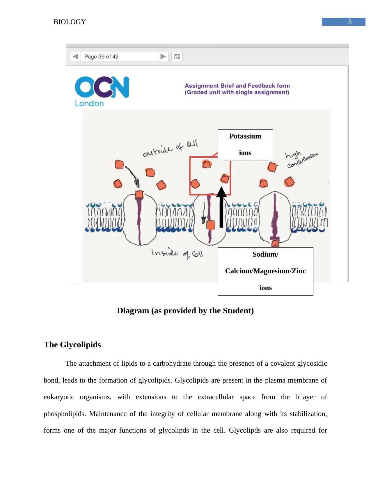
Diagram (as provided by the Student)
The Glycolipids
The attachment of lipids to a carbohydrate through the presence of a covalent glycosidic
bond, leads to the formation of glycolipids. Glycolipids are present in the plasma membrane of
eukaryotic organisms, with extensions to the extracellular space from the bilayer of
phospholipids. Maintenance of the integrity of cellular membrane along with its stabilization,
forms one of the major functions of glycolipds in the cell. Glycolipds are also required for
Potassium
ions
Sodium/
Calcium/Magnesium/Zinc
ions

Paraphrase This Document

Need a fresh take? Get an instant paraphrase of this document with our AI Paraphraser
Document Page
4BIOLOGY
recognition of cells – an essential prerequisite step necessary of initiation of tissue formation
involving cellular connections, as well as in the generation of protective responses by the
immune system (Aigal, Claudinon and Römer 2015).
The Cholesterol
Cholesterol is a substance found abundantly in the membrane of all cells. One of the key
functions of cholesterol is its involvement in the facilitation of signaling processes between cells,
which is required for cells to communicate and interact efficiently amongst each other (Derler et
al. 2016). The maintenance of cell integrity remains one of the most important function of
cholesterol within the cell membrane. A closely knit network of proteins and cholesterol, known
as phospholipids, form the cellular membrane. The amphipathic property of cholesterol, that is
possession of both hydrophobic and hydrophilic properties, contributes to the immobilization of
the cell membrane resulting in its semi-permeability – further preventing the uncontrolled
movement of water-soluble compounds across it (Neuvonen et al. 2014).
The Membrane Proteins
Proteins which form a part of the structure of the cell membrane and are also involved in
interaction with its components are known as membrane proteins. Membrane proteins can be
classified as peripheral membrane proteins and integral membrane proteins. Peripheral
membrane proteins exhibit temporary cell membrane adherence while permanent attachment is
exhibited by integral membrane proteins (Laganowsky et al. 2014). Integral monotropic proteins
attached to a single side and transmembrane proteins travelling across the membrane form
further subtypes of integral membrane proteins. Membrane proteins are involved in a number of
Document Page
5BIOLOGY
functions. These include relaying of cell signals across extracellular and intracellular
environments by membrane receptor proteins (hormones, nutrients, cytokines, growth factors),
movement of substances across membranes by transport proteins (cytochromes, GLUT1),
initiation of cell identification and interaction by cell adhesion molecules (Integrins, Cadherins,
IgSF CAMs, selectins) and enzymatic functions such as those of transferases, oxidoreductases
and hydrolases by membrane enzymes (Liang and Tamm 2016).
Transport Proteins
The function of transporters or transport proteins involve the facilitation of movement of
substances across the plasma membrane, which include macromolecules and ions. Such proteins
initiate the required movement through processes such as active transport and facilitated
diffusion (Lytovchenko and Kunji 2017). Transport proteins can be classified into several types
such as electron carriers (NADPH oxidase, disulfide bond oxidoreductases), group translocators,
electrochemical potential-driven transporters (glucose transporter, excitatory amino acid
transporters, monoamine transporters), primary active transporters (ATP-binding cassette
transporters, decarboxylation driven transporters, oxidoreduction transporters) and pores or
channels (holins, beta-barrel porins, alpha-helical protein channels, nonribosomally synthesized
channels) (Jaehme and Slotboom 2015).
The Carbohydrate Group
Carbohydrates are sugar-based compounds present naturally in the environment and can
be classified as simple (monosaccharides such as fructose and glucose, disaccharides such as
sucrose) and complex (starches such as polysaccharides). Carbohydrates contribute to the
Document Page
6BIOLOGY
structural protection of the cell known as ‘glycocalyx’, which aids in nutrient absorption by gut
microvilli, resistance against high blood pressure and productive enzymes for digestion.
Carbohydrates also aid in cell recognition and form an integral component of cellular plasma
proteins such as enzymes and hormones (Allam et al. 2014).
Bulk Transport
The mechanism of bulk transport involve the movement of large particles across the
plasma membrane. Exocytosis an endocytosis are the major types of bulk transport. Endocytosis
mode of transport involve invagination or inward folding of the plasma membrane surrounding
the concerned compound, further leading to transport through new formation of an internal
vacuole or vesicle. Phagocytosis (used by macrophages), receptor-mediated endocytosis
(performed by receptor proteins) and pinocytosis (for absorption of extracellular fluid) are
further subtypes of endocytosis (O’Kelly 2015). Exocytosis mode of bulk transport involves
extracellular transport of materials from the cell through formation of membrane-enclosed
vesicles. Such vesicles are primarily composed of waste products or proteinaceous substances
formed by the Golgi apparatus (Jiang et al. 2017).

Secure Best Marks with AI Grader

Need help grading? Try our AI Grader for instant feedback on your assignments.
Document Page
7BIOLOGY
References
Aigal, S., Claudinon, J. and Römer, W., 2015. Plasma membrane reorganization: A glycolipid
gateway for microbes. Biochimica et Biophysica Acta (BBA)-Molecular Cell Research, 1853(4),
pp.858-871.
Allam, H., Aoki, K., Benigno, B.B., McDonald, J.F., Mackintosh, S.G., Tiemeyer, M. and
Abbott, K.L., 2014. Glycomic analysis of membrane glycoproteins with bisecting glycosylation
from ovarian cancer tissues reveals novel structures and functions. Journal of proteome
research, 14(1), pp.434-446.
Calero-Cuenca, F.J., Janota, C.S. and Gomes, E.R., 2018. Dealing with the nucleus during cell
migration. Current opinion in cell biology, 50, pp.35-41.
Derler, I., Jardin, I., Stathopulos, P.B., Muik, M., Fahrner, M., Zayats, V., Pandey, S.K., Poteser,
M., Lackner, B., Absolonova, M. and Schindl, R., 2016. Cholesterol modulates Orai1 channel
function. Sci. Signal., 9(412), pp.ra10-ra10.
Hancock, R., 2018. Crowding, entropic forces, and confinement: crucial factors for structures
and functions in the cell nucleus. Biochemistry (Moscow), 83(4), pp.326-337.
Jaehme, M. and Slotboom, D.J., 2015. Diversity of membrane transport proteins for vitamins in
bacteria and archaea. Biochimica et Biophysica Acta (BBA)-General Subjects, 1850(3), pp.565-
576.
Jiang, L.Q., Wang, T.Y., Webster, T.J., Duan, H.J., Qiu, J.Y., Zhao, Z.M., Yin, X.X. and Zheng,
C.L., 2017. Intracellular disposition of chitosan nanoparticles in macrophages: intracellular
Document Page
8BIOLOGY
uptake, exocytosis, and intercellular transport. International journal of nanomedicine, 12,
p.6383.
Laganowsky, A., Reading, E., Allison, T.M., Ulmschneider, M.B., Degiacomi, M.T., Baldwin,
A.J. and Robinson, C.V., 2014. Membrane proteins bind lipids selectively to modulate their
structure and function. Nature, 510(7503), p.172.
Liang, B. and Tamm, L.K., 2016. NMR as a tool to investigate the structure, dynamics and
function of membrane proteins. Nature structural & molecular biology, 23(6), p.468.
Lytovchenko, O. and Kunji, E.R., 2017. Expression and putative role of mitochondrial transport
proteins in cancer. Biochimica et Biophysica Acta (BBA)-Bioenergetics, 1858(8), pp.641-654.
Neuvonen, M., Manna, M., Mokkila, S., Javanainen, M., Rog, T., Liu, Z., Bittman, R.,
Vattulainen, I. and Ikonen, E., 2014. Enzymatic oxidation of cholesterol: properties and
functional effects of cholestenone in cell membranes. PLoS One, 9(8), p.e103743.
Nicolson, G.L., 2014. The Fluid—Mosaic Model of Membrane Structure: Still relevant to
understanding the structure, function and dynamics of biological membranes after more than 40
years. Biochimica et Biophysica Acta (BBA)-Biomembranes, 1838(6), pp.1451-1466.
O’Kelly, I., 2015. Endocytosis as a mode to regulate functional expression of two-pore domain
potassium (K2P) channels. Pflügers Archiv-European Journal of Physiology, 467(5), pp.1133-
1142.
Phillips, M.J. and Voeltz, G.K., 2016. Structure and function of ER membrane contact sites with
other organelles. Nature reviews Molecular cell biology, 17(2), p.69.
Document Page
9BIOLOGY
Raiborg, C. and Stenmark, H., 2016. Plasma membrane repairs by small GTPase Rab3a. J Cell
Biol, 213(6), pp.613-615.
Wang, P.T., Garcin, P.O., Fu, M., Masoudi, M., St-Pierre, P., Panté, N. and Nabi, I.R., 2015.
Distinct mechanisms controlling rough and smooth endoplasmic reticulum contacts with
mitochondria. J Cell Sci, 128(15), pp.2759-2765.
1 out of 10
[object Object]

Your All-in-One AI-Powered Toolkit for Academic Success.

Available 24*7 on WhatsApp / Email

[object Object]