DNA Structure: Genes, Chromosomes, Replication, Mitosis and Meiosis
VerifiedAdded on 2023/05/31
|7
|1725
|433
AI Summary
This article explains the structure of DNA, genes, and chromosomes. It also covers DNA replication, mitosis, and meiosis. The importance of meiosis in generating variation is also discussed. The article cites relevant studies and research to support the information presented.
Contribute Materials
Your contribution can guide someone’s learning journey. Share your
documents today.
DNA Structure 1
DNA Structure 3
DNA Structure
By
Course
Lecturer
University
Department
Date
DNA Structure 3
DNA Structure
By
Course
Lecturer
University
Department
Date
Secure Best Marks with AI Grader
Need help grading? Try our AI Grader for instant feedback on your assignments.
DNA Structure 2
1.1: Structures of DNA, genes and chromosomes
DNA STRUCTURE
According to Saenger (2013) deoxyribonucleic acid is composed of nucleotide
molecules. Each nucleotide molecule contains a sugar group, nitrogen base and phosphate
group. Nitrogen bases include cytosine (C), guanine (G), adenine (A) and thymine (T). The
order of the bases determines genetic code or deoxyribonucleic acid instructions. The human
DNA has about 3 billion bases.
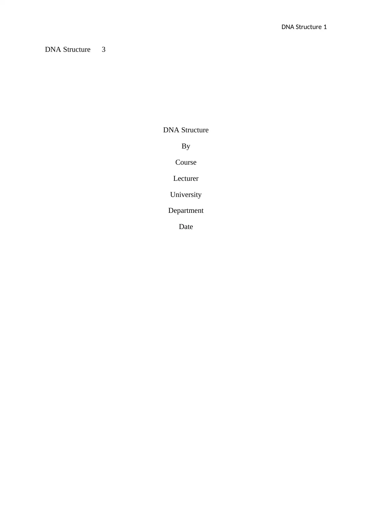
The sequence of DNA creates genes which tell how to make proteins. Molecules of
DNA are long in such that they cannot fit in cells with no proper packing. It contains
organism instructions needed to reproduce, live and develop. Nucleotides attached to create
two spiral strands to form a double helix structure (Saenger 2013).
GENE STRUCTURE
A gene is a section of deoxyribonucleic acid composed of amino acid information
coding for a polypeptide chain and other regulatory sequences that are important for
appropriate expression Hay (2013). A gene is a DNA sequence that is significant for the
manufacture of functional produce (RNA molecule or polypeptide). Genes are discontinuous
in human (eukaryotic genomes). Not all genes are active in cells which show that not all are
expressed.
According to Hay (2013), there are two types of genes: multigene families and
operons. It indicates that cells acquire specialised functions (differentiated). Gene's
expression regulates promoter regions. There are regulatory elements which include silencers,
locus control regions and enhancers. Both regulatory elements and promoters are sites of
genetic disease mutation which alter with regular gene expression. Genes are set along the
chromosomes. Genes have a definite, specific position called gene locus.
DNA Structure 3
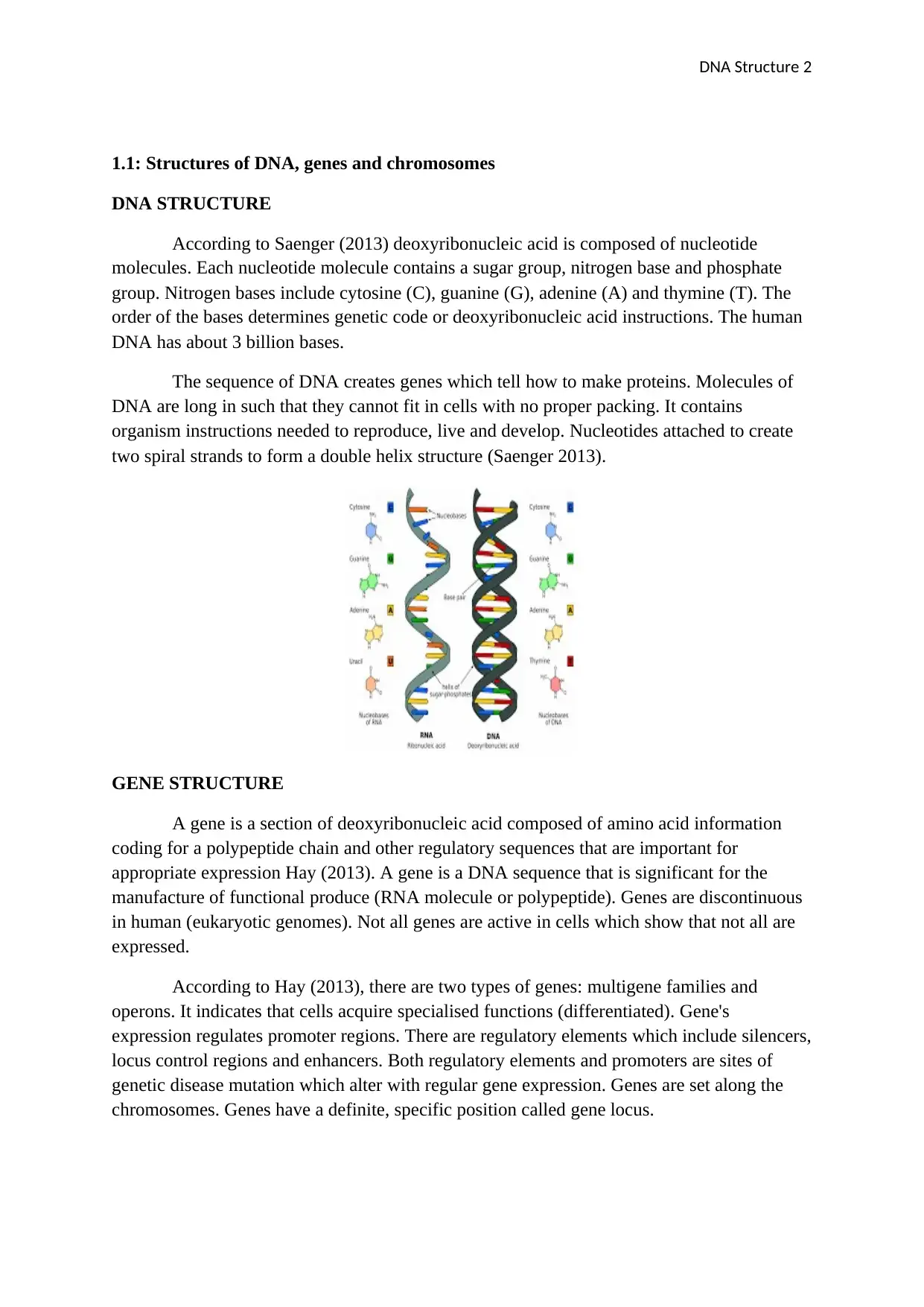
According to Hay (2013),
CHROMOSOMES STRUCTURE
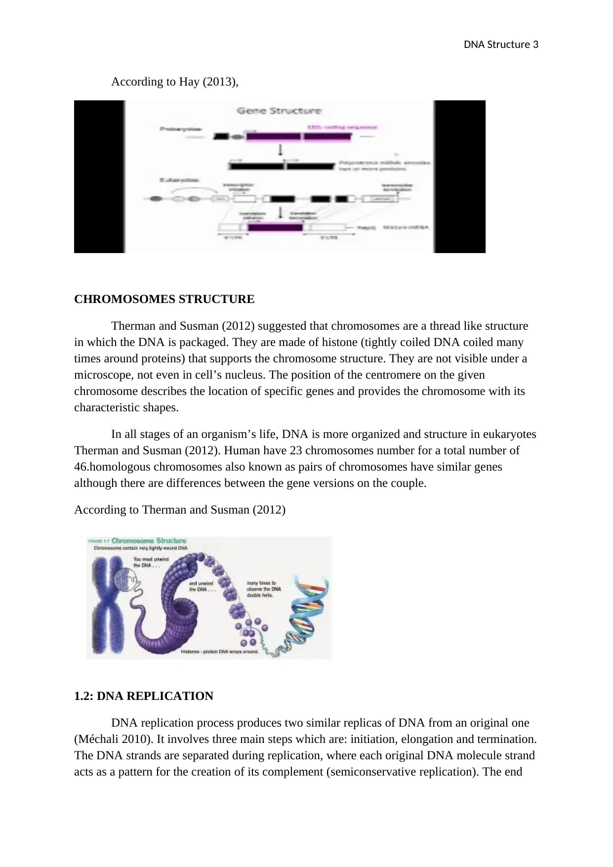
Therman and Susman (2012) suggested that chromosomes are a thread like structure
in which the DNA is packaged. They are made of histone (tightly coiled DNA coiled many
times around proteins) that supports the chromosome structure. They are not visible under a
microscope, not even in cell’s nucleus. The position of the centromere on the given
chromosome describes the location of specific genes and provides the chromosome with its
characteristic shapes.
In all stages of an organism’s life, DNA is more organized and structure in eukaryotes
Therman and Susman (2012). Human have 23 chromosomes number for a total number of
46.homologous chromosomes also known as pairs of chromosomes have similar genes
although there are differences between the gene versions on the couple.
According to Therman and Susman (2012)
1.2: DNA REPLICATION
DNA replication process produces two similar replicas of DNA from an original one
(Méchali 2010). It involves three main steps which are: initiation, elongation and termination.
The DNA strands are separated during replication, where each original DNA molecule strand
acts as a pattern for the creation of its complement (semiconservative replication). The end
DNA Structure 4
product is a new helix which comprises initial DNA strands. In cells, the replication starts at a
particular position.
Méchali (2010) stated that there is the unwinding of the double helix; the strands are
separated easily since the bonds between the strands are so weak compared to chemical
bonds. The replication fork is hen two parent strands are opened up, but partially separated.
In return, A, T would add, C, G would join. It would lead to the automatic formation of
nucleotide sequence. Hence, DNA helix regeneration will occur, with one original helix
strand merging with a newly created compliment to start a DNA strand molecule. This
replication is known as zipper duplication.
2.1: EVENTS OF MITOSIS AND MEIOSIS
a. MITOSIS
It is a eukaryotic cell separation from where parent cells generate two daughter cells
with a similar genetic constituent. It's a process that is continuous and divided into five
events: telophase, anaphase, metaphase, prometaphase and prophase. According to Matos et
al. (2011), the following are the stages;
Prophase
It occupies half of the mitosis. Nucleolus disintegration and small vesicles formed as a
result of nuclear membrane breakdown. Centrosome divides its self-giving two daughter
centrosomes which shift to the opposite of the cell. Microtubules are produced by
centrosomes, and mitotic spindle formed. Sister chromatids formed as a result of the
replicated chromosome and a centromere holds them.
Prometaphase
There is a migration of chromosomes lad by their centromeres. A structure associated
with centromere into which spindle fibres bind in each chromosome referred to as
kinetochore.in this stage there is condensation of chromosomes.
Metaphase
There is an alignment of chromosomes along metaphase plate.
Anaphase
In mitosis, it is the shortest stage. There is disjoining of sister chromatids of each
chromosome and division of centromeres. Daughter chromosomes are separated sister
chromatids.
Telophase
It's the last of mitosis. There is a reformation of the nuclear membrane around
grouped chromosomes on either pole of the cell, uncoiling of chromosomes and turn out to
diffuse, and disappearance of spindle fibres
Secure Best Marks with AI Grader
Need help grading? Try our AI Grader for instant feedback on your assignments.
DNA Structure 5
Cytokinesis
There is the formation of two new cells, then enters interphase.
b. MEIOSIS
Handel and Schimenti (2010) said that haploid sex's cells produced in eukaryotic cell
division from diploid cells. There are two successive cellular divisions (Meiosis I and
Meiosis II). Meiosis headed by DNA replication process which coverts chromosomes to
sister chromatids.
Meiosis I
It's where pairs of homologous chromosomes are separated.it has five main stages: prophase
I, Metaphase I, anaphase I, telophase I. The diploid is reduced to haploid during cell division.
Prophase I: this stage divided into five phase: leptotene, zygotene, pachytene, diplotene,
diakinesis. There is condensation of chromosomes, crossing over of homogeneous
chromosomes and chromosomes recombination.
Metaphase I: chromosomes attachment to spindle fibres and line up at the center of cells
Anaphase I: movement of chromosomes starts, and the spindle fibres shortens.
Telophase I: formation of nuclear membrane as chromosomes reaches the opposite end.
Cytokinesis: occurrence of cell division
Meiosis II
There is a separation of the chromosome into chromatids.
Prophase II: There is condensation of chromosomes, spindle fibres forms and nuclear
membrane dissolves.
Metaphase II: attachment of spindle fibres to chromosomes, lining up of the center of the
cell.
Anaphase II: centromere Division and movement of sister chromatids to opposite ends as a
shortening of spindle fibres.
Telophase II: occurrence of nuclear membrane and chromosomes reach opposite ends.
2.2: COMPARISON OF MEIOSIS AND MITOSIS PRODUCTS
Mitosis products result to two identical daughter cells whereas meiosis gives four gametes as
it involves two fissions of the nucleus.
DNA Structure 6
Meiosis is in all organisms and involves sexual reproduction while mitosis found in the
single-celled organism and aids in tissue growth and reproduction are asexual.
In meiosis, there is crossing over while in mitosis there is no crossing over.
There are four haploid cells in meiosis while in mitosis there are two
The chromosome number reduces by half in meiosis while in mitosis remains the same
Centromeres do separate during anaphase II but do not separate during anaphase I in meiosis,
but in mitosis, splitting occurs during anaphase.
2.3: IMPORTANCE OF MEIOSIS IN GENERATING VARIATION
Webster and Hurst (2012) suggested that meiosis produce variation in young since
the method transposes genes along chromosomes and then splits half of the chromosomes
into gametes. Chromosomal differences act as significant factors in biological diversity and
evolutionary fitness.
Chromosomal differences in a populace of animals show that dissimilar animals have
different weaknesses and strengths (Webster 2012). It is a significant aspect of a species
capability to persist and raise its populace since original predators reveal or food becomes
less thus many animals will die. Due to chromosomal differences, some animals survive since
they perform things like eating different foods and run faster. Consequently, genetic variation
raises opportunities of members persisting.
DNA Structure 7
References
Handel, M.A. and Schimenti, J.C., 2010. Genetics of mammalian meiosis: regulation,
dynamics and impact on fertility. Nature Reviews Genetics, 11(2), p.124.
Hay, E.D. ed., 2013. Cell biology of extracellular matrix. Springer Science & Business Media
Matos, J., Blanco, M.G., Maslen, S., Skehel, J.M. and West, S.C., 2011. Regulatory control
of the resolution of DNA recombination intermediates during meiosis and mitosis. Cell,
147(1), pp.158-172.
Méchali, M., 2010. Eukaryotic DNA replication origins: many choices for appropriate
answers. Nature reviews Molecular cell biology, 11(10), p.728.
Saenger, W., 2013. Principles of nucleic acid structure. Springer Science & Business Media.
Therman, E. and Susman, M., 2012. Human chromosomes: structure, behavior, and effects.
Springer Science & Business Media.
Tiang, C.L., He, Y. and Pawlowski, W.P., 2012. Chromosome organization and dynamics
during interphase, mitosis, and meiosis in plants. Plant physiology, 158(1), pp.26-34.
Webster, M.T. and Hurst, L.D., 2012. Direct and indirect consequences of meiotic
recombination: implications for genome evolution. Trends in genetics, 28(3), pp.101-109.
1 out of 7
Your All-in-One AI-Powered Toolkit for Academic Success.
+13062052269
info@desklib.com
Available 24*7 on WhatsApp / Email
Unlock your academic potential
© 2024 | Zucol Services PVT LTD | All rights reserved.