Genetics Practical: Haemoglobin Variants
VerifiedAdded on 2023/04/11
|15
|3331
|240
AI Summary
This document discusses the different types of haemoglobin variants, their biological importance, and the symptoms and effects of low and high haemoglobin levels. It also includes an analysis of the migration of red blood cells and the results of G banding analysis. Additionally, it explores the diagnosis of gynecomastia and its genetic implications.
Contribute Materials
Your contribution can guide someone’s learning journey. Share your
documents today.

Running Head: Genetics Practical 1
Genetics Practical
Name of the student
Name of the institution
Genetics Practical
Name of the student
Name of the institution
Secure Best Marks with AI Grader
Need help grading? Try our AI Grader for instant feedback on your assignments.
Genetics Practical 2
Haemoglobin Variants Practical
Introduction
Haemoglobin is a red protein usually found attached t the red blood cells that have a high
affinity for oxygen. Oxygen in the lungs is attached to the haemoglobin the blood and
transported to various areas in the body. The body tissues require oxygen to maintain and repair
itself. Haemoglobin also gives the red blood cells their unique function. Haemoglobin is
measured through a blood test. It is usually expressed in grams per deciliter (g/dL).
A patient with a low level of haemoglobin will consequently have low levels of oxygen
in the body. A patient is said to be anaemic if they have a haemoglobin level of 13.5 g/dL for
males and 12 g/dL in females. The haemoglobin levels in children vary with age. Some of the
symptoms of low haemoglobin are weakness, shortness of breath, dizziness, irregular heartbeat,
headache, cold hands and feet, chest pains and pale skin.
On the other hand, having a high haemoglobin level could be a symptom of
polycythemia, a condition where the body produces excess red blood cells. The blood becomes
thicker and the patient is at a higher risk of heart attacks, blood clots and stroke. The condition is
fatal if not treated early. High levels of haemoglobin can also be caused by smoking,
dehydration, living in high altitudes or the presence of an underlying condition (Khoshouei,
Radjainia, Baumeister and Danev 2017).
There are different types of haemoglobin, HbA0, HbA2, HbF, HbS and HbC each
containing a haem group and anion bound to it. HbA is found in normal human beings. It
contains two alpha and two beta chain. It is the most common type of haemoglobin and accounts
for 97% of the total red blood cells. HbA2 is found is some human beings and is considered
normal. It also contains two alpha and two beta chains. HbA2 may be found in high levels in
Genetics Practical 3
people who have beta thalassemia both in the heterozygous and homozygous state. It found in
1.5 to 3.1% of the red blood cells. It occurs in normal levels in patients suffering from sickle cell
anaemia. The biological importance of HbA22 is unknown.
HbF is found in the growing fetus. It contains two alpha and two beta chains. The major
role of HbF is transporting oxygen from the mother to fetus in an efficiently low oxygen
environment. The production of HbF is reduced immediately after birth and by the time the child
is between 1-2 years, there is no more production. However, HbF production may be stimulated
by congenital disorders such as sickle cell anaemia. Individuals who suffer from sickle cell
anaemia and have increase HbF production exhibit mild symptoms of the diseases. HbF inhibits
the precipitation of the sickle cell and makes up for the rapid haemolysis f the unstable HbS red
blood cells. The HbF is also increased in Hereditary Persistence of Fetal Hemoglobin (HPFH).
HbF levels rise without any clinical symptoms. It is a very rare mutation.
HbS arises due to a defect in the beta chain where a mutation occurred in the codons
resulting in translation of valine instead of glutamic acid. The valine is naturally present at
position one while the glutamic position occupied position 6. Presence of the two valine forms a
hydrophobic interaction producing sticky patch when the haemoglobin molecules combine to
form a chain the sticky sites of the HbS precipitate. The precipitated sinks the red blood cells
causing the biconcave shape to become a sickle shape. The lifespan of the HbS is about 30 days.
This is less than half of the life span of the regular red blood cells. In addition, HbS is very
unstable. There is excessive haemolysis of the red blood cell resulting in anaemia. Sickle red
blood cells are very rigid and can easily block the blood vessels causing pain, impaired
circulation and internal bleeding. The psychological changes observed include dizziness,
shortness of breath, weakness and heart murmurs. According to the Center for Disease Control
Genetics Practical 4
and Prevention, 1 in 375 African American babies is born with this condition. There are
individuals who have a HbS that has a single beta chain. This haemoglobin is a recessive trait
and does not cause any symptoms unless it is transferred to the offspring together with mutated
haemoglobin from the other parent (Vinjamur, Bauer and Orkin 2018).
HBC is a recessive trait that is very rare in the homozygous state. The effects of the
haemoglobin are very mild. It results in mild hemolytic anaemia and mild enlargement of the
spleen. When present in heterozygotes, it is asymptomatic.
Results
A practical was conducted to analyze the migration of the red blood cells in the body.
The results obtained were as shown in the table below.
Haemoglobin variants Migration distance/cm
HbA0 9
HbA2 4
HbS 6
HbMIX 1 4, 9
HbMIX 2 6,8
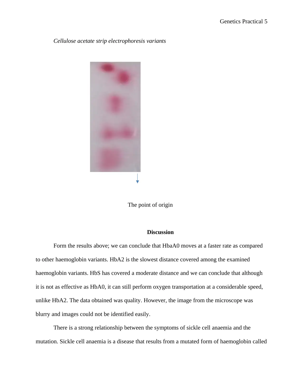
In addition, a blood smear was placed on a microscope slide and the red blood cells
observed. The aim was to analyze some the variant of haemoglobin. The results were as shown
in the diagram below.
Secure Best Marks with AI Grader
Need help grading? Try our AI Grader for instant feedback on your assignments.
Genetics Practical 5
Cellulose acetate strip electrophoresis variants
The point of origin
Discussion
Form the results above; we can conclude that HbaA0 moves at a faster rate as compared
to other haemoglobin variants. HbA2 is the slowest distance covered among the examined
haemoglobin variants. HbS has covered a moderate distance and we can conclude that although
it is not as effective as HbA0, it can still perform oxygen transportation at a considerable speed,
unlike HbA2. The data obtained was quality. However, the image from the microscope was
blurry and images could not be identified easily.
There is a strong relationship between the symptoms of sickle cell anaemia and the
mutation. Sickle cell anaemia is a disease that results from a mutated form of haemoglobin called
Genetics Practical 6
Hb, which causes the red blood cells to become sickle-shaped especially when the individuals in
areas of low oxygen. It is a hereditary disorder that is common among people of African descent.
When the red blood cells are sickle shapes, very little oxygen is transported to the rest of the
body. In addition, the sickle-shaped red blood cells are rigid and cannot easily move around the
boy. They are clogged in some of the blood vessels and blood flow is affected. There is no cure
for the disease however, there are medications that can help relieve the pain and treat the
secondary problems associated.
Some of the symptoms of anaemia vary from one person to another and tend to change
over time. However, the major symptom is anaemia. Sickle-shaped red blood cells are easily
haemolysed because they are unstable. A reduction in the number of red blood cells results in the
body. Another symptom of sickle cell anaemia is a periodic episode of pain. A patient suffering
from this disease stands to be in a lot of pain. This can be attributed to the red blood cells
clogging the blood vessels. Lack of blood flow results in pain in the bones, joints, abdomen and
chest. The intensity of the pain varies from a few hours to several weeks. Some people tend to
have less than five episodes of pain in a year while others have dozen. Some even require
hospitalization in order to survive(Taylor‐Robinson, Maayan, Soares‐Weiser, Donegan and
Garner, 2015)
Another symptom of the diseases is swelling of hands and feet due to the acclamation of
blood in the blocked blood vessels. Frequent infections occur due to the diseases damaging the
spleen. The spleen is the major organ involved in fighting infection. Infants and children
suffering from the sickle cell are usually given all vaccination and are given antibiotics o protect
them from life-threatening illnesses like pneumonia. Delayed growth is observed in children
suffering from the disease. The body requires oxygen among other nutrients in order to grow and
Genetics Practical 7
mature. Lack of organs receiving the appropriate amount of oxygen slows down growth in
infants and delays puberty. Visual problems are common in patients suffering from this disease.
The eyes have the most fragile and smallest blood vessels. The clogging of these vessels with
sickle-shaped anaemia damages blood flow and consequently damages the retina (the part of the
eye responsible for processing visual images).
Sickle cell anaemia has adverse effects at the cellular level, organism level and
population level. At the cellular level, it affects the normal function of other cells. Haemolysis
results in a reduction in the number of available cells, which in turn affect the function of the
cells in general. At the organism level, the disease results in several health complications. The
individual is constantly tired and suffering from one secondary illness to another. It can also
result in the death of the individual. At the population level, the disease reduces the size of the
population through death. The number of individuals who are productive also reduces drastically
affecting the economic status of the population. The economic status of the population reduces
since the individual spends a lot of money on medication to treat the symptoms associated with
the diseases. The diseases are inheritable as a recessive gene. this allows it to be propagated from
one generation to the next (Muñoz et al., 2015)
Paraphrase This Document
Need a fresh take? Get an instant paraphrase of this document with our AI Paraphraser
Genetics Practical 8
G Banding Practical
To the nurse/midwife in charge,
The patent DD was diagnosed with gynecomastia in the last year. The 15-year-old male
was found to have small testes through the physical exams. He had a lot of difficulty with
learning in school especially in spelling and composition. Throughout his childhood, he had
always shown impulsive behaviours and was very irritable. The patient is tall for his age. His
height falls over the 95 the percentile in the height by age for the boys. The student has had no
history of any medical problems. The patient was undergoing an investigation and his blood was
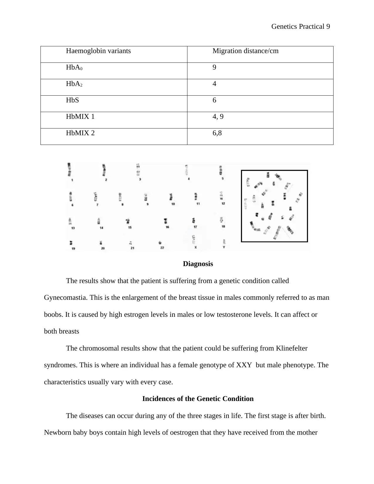
subjected to karyotype analysis. The G banding analysis produced a partially completed
karyotype.
Lab Procedure
G banding is also called Gienma banding. It is a technique that produces a visible
karyotype through the staining and condensing of chromosomes. Blood is obtained and placed on
a slide as a smear. M-FISH is applied to the slides. The assay system used is the Spectra Vision.
It consists of a 5' labelled, 52-probe mixture of WCP. These DNA probes are a representation of
the 24 chromosomes found in humans. The Whole chromosome paint is labelled with a variety
of fluorophores that produce 24 unique colours if a combination of chromosomes occurs. The
fluorophores used to in labelling are Spectrum Far Red, Spectrum Aqua, Spectrum Red,
Spectrum green and spectrum Gold. Addition of Cot 1 DNA to the mixture helps to suppress the
combination of common sequences thus increasing the sensitivity. The slides were allowed to
hybridize. The results were viewed using fluorescent microscopy. M FISH g banding images
were shown by M FISH software (Xie et al.2016)
The results were as follows.
Genetics Practical 9
Haemoglobin variants Migration distance/cm
HbA0 9
HbA2 4
HbS 6
HbMIX 1 4, 9
HbMIX 2 6,8
Diagnosis
The results show that the patient is suffering from a genetic condition called
Gynecomastia. This is the enlargement of the breast tissue in males commonly referred to as man
boobs. It is caused by high estrogen levels in males or low testosterone levels. It can affect or
both breasts
The chromosomal results show that the patient could be suffering from Klinefelter
syndromes. This is where an individual has a female genotype of XXY but male phenotype. The
characteristics usually vary with every case.
Incidences of the Genetic Condition
The diseases can occur during any of the three stages in life. The first stage is after birth.
Newborn baby boys contain high levels of oestrogen that they have received from the mother
Genetics Practical 10
while still developing in the womb. As a result, baby boys are born with enlarged breasts. These
hormones usually balance and the disease clears naturally three weeks after birth. The second
phase is at puberty. Between 12 and 14 years of age, there is a lot of hormone change as the body
undergoes puberty. The varying production of hormones can result in gynecomastia. The
condition clears out by itself six t 24 months after the first onset of puberty symptoms. The last
phase is during midlife. Breast enlargement is very high in men between the ages of 50 and 80. A
recent study conducted showed that one out of every four men has breast enlargement at this age.
There are other conditions that can lead to incidences of the disease. They include obesity, liver
diseases, kidney failure, and lack of a proper diet, hypothyroidism, and tumours in the adrenal
gland s or in the testes, hypogonadism or hypoandrogenism. Certain drugs can also trigger
gynecomastia. For example anabolic steroids that athletes use to enhance performance, human
gonadotrophin used for fertility treatments, estrogens, metronidazole used for bacterial infection,
ketoconazole used for fungal infections, finasteride used to enlarge the prostate, cimetidine used
for heartburn and as a proton pump inhibitor, diazepam used to treat anxiety, spironolactone and
other related calcium channel blockers. In additions, drugs such as heroin, marijuana and alcohol
can also play a role.
Uneven crossing causes the mutation in the karyosomes during meiosis. The X
chromosome is translocated fails to separate. The result is an xxy male. The diseases can occurs
because of autosomal mutation in the X chromosome. The individuals are usually raised as males
and given a male gender identity. However, some may have ambiguous genitalia. The
individuals may need to undergo treatment to induce some of the secondary male characteristics
such as deepening of the voice and masculinity. Hormone treatment is done to prevent the
development of some of the secondary female characteristics (Khattab et al., 2017)
Secure Best Marks with AI Grader
Need help grading? Try our AI Grader for instant feedback on your assignments.
Genetics Practical 11
Causes of the Genetic Condition
The disease is caused by an imbalance of hormones in the body. The two hormones,
estrogen and testosterone, are present in abnormal quantities in the bloodstream. Estrogen is
responsible for the female characteristics while testosterone is responsible for the male
characteristics. Although estrogen is responsible for female characteristics, male produce a small
quantity of the hormone. Consequently, even though testosterone is responsible for male traits;
females also produce a small quantity. Excess production of the estrogen in males results in
gynecomastia
Males have one X and one Y chromosome in their cells. Females have two X
chromosomes. In Klinefelter syndrome, the patient has two XX chromosomes and one Y
chromosomes. The patient has both the male and the female characteristics. The two XX
chromosomes from the mother fail to separate during meiosis, the XY chromosome from the
father separate normally and the result is an XXY baby. On the other hand, the two
chromosomes XX from the mother may separate but the XY chromosomes fail to separate
resulting in an XXY baby (Mohammadpour et al. 2017).
Clinical Presentation of the Condition
Diagnosis is done using a physical exam or through g bands. The illness manifested as
breast pain, tenderness and enlargement of the breast. In a physical examination, a firm rubber
disk found just beneath the nipple area is felt. The disk of tissue is easily movable within the
breast tissue. A mammogram or a breast ultrasound is usually conducted to rule out any cancer-
related issues.
Genetics Practical 12
The appearance of the XXY males can be categorized into three groups. The first is males
that have normal genitalia, males that have external ambiguities and males that have both
genitalia. The external ambiguities may include having a small testis, micropenis, hypospadias or
clitoromegaly. The symptoms of the males usually differ from one person to another. However,
the majority of them are sterile and have a male descent testicular as compared to other males.
Other males may have a low libido while others may exhibit female characteristics like
gynecomastia. Despite the XXY having female genotypes, their behaviours are usually a
representative of masculinity (Link et al., 2015).
Prognosis
The disease usually clears by itself and no treatment is usually required. However, if the
symptoms were triggered through medication, it is usually advised to stop the treatment or
switch to a better alternative. Surgery is not usually recommended for the diseases unless the
gynecomastia is severely painful or the patient feels embarrassed. A visit to an endocrinologist
can help in balancing the hormones produced. Counselling services may be offered to patents
who find it difficult to cope with the issue some usually develop low self-esteem, panic attacks,
anxiety depression and avoids situations that will lead to them appearing bare chest, for example,
swimming or changing clothes in a locker room. A counsellor may help the patient get through
this stage (Berglund et al., 2017)
There is currently no treatment to address this issue. Male sry patients who are XXY are
usually sterile and there is no treatment that can trigger the production of the sperm. Genital
ambiguities like gynecomastia and small testis can be treated through hormone therapy or
through surgery although it is not necessary. Since there is a lot of variation in the presentation of
the disease, the treatment also varied. In some cases, XXY males can undergo surgery to remove
Genetics Practical 13
the female genitalia followed by reconstructive surgery that will help the individual have a male
phenotype. Conversely, if the male wishes to be female, a female genitoplasty can be done and
surgery may be done to make the ambiguous genitalia become female. Some patient may need to
undergo counselling to help rediscover themselves and accept themselves the way they are
(Akinsal Baydilli Demirtas Saatci and Ekmekcioglu 2017).
Paraphrase This Document
Need a fresh take? Get an instant paraphrase of this document with our AI Paraphraser
Genetics Practical 14
References
Akinsal, E.C., Baydilli, N., Demirtas, A., Saatci, C. and Ekmekcioglu, O., 2017. Ten cases with
46, XX testicular disorder of sex development: single center experience. International
braz j urol, 43(4), pp.770-775.
Berglund, A., Johannsen, T.H., Stochholm, K., Aksglæde, L., Fedder, J., Viuff, M.H., Main,
K.M. and Gravholt, C.H., 2017. Incidence, prevalence, diagnostic delay, morbidity,
mortality and socioeconomic status in males with 46, XX disorders of sex development: a
nationwide study. Human Reproduction, 32(8), pp.1751-1760.
Khattab, A., Yau, M., Qamar, A., Gangishetti, P., Barhen, A., Al-Malki, S., Mistry, H., Anthony,
W., Toralles, M.B. and New, M.I., 2017. Long term outcomes in 46, XX adult patients
with congenital adrenal hyperplasia reared as males. The Journal of steroid biochemistry
and molecular biology, 165, pp.12-17.
Khoshouei, M., Radjainia, M., Baumeister, W. and Danev, R., 2017. Cryo-EM structure of
haemoglobin at 3.2 Å determined with the Volta phase plate. Nature communications, 8,
p.16099.
Link, J.C., Chen, X., Prien, C., Borja, M.S., Hammerson, B., Oda, M.N., Arnold, A.P. and Reue,
K., 2015. Increased high-density lipoprotein cholesterol levels in mice with XX versus
XY sex chromosomes. Arteriosclerosis, thrombosis, and vascular biology, 35(8),
pp.1778-1786.
Mohammadpour Lashkari, F., Totonchi, M., Zamanian, M.R., Mansouri, Z., Sadighi Gilani,
M.A., Sabbaghian, M. and Mohseni Meybodi, A., 2017. 46, XX males: a case series
based on clinical and genetics evaluation. Andrologia, 49(7), p.e12710.
Genetics Practical 15
Muñoz, M., Gomez-Ramirez, S., Kozek-Langeneker, S., Shander, A., Richards, T., Pavia, J.,
Kehlet, H., Acheson, A.G., Evans, C., Raobaikady, R. and Javidroozi, M., 2015. ‘Fit to
fly’: overcoming barriers to preoperative haemoglobin optimization in surgical
patients. British Journal of Anaesthesia, 115(1), pp.15-24.
Taylor‐Robinson, D.C., Maayan, N., Soares‐Weiser, K., Donegan, S. and Garner, P., 2015.
Deworming drugs for soil‐transmitted intestinal worms in children: effects on nutritional
indicators, haemoglobin, and school performance. Cochrane Database of Systematic
Reviews, (7).
Vinjamur, D.S., Bauer, D.E. and Orkin, S.H., 2018. Recent progress in understanding and
manipulating haemoglobin switching for the haemoglobinopathies. British journal of
haematology, 180(5), pp.630-643.
Xie, S.C., Dogovski, C., Hanssen, E., Chiu, F., Yang, T., Crespo, M.P., Stafford, C., Batinovic,
S., Teguh, S., Charman, S. and Klonis, N., 2016. Haemoglobin degradation underpins the
sensitivity of early ring stage Plasmodium falciparum to artemisinins. J Cell Sci, 129(2),
pp.406-416.
1 out of 15
Related Documents
Your All-in-One AI-Powered Toolkit for Academic Success.
+13062052269
info@desklib.com
Available 24*7 on WhatsApp / Email
![[object Object]](/_next/static/media/star-bottom.7253800d.svg)
Unlock your academic potential
© 2024 | Zucol Services PVT LTD | All rights reserved.


