Deciphering Ultrastructure of Golgi Apparatus
VerifiedAdded on 2021/04/21
|12
|2003
|241
AI Summary
This assignment requires a comprehensive analysis of the Golgi apparatus using various microscopic techniques. It involves understanding the limitations and advantages of different methods such as cryo-electron microscopy, correlated light and electron microscopy, and plunge freezing. The student needs to discuss the challenges in viewing molecular interactions in Golgi apparatus despite advancements in electron microscopy. A suitable conclusion would be to propose a combination of high-resolution light microscopy with electron and cryo-electron microscopy to decipher the ultrastructure of a functioning Golgi apparatus.
Contribute Materials
Your contribution can guide someone’s learning journey. Share your
documents today.
Running head: MACROMOLECULAR AND CELL BIOLOGY
Macromolecular and Cell Biology
Name of the Student
Name of the University
Author Note
Macromolecular and Cell Biology
Name of the Student
Name of the University
Author Note
Secure Best Marks with AI Grader
Need help grading? Try our AI Grader for instant feedback on your assignments.
1MACROMOLECULAR AND CELL BIOLOGY
Electron Microscopy of sub cellular structure
1.0 Abstract
The discovery of the first electromagnetic lens in 1931 made electron microscopy an
interesting and remarkable tool to study cell structures as it eliminates the barriers to high
resolution imposed by the visible light limitations. It made it easier to study the biological ultra
structures as well as microorganisms smaller than the bacteria. The report briefly presents the
limitations of the light microscope and the principle of the electron microscope. Further, the
report explores the application of the electron microscopy to study the Golgi apparatus which
was discovered in 1898 by light microscope, but its ultra-structure composition was unknown,
subjecting it to electron microscopic investigation.
2.0 Introduction
2.1 Electron microscopy
Max Knoll and Ernst Ruska of Germany discovered electron microscope in 1931 (Pease,
2013). Starting with the theoretical resolution of 10 nm the electron microscopes built in 1944
reduced to 2nm. Between 1930-1960, several modifications were made to finally build the
electron microscope with better lenses, resolution improving and brighter electron guns for
producing higher energy and velocity electrons for sample probing. The period 1960-2000
marks the innovation of different types of electron microscope such as scanning and
Transmission electron microscope (Humphreys, Beanland & Goodhew, 2014).
2MACROMOLECULAR AND CELL BIOLOGY
The fundamental limitations of all the microscopes lay in the difficulty to probe structural
details of cells with a given type of radiation as they are smaller than own wavelength. Optical
microscopy lacks high resolution. It limited its use in many areas of scientific research although
it was the first microscope used for studying structures of mitochondria and bacteria, which was
set at the wavelength of visible light. However, smaller details could not be visualized by light
microscope due to affects of the wave nature of light. Electron microscope eliminates the
limitations of the light microscope by providing high-resolution image of the sample. In light
microscope, optical lenses cannot obtain high magnification. Therefore, it is useful for the
studying the biological ultra structures and discern even macromolecules.. It would not have
been possible for biologists to view the submicroscopic cells organelles like endoplasmic
reticulum, ribosomes, and centrioles or the internal structure of the organelles like mitochondria.
Electron microscopy also made it possible to study virus and viroids (Spence, 2013).
2.2 Principles of Electron microscopy
The general principle of the electron microscope and the light microscope is same.
Electron microscope uses the electron beams, as the source of illumination. Instead of light, high
velocity electrons are allowed to travel in a vacuum tube. Electrons revolve around atomic
nucleus and fly off from atom when excited by the heat energy. High voltage current is used to
heat tungsten to release stream of electrons like light beam. A series of electromagnetic lenses
are used to focus the beam of electrons. It works in contrast to the eye and objective piece lenses
and condenser in the light microscope. The magnified image obtained from the object positioned
between objective and condenser is visible on photographic plate or on fluorescent screen.
However, on light microscope it is observed through eyepiece. The image obtained by electron
3MACROMOLECULAR AND CELL BIOLOGY
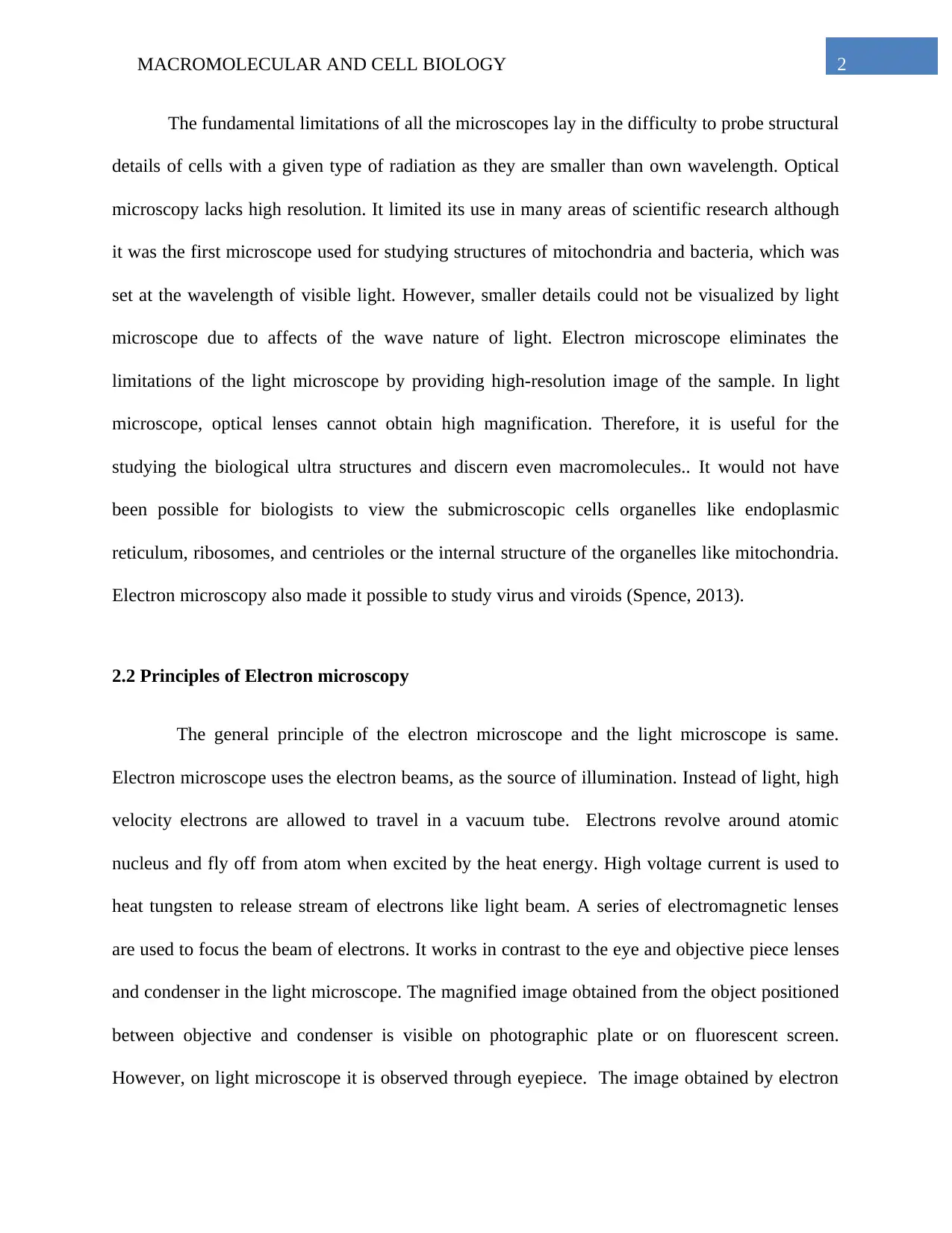
microscope has shades of black, grey or white as electrons are colorless. Fluorescent screen is
used to observe the final image as electrons cannot be viewed by naked eye (De Boer et al.,
2015).
Cross section of electron microscope (Source: http://www.visualdictionaryonline.com)
The wavelengths of the electrons are 100,000 times shorter than the visible light.
Therefore, it has high resolution to detect details of smaller objects, even with small numerical
aperture. The resolving power increases with the decreasing wavelength of light. For instance
Secure Best Marks with AI Grader
Need help grading? Try our AI Grader for instant feedback on your assignments.
4MACROMOLECULAR AND CELL BIOLOGY
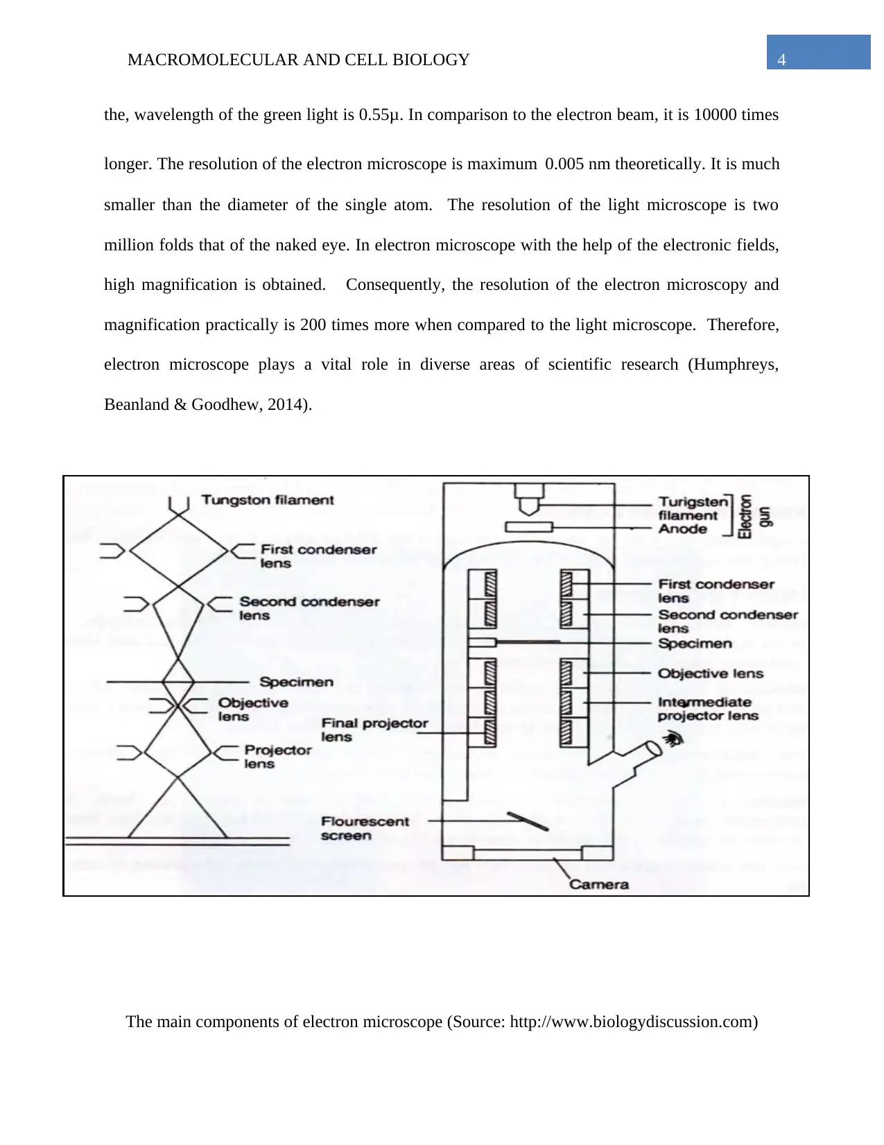
the, wavelength of the green light is 0.55μ. In comparison to the electron beam, it is 10000 times
longer. The resolution of the electron microscope is maximum 0.005 nm theoretically. It is much
smaller than the diameter of the single atom. The resolution of the light microscope is two
million folds that of the naked eye. In electron microscope with the help of the electronic fields,
high magnification is obtained. Consequently, the resolution of the electron microscopy and
magnification practically is 200 times more when compared to the light microscope. Therefore,
electron microscope plays a vital role in diverse areas of scientific research (Humphreys,
Beanland & Goodhew, 2014).
The main components of electron microscope (Source: http://www.biologydiscussion.com)
5MACROMOLECULAR AND CELL BIOLOGY
6MACROMOLECULAR AND CELL BIOLOGY
3.0 Biological Application of Electronic microscopy
3.1. View of Golgi apparatus
The significant advantage of the electron microscopy is its powerful magnification, which was
developed for its use in scientific research fascinated by complex three-dimensional
morphology. One of the applications of the electron microscopy is the study of the Golgi
apparatus by Han et al. (2013).
Visualizing the Golgi apparatus was based on the principle of direct visualization of the
biological material that is frozen hydrated by the electron microscopy. This helped detect the
various structures in the native environment. The preparation method for Golgi apparatus is
delicate as it is prone to fixation artifacts. Cryo-fixation method was chosen to better understand
the morphology of Golgi and its function. Using this method the Guo & Jiang (2014) describes
the possibility of arresting all the cellular processes. The dynamic process inside small cells and
tissues could be arrested with this microscope. Han et al. (2013) analyzed the Golgi apparatus by
the enclosing in the amorphous ice which can be processed by the freeze substitution method. It
can also be analyzed using directly in hydrated state. It can be investigated using the
tomography and the cryo-elctron microscopy. If not exactly like the native structure, the resin
embedded sample can better reveal the previously unknown structural details.
The technique starts with simple negative staining materials on the electron microscope
grids followed by plunge freezing of sample for its stabilization in vitreous ice. Here massive
temperature drops all the processes in the sample, which then looks like glass in high viscosity.
Cooling rate is reduced by the high-pressure freezing or plunged directly into cryogen. Later in
Paraphrase This Document
Need a fresh take? Get an instant paraphrase of this document with our AI Paraphraser
7MACROMOLECULAR AND CELL BIOLOGY
fully hydrate state the sample is examined in cryo-electron microscopic. The resolution of
biological specimen is determined to near atomic level of 0.33–0.46 nm and was considered
recent improvement. However it was limited to only examination of viral capsid proteins.
Similarly, single-electron counting detectors were successful for ribosomes and proteomes.
However, it was not completely successful for Golgi apparatus. Yet the single particle analysis
facilitated the understanding of the architecture of the membrane bound vesicles containing coats
on their interaction with electron beam (such as Clatharin) as well as cisternal buds. This
technique helped to understand the Sec13/31 COPII at (3 nm resolution) coat architecture which
contains cytoplasmic coat proteins. This mediates the intracellular transportation of cargo and
lipids. Further, the flexibility during formation of coat and its assembly was better determined by
combining mass spectroscopy and cryo-electron microscopy (at 1.2 nm resolution). In short the
cryo-fixation technique including the high-pressure freeze fixation combined with the freeze
substitution allowed observation of the constricted tubules by COPII like beads on string (Han et
al., 2013). Overall, it was possible by the electron microscopy to detect Golgi saccules after
inducing the secretion of procollagen in vitreous ice-embedded samples
8MACROMOLECULAR AND CELL BIOLOGY
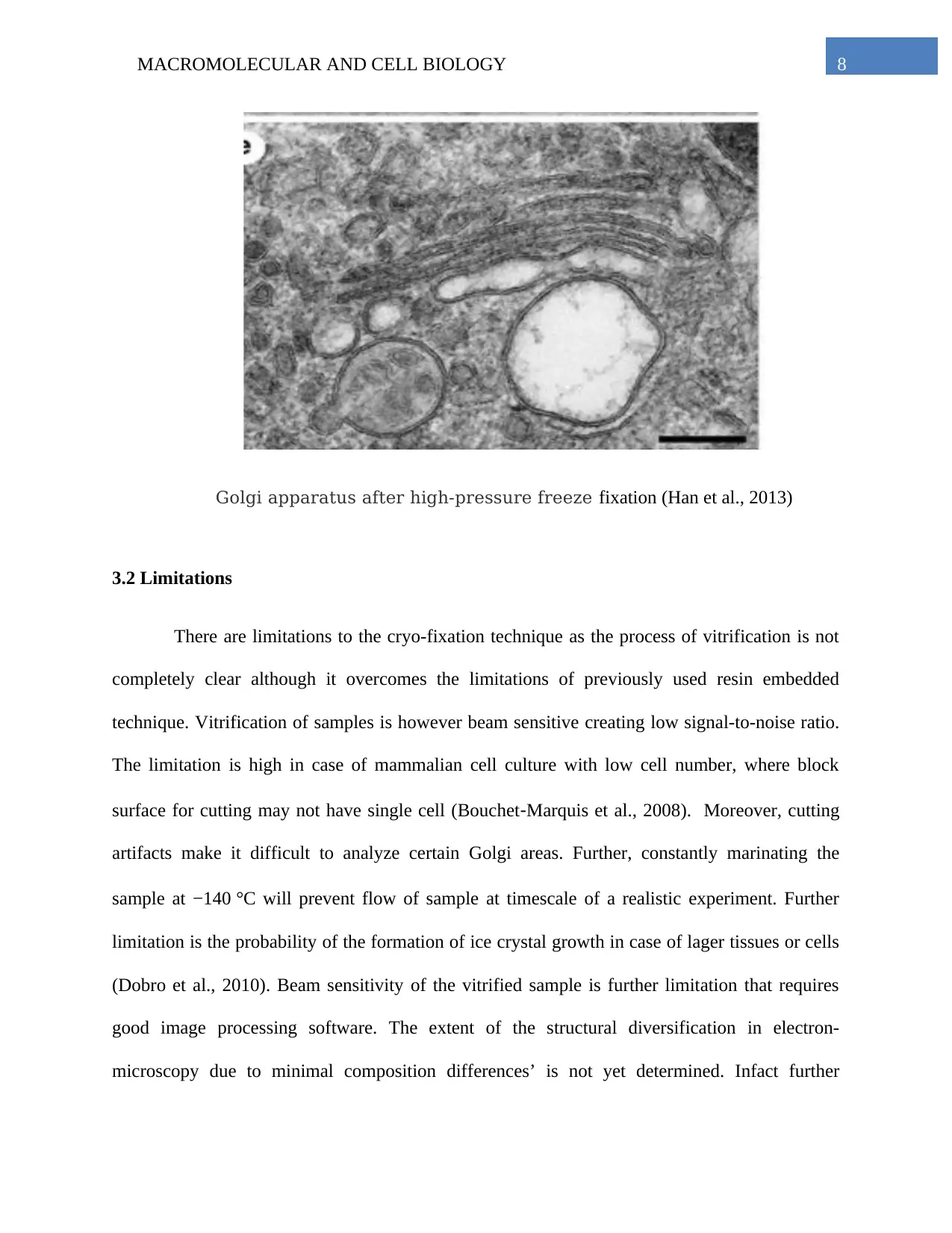
Golgi apparatus after high-pressure freeze fixation (Han et al., 2013)
3.2 Limitations
There are limitations to the cryo-fixation technique as the process of vitrification is not
completely clear although it overcomes the limitations of previously used resin embedded
technique. Vitrification of samples is however beam sensitive creating low signal-to-noise ratio.
The limitation is high in case of mammalian cell culture with low cell number, where block
surface for cutting may not have single cell (Bouchet‐Marquis et al., 2008). Moreover, cutting
artifacts make it difficult to analyze certain Golgi areas. Further, constantly marinating the
sample at −140 °C will prevent flow of sample at timescale of a realistic experiment. Further
limitation is the probability of the formation of ice crystal growth in case of lager tissues or cells
(Dobro et al., 2010). Beam sensitivity of the vitrified sample is further limitation that requires
good image processing software. The extent of the structural diversification in electron-
microscopy due to minimal composition differences’ is not yet determined. Infact further
9MACROMOLECULAR AND CELL BIOLOGY
modifications are needed to fixation and labeling technique to view the molecular interactions in
Golgi despite so much advancement in electron microscopy, for better describing the secretory
pathway in its native state (Li et al., 2013, Han et al., 2013). In conclusion, the decipher of
ultrastructure of a functioning Golgi may be way to go as it may require good combination of
high resolution light microscope with electron and cryo-electron microscopy.
Secure Best Marks with AI Grader
Need help grading? Try our AI Grader for instant feedback on your assignments.
10MACROMOLECULAR AND CELL BIOLOGY
4.0 References
BOUCHET‐MARQUIS, C., Starkuviene, V., & Grabenbauer, M. (2008). Golgi apparatus
studied in vitreous sections. Journal of microscopy, 230(2), 308-316.
De Boer, P., Hoogenboom, J. P., & Giepmans, B. N. (2015). Correlated light and electron
microscopy: ultrastructure lights up!. Nature methods, 12(6), 503.
Dobro, M. J., Melanson, L. A., Jensen, G. J., & McDowall, A. W. (2010). Plunge freezing for
electron cryomicroscopy. In Methods in enzymology (Vol. 481, pp. 63-82). Academic
Press.
Guo, F., & Jiang, W. (2014). Single particle cryo-electron microscopy and 3-D reconstruction of
viruses. In Electron Microscopy (pp. 401-443). Humana Press, Totowa, NJ.
Han, H. M., Bouchet-Marquis, C., Huebinger, J., & Grabenbauer, M. (2013). Golgi apparatus
analyzed by cryo-electron microscopy. Histochemistry and cell biology, 140(4), 369-381.
Humphreys, J., Beanland, R., & Goodhew, P. J. (2014). Electron microscopy and analysis. Third
Edition. London: CRC Press. ISBN: 9781482289343
Li, X., Mooney, P., Zheng, S., Booth, C. R., Braunfeld, M. B., Gubbens, S., ... & Cheng, Y.
(2013). Electron counting and beam-induced motion correction enable near-atomic-
resolution single-particle cryo-EM. Nature methods, 10(6), 584.
Pease, D. C. (2013). Histological techniques for electron microscopy. Elsevier. Retrieved from:
https://books.google.co.in/books?
hl=en&lr=&id=LCzLBAAAQBAJ&oi=fnd&pg=PP1&dq=Histological+techniques+for+
11MACROMOLECULAR AND CELL BIOLOGY
electron+microscopy&ots=xM_9vDuPaF&sig=boRiSpzr0U_6SP0h-
68uvf31GhY&redir_esc=y#v=onepage&q=Histological%20techniques%20for
%20electron%20microscopy&f=false
Spence, J. C. (2013). High-resolution electron microscopy. OUP Oxford. Retrieved from:
https://books.google.co.in/books?
hl=en&lr=&id=PitoAgAAQBAJ&oi=fnd&pg=PP1&dq=electron+microscopy&ots=ecgz
cU-1U8&sig=iBTIxl92p9_zky3cDg22jWLSQ10&redir_esc=y#v=onepage&q=electron
%20microscopy&f=false
1 out of 12
Related Documents


Your All-in-One AI-Powered Toolkit for Academic Success.
+13062052269
info@desklib.com
Available 24*7 on WhatsApp / Email
![[object Object]](/_next/static/media/star-bottom.7253800d.svg)
Unlock your academic potential
© 2024 | Zucol Services PVT LTD | All rights reserved.