Virology Practical Assignment: PCR Analysis of Viral DNA in HeLa Cells
VerifiedAdded on 2022/08/21
|11
|2156
|17
Practical Assignment
AI Summary
This practical assignment focuses on the identification of viruses in infected cells using Polymerase Chain Reaction (PCR) and agarose gel electrophoresis. The experiment aims to detect the presence of Human Papillomavirus (HPV) DNA in HeLa cells, derived from cervical cancer patients, and compare the results with a control sample (MRC5 cells). The methodology involves extracting genomic DNA, amplifying HPV DNA using specific primers, and visualizing the amplified DNA fragments through gel electrophoresis. The results indicate the presence of HPV DNA in HeLa cells, confirming the theory that cervical cancer patients have HPV genome in their cell lines. The study concludes by emphasizing the importance of these techniques in virology for identifying and quantifying viral DNA, and suggests potential improvements for future studies, such as using quantitative RT-PCR.
Running head: VIROLOGY
PCR AND AGAROSE GEL ELECTROPHORESIS (VIRUS)
Name of the Student
Name of the University
Author Note
PCR AND AGAROSE GEL ELECTROPHORESIS (VIRUS)
Name of the Student
Name of the University
Author Note
Paraphrase This Document
Need a fresh take? Get an instant paraphrase of this document with our AI Paraphraser
1
VIROLOGY
Abstract
One of the most significantly powerful technique for the detection of viral genomes is PCR
(Polymerase chain reaction). PCR has been found to be sensitive and specific for the
detection of new viral subtypes. (). HeLa cell lines were collected from patients of cervical
cancer. These cells contained the DNA of HPV virus which was amplified by PCR for
visualization. This experiment was based on the above-stated theory. However, this
experiment was performed to prove the theory that human patients affected by cervical
cancer consist of HPV genome in their cell lines. On a concluding note, it can be stated that
the human DNA (HeLa) cells collected for the experiment were infected with HPV.
Table of Content
2
VIROLOGY
s
Introduction................................................................................................................................3
Methods......................................................................................................................................4
Part 1 (PCR)...........................................................................................................................4
Part 2 (Agarose gel electrophoresis)......................................................................................4
Results........................................................................................................................................5
Discussion..................................................................................................................................6
Conclusion..................................................................................................................................7
References..................................................................................................................................8
⊘ This is a preview!⊘
Do you want full access?
Subscribe today to unlock all pages.
Trusted by 1+ million students worldwide
3
VIROLOGY
Introduction
Viruses are very minute and infectious agents which replicate inside living cells only.
Virology is defined as the main branch of science which deals with the study of genetic
materials associated with viral particles. This study focusses on the structure, evolution,
classification, modes of infection and the reproduction procedure of viruses (Richman,
Whitley and Hayden 2016). Viruses are both lysogenic and lytic in nature. Since viral
infections are very hard to treat, the infections have been found to have a terrible impact on
human beings. Thus, virology has been found to be an important field of study nowadays.
These minute organisms have been found to infect all the life forms including animals, plants,
human beings and bacteria, archaea also. One of the most significantly powerful technique
for the detection of viral genomes is PCR (Polymerase chain reaction) (Liu et al. 2017). PCR
has been found to be sensitive and specific for the detection of new viral subtypes. PCR is
defined as a method which rapidly forms a large number (millions) of copies of a specific
DNA In the general procedure, viral DNA is first extracted by RNA extraction procedure
from a biological sample and after its conversion to cDNA, PCR is run (Souiriet al. 2017).
Three significant steps are followed for a PCR which involves heat denaturation, annealing,
extension and end of the first PCR cycle. Finally, the amplified DNA is detected by agarose
gel electrophoresis. Papillomavirus has been found to cause cervical cancer which has been
considered as a major problem to public health (Schiffman et al. 2016). HeLa cells have been
derived from cells of cervical cancer. HeLa cells have been found to be an immortal cell line
which has been used in various scientific research studies (Thierry and Yaniv, 2018). This
cell line has been considered to be the oldest and has been derived from cervical cancer cells
which have been first taken from a patient of cervical cancer. These cells have been found to
be durable and highly prolific in nature that gives rise to their extensive use in scientific
research studies (Ma and Poon, 2017). A very small amount of sample DNA has been found
Paraphrase This Document
Need a fresh take? Get an instant paraphrase of this document with our AI Paraphraser
4
VIROLOGY
to be amplified into a very large amount for an in-detail study. This experiment will also use
the same process of PCR followed by agarose gel electrophoresis in order to identify the
presence of viral DNA in the sample.
Methods
Part 1 (PCR)
Genomic DNA was extracted from HeLa cells and MRC5 cells. Oligonucleotide
primers against HPV-18 genome are then generated. PCR buffer was prepared followed by
the use of Taq polymerase and sterilized water for the preparation of master mix. After the
preparation of master mix, PCR was run and after 30 rounds of PCR cycle, the whole
apparatus was allowed to cool at four-degree centigrade.
HPV forward primer ; 'HPV 18F'
5' ATG GCG CGC TTT GAG GAT CC-3'
HPV reverse primer :'HPV 18R'
5' GCA TGC GGT ATA CTG TCT CT-3'
Part 2 (Agarose gel electrophoresis)
The three tubes which contain the PCR products were analysed by agarose gel
electrophoresis. After the electrophoresis was completed the gel was removed and observed
under a UV transilluminator. The images have been given under the results section.
5
VIROLOGY
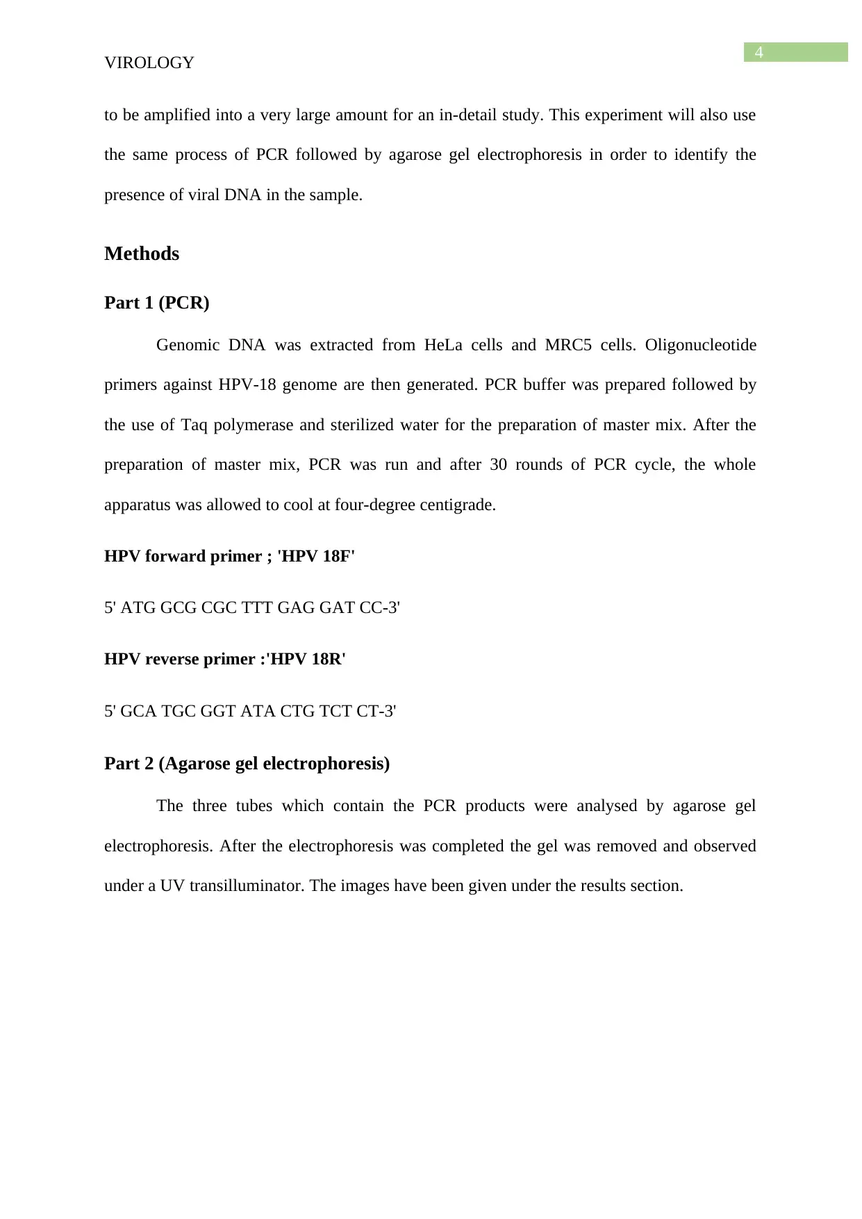
Results
Fig 1: PCR gel
Source: (Practical week 2)
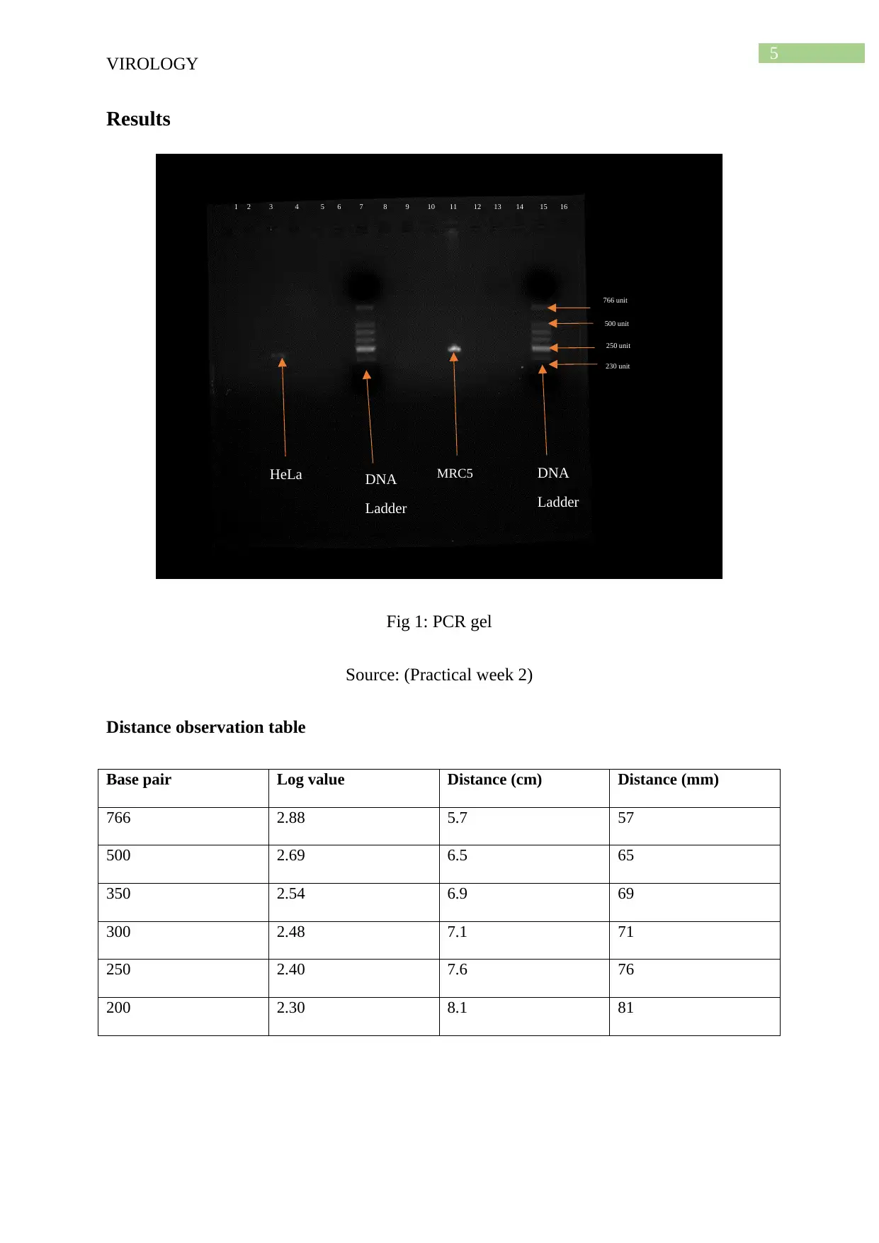
Distance observation table
Base pair Log value Distance (cm) Distance (mm)
766 2.88 5.7 57
500 2.69 6.5 65
350 2.54 6.9 69
300 2.48 7.1 71
250 2.40 7.6 76
200 2.30 8.1 81
DNA
Ladder
DNA
Ladder
766 unit
500 unit
250 unit
230 unit
MRC5HeLa
1 2 3 4 5 6 7 8 9 10 11 12 13 14 15 16
VIROLOGY
Results
Fig 1: PCR gel
Source: (Practical week 2)
Distance observation table
Base pair Log value Distance (cm) Distance (mm)
766 2.88 5.7 57
500 2.69 6.5 65
350 2.54 6.9 69
300 2.48 7.1 71
250 2.40 7.6 76
200 2.30 8.1 81
DNA
Ladder
DNA
Ladder
766 unit
500 unit
250 unit
230 unit
MRC5HeLa
1 2 3 4 5 6 7 8 9 10 11 12 13 14 15 16
⊘ This is a preview!⊘
Do you want full access?
Subscribe today to unlock all pages.
Trusted by 1+ million students worldwide
6
VIROLOGY
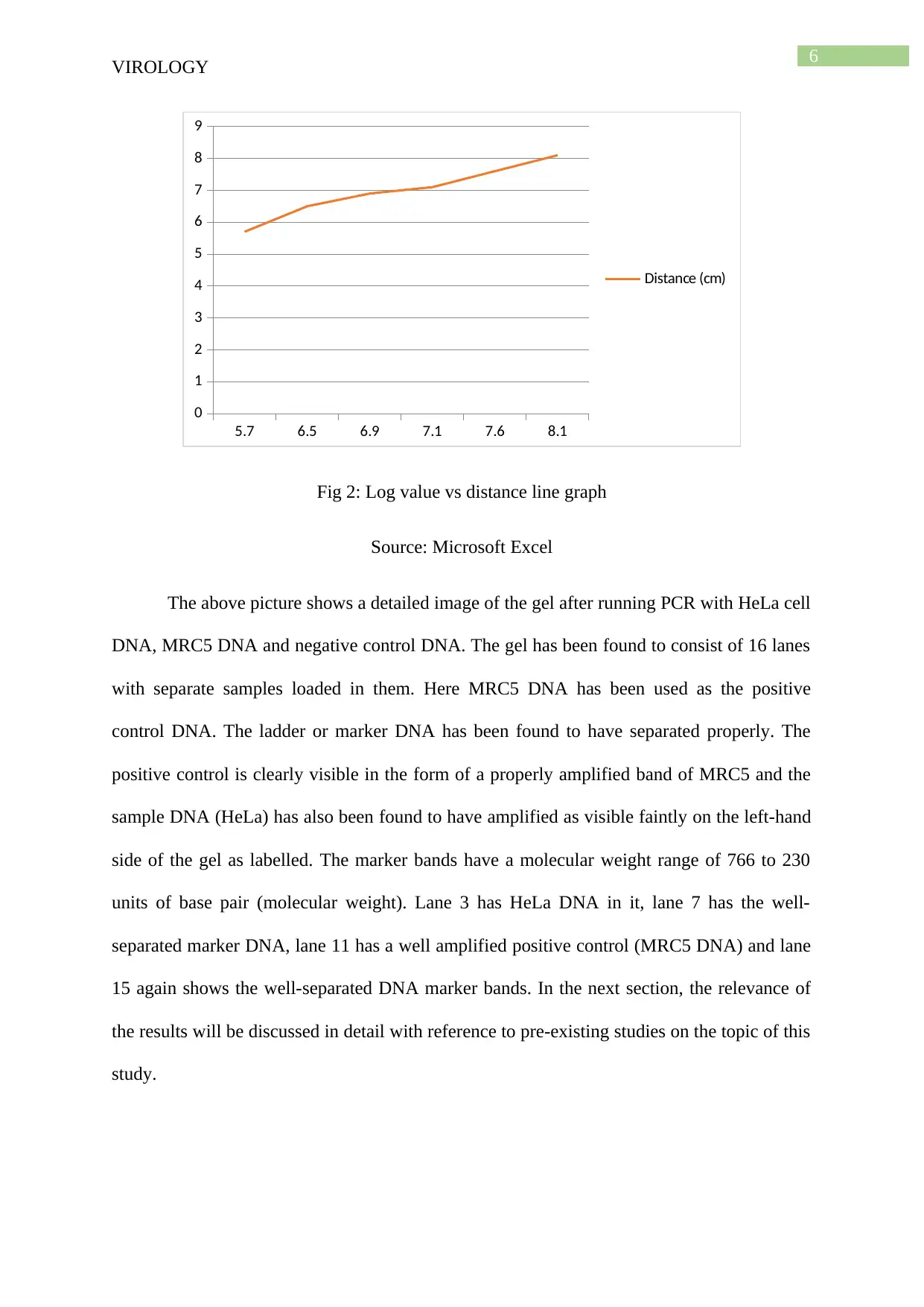
5.7 6.5 6.9 7.1 7.6 8.1
0
1
2
3
4
5
6
7
8
9
Distance (cm)
Fig 2: Log value vs distance line graph
Source: Microsoft Excel
The above picture shows a detailed image of the gel after running PCR with HeLa cell
DNA, MRC5 DNA and negative control DNA. The gel has been found to consist of 16 lanes
with separate samples loaded in them. Here MRC5 DNA has been used as the positive
control DNA. The ladder or marker DNA has been found to have separated properly. The
positive control is clearly visible in the form of a properly amplified band of MRC5 and the
sample DNA (HeLa) has also been found to have amplified as visible faintly on the left-hand
side of the gel as labelled. The marker bands have a molecular weight range of 766 to 230
units of base pair (molecular weight). Lane 3 has HeLa DNA in it, lane 7 has the well-
separated marker DNA, lane 11 has a well amplified positive control (MRC5 DNA) and lane
15 again shows the well-separated DNA marker bands. In the next section, the relevance of
the results will be discussed in detail with reference to pre-existing studies on the topic of this
study.
VIROLOGY
5.7 6.5 6.9 7.1 7.6 8.1
0
1
2
3
4
5
6
7
8
9
Distance (cm)
Fig 2: Log value vs distance line graph
Source: Microsoft Excel
The above picture shows a detailed image of the gel after running PCR with HeLa cell
DNA, MRC5 DNA and negative control DNA. The gel has been found to consist of 16 lanes
with separate samples loaded in them. Here MRC5 DNA has been used as the positive
control DNA. The ladder or marker DNA has been found to have separated properly. The
positive control is clearly visible in the form of a properly amplified band of MRC5 and the
sample DNA (HeLa) has also been found to have amplified as visible faintly on the left-hand
side of the gel as labelled. The marker bands have a molecular weight range of 766 to 230
units of base pair (molecular weight). Lane 3 has HeLa DNA in it, lane 7 has the well-
separated marker DNA, lane 11 has a well amplified positive control (MRC5 DNA) and lane
15 again shows the well-separated DNA marker bands. In the next section, the relevance of
the results will be discussed in detail with reference to pre-existing studies on the topic of this
study.
Paraphrase This Document
Need a fresh take? Get an instant paraphrase of this document with our AI Paraphraser
7
VIROLOGY
Discussion
The main purpose of this experiment was to check whether there were any viral
materials present in the collected DNA or not. For analysing this fact, gel electrophoresis was
used to visualize the viral DNA associated with HeLa cells collected from the patients of
cervical cancer. Various studies associated with this topic has been studied previously.DNA
ladder has been used in order to measure the molecular weight of the target DNA which has
amplified after the PCR reaction (Lertworapreecha and Thongnan 2018). The amplification
has been found to be similar to the positive control used for this experiment. A positive
control is used to verify the result of an experiment. The negative control taken for this study
has not amplified since the viral DNA primers failed to bind it and amplify the strands. The
positive control used for this experiment is MRC-5 DNA obtained from a cell culture line
which is composed of fibroblasts. This DNA has been obtained from 14-week old male fetus.
There are various research studies which sued Q-RTPCR in order to measure the amount of
contained viral DNA in the sample cells (Barr et al. 2017). This process further makes the
process of quantification easier than it is for this experiment. The amplification size of MRC5
has been found to be similar to HeLa cells. This factor has been found to be studied in
various research studies (Huang et al. 2016). HeLa cell lines were collected from patients of
cervical cancer. These cells contained the DNA of HPV virus which was amplified by PCR
for visualization. This experiment was based on the above-stated theory. However, this
experiment was performed to prove the theory that human patients affected by cervical
cancer consist of HPV genome in their cell lines. This theory has been proved to be
applicable for this experiment because it has been proved that the collected HeLa cells
contained viral DNA. From the results section, it is evident that the viral DNA has amplified
to 230 units base-pair length starting from the top of the marker. This amplification has been
found to be similar for the positive control taken for this gel experiment. The primers used for
8
VIROLOGY
this experiment were against the viral genome (Tianet al. 2018). Thus, it can be stated that
these primers attached themselves with the complementary region inside the HeLa DNA and
amplified the whole DNA as visible in the gel. Future studies on this topic can be improved
by performing a quantitative RT PCR before performing the final PCR reaction for the
visualization of bands.
Conclusion
After the long discussion, it can be stated that the human DNA (HeLa) cells collected
for the experiment were infected with HPV. This experiment proved that these cells contain
the viral DNA segment which got amplified and was visualized in the gel. Further
experiments with this can be done by slicing out the viral DNA carefully with a gel cutter and
performing several quantification experiments. These experiments are important because of
the fact that the amount of viral DNA plays a major role during the measurement of viral load
in the blood. This experiment followed the preliminary procedure of viral DNA identification
in a particular cell line. The main strength of this experiment is that it successfully identified
the fact that HeLa cells collected from cervical cancer patients contain the DNA of HPV.
Both the PCR reaction and agarose gel electrophoresis was successful in supporting the
previous statement.
⊘ This is a preview!⊘
Do you want full access?
Subscribe today to unlock all pages.
Trusted by 1+ million students worldwide
9
VIROLOGY
References
Barr, J.L., Thomas, T.W., Gibson, R.S., Dubick, M.A. and Bowman, P.D., 2017. Quantitative
Real Time PCR (qRT-PCR) Evaluation of Pig Mitochondrial DNA Damage. The FASEB
Journal, 31(1_supplement), pp.470-6.
Huang, M., Yang, H., Zhu, L., Li, H., Zhou, J. and Zhou, Z., 2016. Inhibition of connective
tissue growth factor attenuates paraquat‐induced lung fibrosis in a human MRC‐5 cell
line. Environmental toxicology, 31(11), pp.1620-1626.
Lertworapreecha, M. and Thongnan, J., 2018. Construction of Recombinant DNA Plasmid
for Production 100 Base Pair DNA Ladder for Endless Usage Based on PCR
Technique. Genomics and Genetics, 11(1&2), pp.22-25.
Liu, C., Chang, L., Jia, T., Guo, F., Zhang, L., Ji, H., Zhao, J. and Wang, L., 2017. Real-time
PCR assays for hepatitis B virus DNA quantification may require two different
targets. Virology journal, 14(1), p.94.
Ma, H.T. and Poon, R.Y., 2017. Synchronization of HeLa cells. In Cell Cycle
Synchronization (pp. 189-201). Humana Press, New York, NY.
Richman, D.D., Whitley, R.J. and Hayden, F.G. eds., 2016. Clinical virology. John Wiley &
Sons.
Schiffman, M., Doorbar, J., Wentzensen, N., De Sanjosé, S., Fakhry, C., Monk, B.J., Stanley,
M.A. and Franceschi, S., 2016. Carcinogenic human papillomavirus infection. Nature
reviews Disease primers, 2(1), pp.1-20.
Souiri, A., Zemzami, M., Khataby, K., Laatiris, H. and Amzazi, S., 2017. A Simple, Rapid
and Efficient Method of Pepino mosaic Virus RNA Isolation from Tomato Fruit. J Plant
PatholMicrobiol, 8(395), p.2.
Paraphrase This Document
Need a fresh take? Get an instant paraphrase of this document with our AI Paraphraser

10
VIROLOGY
Thierry, F. and Yaniv, M., 2018. The control of human papillomavirus transcription. In Viral
Regulatory Structures And Their Degeneracy (pp. 53-70). CRC Press.
Tian, C.E., Hong, T.W., Zhou, Y.P., Chen, Q.H., Huang, X.L. and Guo, X.Y., 2018. The
Length Limit of 5′ Nucleotide Additions to PCR Primers. National Academy Science
Letters, 41(4), pp.207-210.
1 out of 11
Related Documents


Your All-in-One AI-Powered Toolkit for Academic Success.
+13062052269
info@desklib.com
Available 24*7 on WhatsApp / Email
![[object Object]](/_next/static/media/star-bottom.7253800d.svg)
Unlock your academic potential
Copyright © 2020–2026 A2Z Services. All Rights Reserved. Developed and managed by ZUCOL.